
Role of the IGFBP Family in IGF Signaling Regulation and Disease Biomarker Research
Role of the IGFBP Family in IGF Signaling Regulation and Disease Biomarker Research
The IGFBP family regulates the stability, tissue distribution, and receptor accessibility of IGFs, participates in metabolism, fibrosis, cardiovascular diseases, and tumor progression, and has potential value as disease biomarkers.
Keywords: IGFBP; IGF-I; IGF-II; IGF1R; bioavailability; PAPP-A; autoimmune diseases; cardiovascular diseases; tumor biomarkers; treatment response
1 Composition and Molecular Features of the IGFBP Family
1.1 Classical IGFBP Family Members
The classical insulin-like growth factor binding protein family includes IGFBP1, IGFBP2, IGFBP3, IGFBP4, IGFBP5, and IGFBP6. All six proteins can bind IGF-I or IGF-II with high affinity, but they differ in tissue source, ligand preference, proteolytic sensitivity, and extracellular matrix binding capacity. IGFBP7 is commonly used in tumor, cardiovascular disease, and organ injury biomarker research, but its binding affinity for IGF is markedly lower than that of IGFBP1-6. It belongs to the IGFBP-related protein group and should not be fully equated with the six classical high-affinity IGFBPs.
1.2 Structural Composition of IGFBPs
Classical IGFBPs usually consist of a conserved N-terminal domain, a variable central linker region, and a conserved C-terminal domain.
(1) N-terminal domain
The N-terminal domain is rich in conserved cysteines and is an important region for forming the high-affinity IGF-binding interface.
(2) Central linker region
The central linker region shows greater sequence variation and contains phosphorylation, glycosylation, and protease cleavage sites, which can regulate IGFBP stability, ligand affinity, and tissue distribution.
(3) C-terminal domain
The C-terminal domain participates in IGF binding and interactions with cell surfaces, extracellular matrix, and other proteins. Some members also contain integrin-binding sequences or nuclear localization-related structures.
1.3 Functional Differences Among IGFBP Family Members
Family Member | Main Molecular Features | IGF Regulatory Features | Key Research Directions |
IGFBP1 | Mainly produced by the liver and rapidly regulated by insulin and nutritional status | Phosphorylation can enhance its binding to IGF-I and regulate free IGF levels | Insulin resistance, pregnancy, and metabolic diseases |
IGFBP2 | Contains an RGD sequence and is highly expressed in embryonic tissues and many tumors | Can restrict IGF binding to receptors and may also promote local IGF delivery | Tumor invasion, metabolic abnormalities, and nervous system diseases |
IGFBP3 | Highly abundant in circulation and can form a ternary complex with IGF and ALS | Stabilizes circulating IGF and also regulates cell cycle and apoptosis | Growth evaluation, tumors, and cardiovascular diseases |
IGFBP4 | Locally expressed in multiple tissues and is an important substrate of PAPP-A | Intact protein usually restricts IGF1R activation, while cleavage releases IGF | Vascular remodeling, bone metabolism, reproduction, and tumors |
IGFBP5 | Has strong extracellular matrix binding capacity | Participates in local IGF storage, tissue localization, and release | Fibrosis, aging, muscle, and tumor stroma |
IGFBP6 | Has relatively high binding preference for IGF-II | Mainly restricts IGF-II-dependent cell growth | Development, tumors, and vascular inflammation |
2 IGFBP-Mediated IGF Transport and Bioavailability Regulation
2.1 Stable Transport of Circulating IGF
Most IGF in blood is bound to IGFBPs rather than existing in a free form. After IGFBP3 binds IGF, it can further recruit the acid-labile subunit (ALS) to form a large ternary complex. IGFBP5 can also participate in similar complex formation under certain conditions. The ternary complex can prolong the circulating half-life of IGF and restrict its rapid passage across vascular endothelium, whereas free IGF and smaller IGF-IGFBP binary complexes more easily enter the interstitial space. Therefore, serum total IGF levels cannot directly represent bioactive IGF that can immediately activate IGF1R.
2.2 IGFBP Proteolysis and Local IGF Release
After IGFBPs are cleaved by proteases, their binding capacity for IGF usually decreases, allowing IGF to be released locally in tissues and approach IGF1R.
(1) PAPP-A
PAPP-A mainly cleaves IGFBP4. Its proteolytic activity usually depends on formation of the IGF-IGFBP4 complex and can increase local IGF bioavailability.
(2) PAPP-A2
PAPP-A2 mainly cleaves IGFBP3 and IGFBP5 and participates in regulation of IGF release in circulation and tissues.
(3) Other proteases
Matrix metalloproteinases, serine proteases, and cathepsins can cleave IGFBPs in inflammatory, tumor, and tissue injury environments, altering the ratio of intact proteins to cleaved fragments.
2.3 IGF Localization in the Extracellular Matrix
IGFBP2, IGFBP3, and IGFBP5 can bind cell surface molecules or extracellular matrix components, enriching IGF in specific tissue regions. Matrix binding can temporarily sequester IGF and limit receptor activation, but it can also form a local IGF release reservoir after proteolysis or matrix remodeling. The effect of IGFBPs on IGF signaling depends on the ratio of intact protein to cleaved fragments, IGF concentration, protease activity, extracellular matrix status, and IGF1R expression. It cannot be uniformly classified as promoting or inhibiting IGF signaling.
3 IGF-Dependent and IGF-Independent Cellular Effects of IGFBPs
3.1 IGF-Dependent Signal Regulation
IGFBPs alter IGF1R and downstream PI3K-AKT-mTOR and RAS-RAF-MEK-ERK signaling by regulating IGF stability, tissue localization, and release.
(1) When intact IGFBPs form stable complexes with IGF, they can reduce free IGF concentration and restrict IGF1R activation.
(2) When IGFBPs transport IGF to the cell surface or extracellular matrix, local IGF concentration may increase.
(3) After IGFBPs are cleaved by proteases, IGF can be released and receptor signaling can be enhanced.
(4) Insufficient IGFBP expression may also reduce IGF stability and long-distance transport capacity.
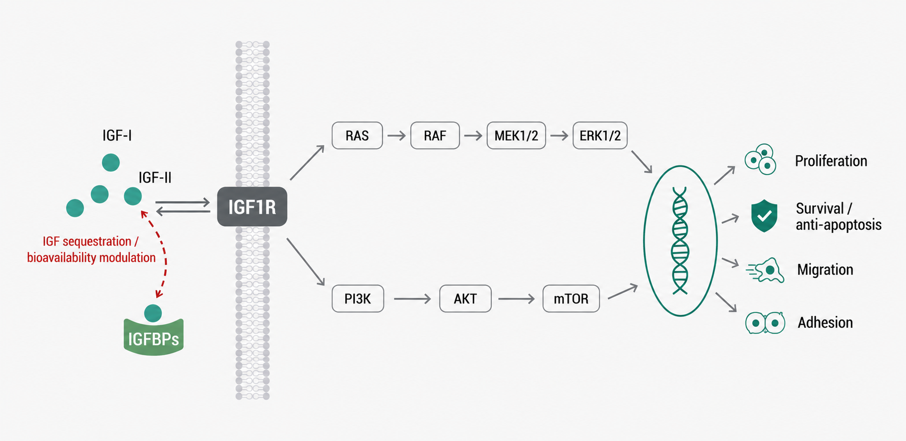
Figure 1. Schematic of IGFBP-Mediated Regulation of IGF–IGF1R Signaling and Downstream Cellular Effects
3.2 IGF-Independent Effects
(1) IGFBP2
IGFBP2 can interact with integrins through its RGD sequence and regulate adhesion, migration, PI3K-AKT signaling, and therapy tolerance in some tumor cells.
(2) IGFBP3
IGFBP3 can interact with cell surface proteins, nuclear molecules, and apoptosis regulatory networks, affecting the cell cycle, DNA damage response, and apoptosis.
(3) IGFBP5
IGFBP5 can bind the extracellular matrix and undergo nuclear translocation, participating in cell migration, senescence, fibrosis, and tissue remodeling.
(4) IGFBP6
In addition to preferentially binding IGF-II, IGFBP6 can also affect migration, differentiation, and vascular inflammation in some cells.
3.3 Influence of Cellular Context on IGFBP Effects
The same IGFBP may produce different or even opposite effects in different cells and diseases. IGFBP3 can restrict IGF signaling and promote apoptosis in some tumors, but may also support cell survival through matrix binding or nuclear actions in specific microenvironments. IGFBP5 can participate in normal tissue repair, but sustained expression may promote extracellular matrix deposition and fibrosis.
4 Abnormal IGFBP Expression and Disease Development
4.1 Growth and Metabolic Abnormalities
IGFBP1 is rapidly inhibited by insulin, and its circulating level can reflect hepatic insulin action, nutritional status, and free IGF regulation. Insulin resistance and hyperinsulinemia are often accompanied by decreased IGFBP1, but liver function, inflammation, and pregnancy status can also affect detection results.
IGFBP2 is associated with fat distribution, insulin sensitivity, and hepatic metabolism. Some obese and metabolically abnormal populations may show decreased circulating IGFBP2, while increased levels may also appear in aging, wasting diseases, and chronic organ dysfunction.
4.2 Chronic Inflammation and Tissue Fibrosis
Chronic inflammation can alter IGFBP expression, secretion, and proteolysis, shifting IGF signaling from physiological growth regulation toward sustained repair and matrix deposition. IGFBP3 and IGFBP5 can participate in fibroblast activation, extracellular matrix binding, and tissue remodeling. IGFBP7 is often associated with cellular senescence, endothelial stress, and fibrotic states.
4.3 Organ Injury and Cellular Stress
Changes in IGFBP levels can reflect cell cycle arrest, endothelial injury, metabolic stress, and tissue repair. Combined detection of IGFBP7 and TIMP-2 can be used for acute kidney injury risk assessment, but this combination mainly reflects tubular cell stress and cannot directly represent overall enhancement or weakening of classical IGF signaling.
5 Biomarker Value of IGFBPs in Autoimmune and Cardiovascular Diseases
5.1 Autoimmune Diseases
Systemic lupus erythematosus, lupus nephritis, rheumatoid arthritis, systemic sclerosis, and other diseases can be accompanied by altered IGFBP expression. These changes may originate from immune inflammation, kidney injury, endothelial stress, or fibrosis. In lupus nephritis research, serum or plasma IGFBP4 can be investigated as an exploratory biomarker of renal involvement, disease activity, or pathological changes. IGFBP2, IGFBP3, and IGFBP7 can also be used for stratification of inflammation, tissue injury, and fibrosis, but at this stage they are more suitable as combined biomarkers and cannot replace autoantibodies, complement, urinary protein, and histopathological evaluation.
5.2 Cardiovascular Diseases
IGFBP1 and IGFBP2 can reflect metabolic status and systemic stress. IGFBP3, together with IGF-I, reflects the growth hormone-IGF axis. IGFBP4 and its cleavage fragments can reflect PAPP-A-related proteolysis and local IGF release. IGFBP7 is commonly studied in heart failure, coronary artery disease, atrial remodeling, and cardiorenal dysfunction. Its elevation may simultaneously reflect myocardial stress, endothelial dysfunction, inflammation, and renal function decline, making it unsuitable as a specific indicator of a single pathological process.
5.3 Interpretation of Biomarker Results
IGFBP levels are affected by age, sex, nutritional status, liver and kidney function, growth hormone, insulin, and systemic inflammation. Disease biomarker research should combine IGF-I, IGF-II, related IGFBPs, protease activity, and organ function indicators, avoiding direct interpretation of a single IGFBP increase or decrease as enhanced or weakened IGF signaling.
6 Applications of IGFBPs in Tumor Diagnosis, Prognosis, and Treatment Response Evaluation
6.1 Tumor Diagnosis and Stratification
IGFBPs can be produced by tumor cells, stromal cells, endothelial cells, and immune cells. Their blood levels and tissue expression can reflect tumor burden, stromal remodeling, nutritional status, and systemic inflammation. IGFBP2 is often elevated in glioma, ovarian cancer, lung cancer, and some digestive system tumors and can be associated with invasiveness, immunosuppression, and poor prognosis. However, its expression is also affected by age, metabolism, and organ function, so it cannot serve as a tumor-specific diagnostic marker.
6.2 Tumor Prognostic Evaluation
(1) IGFBP2
In some gliomas and solid tumors, IGFBP2 is associated with tumor grade, recurrence risk, and poorer survival.
(2) IGFBP3
IGFBP3 can exert tumor-suppressive effects by restricting IGF signaling, promoting apoptosis, or regulating nuclear responses. Its prognostic direction is influenced by cancer type, expression location, and treatment background.
(3) IGFBP5
IGFBP5 can participate in extracellular matrix remodeling, metastasis, and therapy tolerance, but its prognostic significance differs across tumor types.
(4) IGFBP7
In some tumors, IGFBP7 is associated with cellular senescence and growth inhibition. In other tumors, high expression may be accompanied by stromal activation, immune infiltration, and poor prognosis.
6.3 Treatment Response and Resistance Evaluation
Chemotherapy, radiotherapy, endocrine therapy, and targeted therapy can all alter IGFBP expression. IGFBP2 can enhance integrin signaling, DNA damage tolerance, and cell survival in some tumors. Changes in IGFBP3, IGFBP4, and IGFBP5 may reflect IGF1R signaling reconstruction, stromal changes, or drug-induced cellular state transitions. Treatment response research should distinguish pretreatment predictive biomarkers from post-treatment pharmacodynamic biomarkers and should combine paired samples, functional intervention, and independent cohort validation. A single IGFBP test cannot determine whether it is a driver of resistance or a concomitant result of tumor burden changes.
7 IGFBP-Related Signaling Networks and Targeted Research Progress
7.1 IGF Ligand and IGF1R Intervention
Neutralizing IGF-I or IGF-II, inhibiting IGF1R, and simultaneously regulating IGF1R and insulin receptor-related signaling can reduce activation of the PI3K-AKT and MAPK pathways. The IGF system has ligand redundancy, receptor cross-talk, and metabolic compensation. When IGF1R is blocked alone, survival signaling may be re-established through the insulin receptor, other receptor tyrosine kinases, or downstream pathways.
7.2 Regulation of IGFBP Proteolysis
Inhibition of PAPP-A can reduce IGFBP4 cleavage and restrict local IGF release. Regulation of PAPP-A2 can affect IGF availability in circulation and tissues mediated by IGFBP3 and IGFBP5. Endogenous regulatory proteins such as STC1 and STC2 can inhibit PAPP-A family proteases, providing new intervention nodes for controlling local IGF activity.
7.3 Direct IGFBP Intervention
(1) Using recombinant IGFBPs or protease-resistant IGFBPs to restrict IGF binding to IGF1R.
(2) Blocking interactions between IGFBPs and integrins, extracellular matrix, or other binding proteins through antibodies, peptides, or small molecules.
(3) Reducing abnormal IGFBP expression using siRNA, antisense oligonucleotides, or gene editing.
(4) Regulating promoter methylation, transcription factors, or non-coding RNAs to alter IGFBP transcription and stability.
Most of the above strategies remain at the mechanistic research or early translational stage. Currently, IGFBPs are more suitable to be combined with IGF ligands, IGF1R phosphorylation, protease activity, and disease phenotypes for patient stratification and pharmacodynamic evaluation.
8 Products Related to IGFBP-IGF Signaling and Disease Biomarker Research
Cat. No. | Product Name | Grade & Purity | Research Stage | Main Application |
IGFBP1 Human Pre-designed siRNA Set A |
| IGFBP1 gene intervention | Knocks down IGFBP1 and analyzes its effects on IGF bioavailability, cell proliferation, and metabolic phenotypes | |
Recombinant IGFBP1 Antibody | Recombinant, ExactAb™, validated, see COA | IGFBP1 expression detection | Used for IGFBP1 protein expression and cell or tissue sample detection | |
Recombinant Mouse IGFBP1 Protein | ≥90%(SDS-PAGE) | IGFBP1 functional research | Studies IGFBP1-IGF binding and its regulation of IGF1R signaling | |
IGFBP7 Human Pre-designed siRNA Set A |
| IGFBP7 gene intervention | Analyzes the role of IGFBP7 in cellular stress, fibrosis, and tumor-related phenotypes | |
Recombinant IGFBP7 Antibody | Recombinant, ExactAb™, validated, see COA | IGFBP7 expression detection | Used for IGFBP7 protein expression, tissue localization, and disease biomarker research | |
Recombinant IGFBP7 Antibody | KD Validation | IGFBP7-specific detection | Used to validate IGFBP7 knockdown efficiency and detect endogenous IGFBP7 | |
Recombinant Human IGF-I/IGF-1 Protein | Carrier-free, bioactive, ActiBioPure™, high performance, PBS Only, ≥95%(SDS-PAGE), see COA | IGF-I ligand research | Activates IGF1R and establishes models of cell proliferation, survival, and IGFBP regulation | |
Recombinant Human IGF-II/IGF2 Protein | Carrier-free, bioactive, ActiBioPure™, high performance, PBS Only, ≥95%(SDS-PAGE), see COA | IGF-II ligand research | Studies IGFBP regulation of IGF-II stability, receptor accessibility, and cellular effects | |
Recombinant Human LR3 IGF-I/IGF-1 Protein | Animal-free, carrier-free, bioactive, ActiBioPure™, high performance, sterile, His tag, PBS Only, ≥98%(SDS-PAGE&HPLC) | IGFBP dependence validation | Uses the reduced IGFBP binding property of LR3 IGF-I to help distinguish IGFBP-dependent from IGFBP-independent effects | |
Recombinant Human IGF-I R/IGF1R Protein | Carrier-free, His tag, ≥90%(SDS-PAGE), see COA | IGF1R receptor research | Used for ligand binding, antibody screening, and IGF-IGFBP-IGF1R interaction research | |
IGF1R Human Pre-designed siRNA Set A |
| IGF1R gene intervention | Knocks down IGF1R to determine whether IGFBP-related cellular effects depend on IGF1R | |
BMS-536924 | Moligand™, ≥98% | IR/IGF1R dual inhibition | Evaluates cross-talk and compensatory signaling between IGF1R and insulin receptor | |
NVP-ADW742 | ≥99% | IGF1R kinase inhibition | Blocks IGF1R signaling and analyzes IGFBP regulation of AKT and ERK pathways | |
PQ 401 | Moligand™, ≥99% | IGF1R pharmacological intervention | Used for IGF1R functional validation in cell proliferation, migration, and treatment response research | |
Cixutumumab (anti-IGF1R) | Animal-free, carrier-free, recombinant, ExactAb™, low endotoxin, azide-free, validated, ≥95%(SDS-PAGE&SEC-HPLC), see COA | IGF1R antibody blockade | Studies the effects of IGF1R blockade on tumor cell proliferation, survival, and treatment response | |
Dalotuzumab (anti-IGF1R) | Animal-free, carrier-free, recombinant, ExactAb™, low endotoxin, azide-free, validated, ≥95%(SDS-PAGE&SEC-HPLC), see COA | IGF1R antibody blockade | Establishes IGF1R-targeted intervention and combination therapy research models | |
Figitumumab (anti-IGF1R) | Animal-free, carrier-free, recombinant, ExactAb™, low endotoxin, azide-free, validated, ≥95%(SDS-PAGE&SEC-HPLC), see COA | IGF1R antibody blockade | Evaluates cellular signaling and tumor-related phenotypes after IGF1R inhibition | |
Ganitumab (anti-IGF1R) | Animal-free, carrier-free, recombinant, ExactAb™, low endotoxin, azide-free, validated, ≥95%(SDS-PAGE&SEC-HPLC), see COA | IGF1R antibody blockade | Used for IGF ligand-dependent growth and therapy sensitivity research | |
Robatumumab (anti-IGF1R) | Animal-free, carrier-free, recombinant, ExactAb™, low endotoxin, azide-free, validated, ≥95%(SDS-PAGE&SEC-HPLC), see COA | IGF1R antibody blockade | Analyzes the effects of IGF1R-targeted inhibition on tumor cell function | |
Teprotumumab (anti-IGF1R) | Animal-free, carrier-free, recombinant, ExactAb™, low endotoxin, azide-free, validated, ≥95%(SDS-PAGE&SEC-HPLC), see COA | IGF1R antibody intervention | Used for IGF1R-related cellular signaling and disease mechanism research | |
Recombinant Human Pappalysin-2/PAPP-A2 Protein | Animal-free, carrier-free, His tag, ≥90%(SDS-PAGE), expressed in HEK293, see COA | IGFBP proteolysis research | Used for PAPP-A2-related detection and interaction research; protein activity should be confirmed before cleavage experiments | |
Human Pregnancy-associated Plasma Protein A(PAPP-A) ELISA Kit | BioReagent | PAPP-A quantification | Detects PAPP-A protein levels in human samples; evaluation of IGFBP4 proteolysis additionally requires assessment of PAPP-A activity and IGFBP4 cleavage fragments | |
Recombinant AKT1/AKT2/AKT3 Antibody | Recombinant, ExactAb™, validated, see COA | AKT signal detection | Detects total AKT protein expression downstream of IGF1R | |
Human Phosphorylated AKT Protein (p-AKT) ELISA Kit | BioReagent | AKT activation detection | Evaluates AKT phosphorylation levels after IGF, IGFBP, and IGF1R intervention | |
GDC-0068 | Moligand™, ≥98% | AKT functional validation | Blocks AKT signaling and analyzes downstream dependence of IGFBP-related cellular effects | |
Recombinant ERK1/2 Antibody | KD Validation | ERK signal detection | Detects ERK1/2 expression downstream of IGF1R and assists in evaluating the MAPK pathway | |
ERK1/2 inhibitor 1 | ≥99% | ERK functional validation | Inhibits ERK1/2 to determine whether IGFBP-related proliferation and migration effects depend on MAPK signaling |
IGFBP research needs to distinguish different family members, intact proteins and cleaved fragments, and IGF-dependent and IGF-independent effects. IGF ligands, IGF1R activity, protease regulation, and downstream AKT and ERK signaling should be evaluated together to accurately determine the value of IGFBPs in disease biomarker and targeted research.
For more related articles, please see below:
[1] Composition, Activation Mechanisms, and Biological Effects of the AKT Pathway


 危险品化学品经营许可证(带存储)