细胞凋亡Hoechst 33342/PI双染试剂盒为您提供了一种经典而又快速简便的细胞凋亡与细胞坏死检测方法。本试剂盒采用Hoechst 33342和碘化丙啶(Propidium Iodide,PI)双染的方法。细胞核荧光染料Hoechst 33342可以穿透细胞膜,嵌入双链DNA后释放蓝色荧光。对于正常细胞,其可少许进入细胞膜使其染上低蓝色。而凋亡细胞由于细胞膜通透性增强,从而使得进入凋亡细胞内的Hoechst 33342明显多于正常细胞,荧光强度比正常细胞要高。另外,细胞发生凋亡时染色质会固缩,从而使得染料能更有效和聚集的结合于DNA,并且凋亡细胞膜上的p-糖蛋白泵功能受损而不能有效的将Hoechst 33342排除到细胞外使其在细胞内的积累增加,这些特征都使得凋亡细胞经染色后荧光强度会比正常细胞明显增大。但细胞核荧光染料PI不能穿透细胞膜完整的正常细胞或凋亡细胞,即活细胞对PI染料拒染。而对于坏死细胞,其细胞膜的完整性在早期即已丧失,可被PI染色。
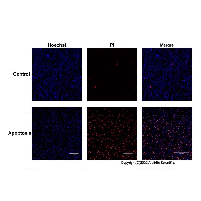
当使用这两种染料双染时,正常细胞主要被Hoechst 33342染成弱蓝色;当细胞被诱导发生凋亡时,凋亡细胞可被Hoechst 33342染成强蓝色,坏死细胞被PI染成强红色。
产品组分
H1373483 | Component | 100T | 500T | Storage | Quantity Per Test |
H1373483A | Hoechst 33342 Staining Solution | 1 mL | 5 mL | -20℃, Store in the dark. | 10 μL per 0.5–1.0×10⁶ cells/mL. |
H1373483B | Propidium iodide Staining Solution (PI) | 1 mL | 5 mL | -20℃, Store in the dark. | 10 μL per 0.5–1.0×10⁶ cells/mL. |
注:每个Test推荐染色的细胞数量为 0.5–1.0×10⁶ cells.
使用方法
1. 细胞准备
贴壁细胞:6孔板培养细胞至对数生长期,PBS清洗两次,加入1 mL的PBS
悬浮细胞:预冷PBS清洗细胞两次后,按照 0.5–1.0×10⁶/mL的密度用PBS重悬。
2. 染色
贴壁细胞:在6孔板中加入10 μL Hoechst 33342和10 μL PI,4℃,避光孵育20-30分钟。
悬浮细胞: 取1 mL细胞悬液,加入10 μL Hoechst 33342和10 μL PI,4℃,避光孵育20-30分钟。
注意:染色结束后尽快进行后续荧光检测,每个检测细胞数不超过 1.0×10⁶个。
3. 荧光显微镜检测与分析
贴壁细胞:弃染色液,PBS洗涤两次,加入适量PBS,然后在荧光显微镜下观察。
悬浮细胞:PBS清洗两次。用PBS重新悬浮细胞,置于培养板中观察红色和蓝色荧光。
注意事项
1. 染色完成后需要尽快进行检测。
2. Hoechst 33342和PI对人体有害,使用时请注意防护。
3. 为了您的安全和健康,请穿实验服并戴一次性手套操作。
4. 本产品仅限于科研使用,不得用于临床诊断或治疗。
The Hoechst 33342/PI Double Staining Apoptosis Detection Kit provides a classic, rapid, and straightforward method for distinguishing between apoptotic and necrotic cells. This kit utilizes a dual-staining approach with Hoechst 33342 and propidium iodide (PI). The nuclear dye Hoechst 33342 permeates cell membranes and emits blue fluorescence upon binding to double-stranded DNA. In normal cells, limited dye entry results in weak blue fluorescence. In apoptotic cells, however, increased membrane permeability allows significantly more Hoechst 33342 to enter, leading to higher fluorescence intensity. Additionally, chromatin condensation during apoptosis enhances the efficiency and concentration of DNA binding by the dye. Impaired function of the P-glycoprotein pump on the membrane of apoptotic cells further prevents effective efflux of Hoechst 33342, resulting in its intracellular accumulation. These characteristics collectively contribute to a markedly stronger fluorescence in apoptotic cells compared to normal cells. In contrast, propidium iodide (PI), another nuclear dye, cannot penetrate the intact membranes of normal or apoptotic cells, making viable cells impermeable to PI staining. Necrotic cells, which lose membrane integrity at an early stage, are readily stained by PI.
When dual-stained with these dyes, normal cells exhibit weak blue fluorescence primarily from Hoechst 33342. Apoptotic cells display intense blue fluorescence due to enhanced Hoechst 33342 staining, while necrotic cells show strong red fluorescence from PI.
Component
H1373483 | Component | 100T | 500T | Storage | Quantity Per Test |
H1373483A | Hoechst 33342 Staining Solution | 1 mL | 5 mL | -20℃, Store in the dark. | 10 μL per 0.5–1.0×10⁶ cells/mL. |
H1373483B | Propidium iodide Staining Solution (PI) | 1 mL | 5 mL | -20℃, Store in the dark. | 10 μL per 0.5–1.0×10⁶ cells/mL. |
Note: The recommended number of cells to stain per test 0.5–1.0×10⁶ cells.
Usage method:
1. Cell Preparation
Adherent cells: Grow cells in 6-well plates to the logarithmic growth phase. Wash twice with PBS and add 1 mL of PBS.
Suspension cells: Wash cells twice with pre-cooled PBS and resuspend in PBS at a density of 0.5–1.0×10⁶ cells/mL.
2. Staining
Adherent cells: Add 10 µL of Hoechst 33342 and 10 µL of PI directly to each well of the 6-well plate. Incubate at 4 °C in the dark for 20–30 minutes.
Suspension cells: Take 1 mL of the cell suspension, add 10 µL of Hoechst 33342 and 10 µL of PI, and incubate at 4 °C in the dark for 20–30 minutes.
Note: After staining, proceed with fluorescence detection as soon as possible. The number of cells for each detection should not exceed 1×10⁶.
3. Fluorescence Microscopy Detection and Analysis
Adherent cells: Remove the staining solution, wash twice with PBS, add an appropriate amount of PBS, and observe under a fluorescence microscope.
Suspension cells: Wash twice with PBS, resuspend the cells in PBS, transfer to a culture dish, and observe for red and blue fluorescence.
Matters needing attention:
1. After the staining process is completed, the detection should be carried out as soon as possible.
Hoechst 33342 and PI are harmful to the human body. Please take protective measures when using them.
2. For your safety and health, please wear a laboratory coat and put on disposable gloves when operating.
3. This product is exclusively for scientific research purposes and must not be used for clinical diagnosis or treatment.

 ZJ25F1230559


 危险品化学品经营许可证(带存储)