
计算溶液所需的质量、体积或浓度。
慢病毒包装试剂盒 (10T)
有货
BioReagent,分子生物学级,无菌,用于细胞培养 级 ,适用于对基线干扰要求严格的色谱和分析工作流程。
-20°C储存,避免反复冻融。超低温运输 。请查阅批次 COA 获取详细规格。
SDS、COA、产品数据表及规格说明书均可下载。可通过批号查询获取批次 COA。
在色谱分析、有机合成和交叉偶联反应领域已被 0 篇同行评审文献引用。
慢病毒是一种有包膜的RNA病毒,直径为80-120 nm,呈二十面体对称结构、球形。病毒颗粒最外层是包膜(包膜蛋白决定了感染细胞的类型),再往里依次为基质蛋白和衣壳,最里面是两条相同的正股RNA链和酶(逆转录酶、整合酶和蛋白酶)。
慢病毒表达载体包含了包装、转染、稳定整合所需要的遗传信息。慢病毒包装质粒可提供包装重组慢病毒载体所需的所有辅助蛋白。为产生高滴度的病毒颗粒,需要利用表达载体和包装质粒同时共转染细胞,在细胞中进行病毒包装,包装好的假病毒颗粒分泌到细胞外的培养基中,离心取得上清液后,可以直接或浓缩后用于宿主细胞的感染。
慢病毒感染细胞的第一步是通过包膜蛋白与细胞表面受体结合。慢病毒与宿主细胞膜融合后释放结构蛋白、酶蛋白和病毒核心。病毒RNA在逆转录酶的作用下逆转录并与整合酶形成整合前复合物。整合前复合物进入细胞核后,整合酶催化其整合至宿主基因组。整合后的DNA转录为mRNA,回到细胞浆中,表达目的蛋白或产生RNAi干扰(下图)。

Lentiviral Packaging Kit (10T) 属于第二代慢病毒包装体系,同时可兼容第二代和第三代慢病毒包装质粒。慢病毒中的毒性基因已经被剔除并被外源性目的基因所取代,属于假型病毒,即产生的慢病毒感染目的细胞后不会再感染其他细胞,也不会利用宿主细胞产生新的病毒颗粒。Lentiviral Packaging Kit (10T) 包装的慢病毒具有感染效率高、感染谱广等特点,可实现外源基因在宿主细胞中的稳定持续表达。
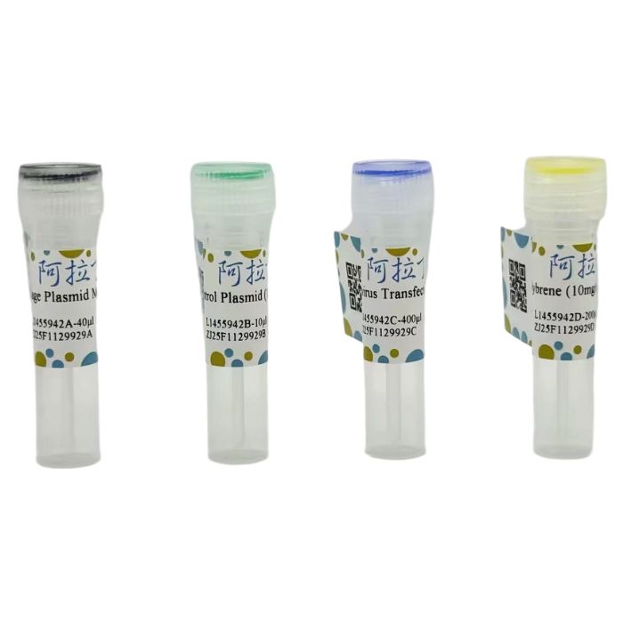
Lentiviral Packaging Kit (10T) 包含如下组分:
(1) 优化配比的慢病毒包装辅助质粒混合物(Package Plasmid Mix),经过设计调整,可兼容大多数慢病毒表达载体,确保将重组子包装成具有高感染率的慢病毒颗粒。
(2) 表达GFP 和 Puro标记的对照质粒(Control Plasmid),便于观察病毒包装效率及测定病毒滴度。
(3) 高效转染试剂(Lentivirus Transfection Reagent),优化的配方特别适合于慢病毒包装细胞系(HEK293T、HEK293FT等)的病毒包装,具有高效低毒、操作简单、重复性好等优点,能显著提高慢病毒载体在慢病毒包装细胞系中的包装效率,并能产生更高滴度的重组慢病毒。
(4) 常用病毒感染增强试剂Polybrene,能够显著提高病毒与细胞的接触及感染效率。
产品组分及储存条件:
| L1455942 | Component | 10T | 20T | 40T | Storage |
| L1455942A | Package Plasmid Mix | 40 μL | 80 μL | 160 μL | -20℃ |
| L1455942B | Control Plasmid (1μg/μL) | 10 μL | 20 μL | 40 μL | -20℃ |
| L1455942C | Lentivirus Transfection Reagent | 400 μL | 800 μL | 1600 μL | -20℃ |
| L1455942D | Polybrene (10mg/mL) | 200 μL | 400 μL | 800 μL | -20℃ |
产品特点:
⁕ 简单快速——操作简单,无需对包装辅助质粒进行抽提,也无需对包装体系进行优化
⁕ 病毒滴度高——可包装出高滴度、高表达水平的慢病毒
⁕ 应用范围广——经过设计及优化配比,兼容大多数慢病毒表达载体
实验流程概要:

图1. Lentiviral Packaging Kit (10T) 慢病毒包装试剂盒实验流程图
实验步骤:
一、重组慢病毒制备
1.细胞准备
转染前一天,将4-6×10⁶个慢病毒包装细胞(HEK293T、HEK293FT等)接种到10 cm细胞培养皿中,放置于37℃,5% CO₂培养箱培养16-24小时,使其在包装时细胞密度为70%-80%。
⁕ 细胞状态会极大影响病毒包装效率,包装细胞应处于良好生长状态,建议使用生长处于指数期、存活率大于90% 的细胞进行慢病毒包装。
⁕ 该包装系统与 Lentivirus Packaging Cell Line 配合使用,可包装出更高滴度、更高表达水平的慢病毒。
2.慢病毒包装
(1) 取4 μL慢病毒包装辅助质粒混合物(Package Plasmid Mix)加入到2 mL离心管中,加入4 μg含有目的基因的慢病毒载体质粒,轻轻混合,加入40 μL Lentivirus Transfection Reagent与质粒混合,室温孵育3分钟。
⁕ 慢病毒载体质粒需根据研究基因自行构建,应使用无内毒素质粒抽提试剂盒进行质粒提取,保证质粒A₂₆₀/A₂₈₀ 比值为1.8-2.0,浓度不低于500 ng/μL。溶解后的质粒切忌反复冻融,需按照使用量进行分装并于-20℃保存。
(2) 往上述混合物中加入1.8 mL无血清无双抗的基础培养基,轻轻混匀,在室温下静置30分钟,形成转染试剂-核酸复合物。
⁕ 转染试剂-核酸复合物在室温下4个小时内保持稳定。
(3) 将上述1.8 mL转染试剂-核酸复合物轻轻地逐滴均匀加入10 cm皿的包装细胞细胞中,轻轻摇动培养皿混匀,置于37℃,5% CO₂培养箱中培养产毒。
3.收获病毒
(1) 转染24 小时后,可选择弃去初始病毒上清液,加入10-15 mL新鲜的完全培养基(含血清)至10 cm细胞培养皿中,置于37℃,5% CO₂培养箱继续培养(此步骤可选)。
(2) 转染48 小时后,收集病毒上清液,再加入10-15 mL新鲜的完全培养基(含血清)至10 cm细胞培养皿中,置于37℃,5% CO₂培养箱继续培养。
(3) 转染72 小时后,观察细胞状态并拍照,进行二次病毒上清液收集,与48 小时收集的上清液混合,800xg离心10分钟以除去细胞碎片,使用0.45 μm滤器过滤,过滤后的上清液可直接感染目的细胞,或使用慢病毒浓缩试剂盒(S1506239)浓缩后感染目的细胞。
⁕切忌反复冻融病毒,冻融一次病毒滴度将降低10-20%。-80℃保存的慢病毒最好在半年内使用。
二、病毒滴度测定
1. 孔稀释法标定带荧光的重组慢病毒滴度
(1) Day1:准备细胞
对生长状态良好的293T细胞消化计数后稀释至 1-3x10⁵个/mL ,加入96孔板,100 μL/孔(1-3x10⁴个/孔),每⼀种慢病毒设6个梯度孔,置于37℃,5% CO₂培养箱中培养过夜。
(2) Day2:病毒的稀释与感染
在EP管中做10倍梯度稀释,连续6个稀释梯度。稀释方法如下:准备6个⽆菌EP管,每个EP管中加⼊90μL 新鲜的完全培养基(含10 μg/mL polybrene),取待测定病毒液10 μL加⼊到第⼀个EP管中(标记10 μL),混匀后取10 μL病毒稀释液加⼊到第⼆个EP管中(标记1 μL),以此类推,直到稀释到最后⼀管。弃去96孔板中原有的培养基,将稀释好的病毒依次加入孔中,并做好标记,⼩⼼操作,不要吹起细胞,置于培养箱中培养16 小时。如需设置复孔,则按复孔的数量倍数增加以上用量。

(3) Day3:弃病毒,换液
弃去带有病毒的培养基,在每个孔中再加入 100 μL 预热的新鲜完全培养基。
(4) Day4:荧光计数与滴度计算
方法 1:末位稀释计数法
感染 48 小时后,用荧光显微镜对荧光阳性细胞进行计数。数出最后两孔的荧光细胞数(若设置了复孔,则计算复孔内的总数之和并计算出平均数),假设为 A(倒数第二孔的荧光细胞数)和 B(倒数第一孔的荧光细胞数)。
慢病毒滴度计算公式 1:病毒滴度 (TU/mL) =(A+B×10)×1000/2/A 孔病毒量(μL)
举例 1:5 号管对应的荧光阳性细胞数为 10 个,6 号管对应的荧光阳性细胞数为 2 个,则病毒滴度为:
滴度(TU/mL)= ( 10+2x10)x1000/2/0.001 = 1.5x10⁷
如果需要感染 2x10⁵个细胞,MOI=5(1 个细胞对应 5 个病毒颗粒),则需病毒体积为:所需病毒体积 = 2 x10⁵x 5/1.5x10⁷(mL)= 0.067 mL = 67 μL
方法 2:适量荧光计数法
感染 48 小时后,用荧光显微镜或流式细胞术对荧光比例合适(10%-50%)的连续两个梯度进行统计,数出两孔的荧光细胞数(若设置了复孔,则计算复孔内的总数之和并计算出平均数),假设为 C(较高浓度孔的荧光细胞数)和 D(较低浓度孔的荧光细胞数)。
慢病毒滴度计算公式 2:病毒滴度 (TU/mL) =(C+D×10)×1000/2/C 孔病毒量(μL)
举例 2:3 号管对应的荧光阳性细胞数为 10000 个,4 号管对应的荧光阳性细胞数为 2000个,则病毒滴度为:
滴度(TU/mL)= ( 10000+2000x10)x1000/2/0.1 = 1.5x10⁸
如果需要感染 2x10⁵个细胞,MOI=30(1 个细胞对应 30 个病毒颗粒),则需病毒体积为:
所需病毒体积 = 2 x10⁵x 30/1.5x10⁸(mL)= 0.04 mL = 40 μL
2. 相对定量 PCR 标定非荧光的重组慢病毒滴度
(1) Day1:准备细胞
对生长状态良好的 293T 细胞消化计数后稀释至 1-3x10⁵个/mL ,加入 24 孔板,500 μL/孔( 0.5-1.5x10⁵个 /孔),每一种慢病毒设 6 个梯度孔,置于 37℃,5% CO₂培养箱中培养过夜。
(2) Day2:病毒的稀释与感染
在 EP 管中做 10 倍梯度稀释,连续 6 个稀释梯度。稀释方法如下:准备 6 个无菌 EP 管,每个 EP 管中加入450μL 新鲜的完全培养基(含 10 μg/mL polybrene),取待测定病毒液50 μL 加入到第一个 EP 管中(标记 50 μL),混匀后取 50 μL 病毒稀释液加入到第二个 EP管中(标记 5 μL),以此类推,直到稀释到最后一管。弃去 24 孔板中原有的培养基,将稀释好的病毒依次加入孔中,并做好标记,小心操作,不要吹起细胞,置于培养箱中培养 16 小时 。如需设置复孔,则按复孔的数量倍数增加以上用量。

(3) Day3:弃病毒,换液
吸去带有病毒的培养基,在每个孔中再加入 500 μL 预热的新鲜完全培养基。
(4) Day4:相对定量 PCR 与滴度计算
用 PBS 清洗并将细胞吹打下来,离心收集细胞,进行基因组 DNA 的提取,qPCR 检测。以被测慢病毒载体质粒梯度稀释为标准品,慢病毒载体上的通用引物 RRE 进行 qPCR 以获得病毒整合拷贝数。以 ACTB 质粒梯度稀释为标准品,ACTB 引物进行 qPCR 以得到基因组拷贝数。
每个细胞中慢病毒拷贝数=2*慢病毒拷贝数/ACTB 拷贝数
慢病毒滴度计算公式 3:滴度(TU/mL)=(E×N×1000)/对应孔病毒量(μL)
其中 E=每个细胞中慢病毒拷贝数,N=感染时细胞的数目(约为 1.5×10⁵)。
⁕ 测定时,设置一组带荧光的已知 TU 的慢病毒作为对照,以校验检测出的数值。
三、慢病毒感染目的细胞
VSVG 包膜蛋白对不同的细胞和组织的亲嗜性有很大差异,在使用慢病毒感染目的细胞之前,需要查阅相关文献或进行预实验确定目的细胞的复感染指数(MOI,multiplicity of infection)。
1. 按照上述步骤包装慢病毒及确定慢病毒滴度,通过病毒滴度和 MOI 计算所需病毒量(计算公式参考 06/实验步骤/病毒滴度测定)。
2. 实验前一天,将生长状态良好的目的细胞消化计数后稀释至 1-3×10⁵个/mL,加入 24 孔板,500μL/孔(0.5-1.5x10⁵ 个/孔),放入 37℃,5% CO₂培养箱中培养过夜,保证第二天进行病毒感染时细胞汇合度约为 30%-50%。
⁕ 悬浮细胞不需要提前一天接种,实验前将细胞计数接种后,直接加入病毒混合液即可。
3. 配制慢病毒+polybrene+培养基混合液。polybrene 为常用的促感染试剂,能够显著提高病毒与细胞的接触及感染效率,一般使用浓度为 2-10μg/mL,但对某些细胞(如末端分化的神经元,DC 细胞,免疫细胞,干细胞等)毒性较大,初次使用建议先做毒性测试。相比于添加 polybrene 所产生的细胞毒性来说,Viral Transfection Enhancer (V1501911) 对细胞的毒性极低,几乎对细胞生长无任何损伤,因此,针对不能使用 polybrene 的细胞,Viral Transfection Enhancer (V1501911) 是首选的病毒感染增强剂。
4. 吸去 24 孔板中目的细胞的培养基,加入 3 中配制的慢病毒混合液,放入 37℃,5% CO₂培养箱中继续培养。
⁕ 感染悬浮细胞或半悬浮细胞,则需采用平角离心转染法,即将适量的病毒液加入细胞培养板后,封好口,放入平角离心机中,800xg 离心 30 分钟(因细胞而异),然后放入培养箱中继续培养。
5. 病毒感染 16 小时后更换为新鲜培养基继续培养。
6. 病毒感染 48 小时后,根据实际情况用荧光检测法、Western Blot、RT-PCR、ELISA、流式细胞术、报告基因等检测表达效果,或加入筛选药物进行稳定细胞株的筛选。
7. 若表达效果不理想,可调整 MOI(例如,设置 MOI 梯度)再次检测。
适用范围:
兼容大多数慢病毒表达载体,经过设计及优化配比,确保将重组子包装成具有高感染率的慢病毒颗粒,同时防止病毒的自我复制。
注意事项:
1. 该系统与 Lentivirus Packaging Cell Line 配合使用,可包装出更高滴度、更高表达水平的慢病毒。
2. 在收获粗病毒后,建议额外分装几支 20-100 µL 小体积管,和其他分装的大体积病毒一并置于-80℃保存。待这些小体积病毒结冰后,取出来融化,再测定滴度。这一步骤可以模拟一次冻融带来的活性滴度下降,这样测得的滴度才是真正具有参考价值的。
3. 为了您的健康安全,请规范操作,穿戴实验服与手套开展实验。
4. 所有慢病毒操作均需在 BSL2 级生物安全柜中进行。
5. 本产品仅供科研使用,请勿用于临床诊断及治疗。
实验案例分析:
1. 提前一天使用10 cm 细胞培养皿培养Lentivirus Packaging Cell Line,待细胞密度为 75% 左右开始包装慢病毒。
2. 使用 Lentiviral Packaging Kit (L1455942) 进行慢病毒包装。取 4 μL 慢病毒包装辅助质粒混合(Package Plasmid Mix)加入到 2 mL 离心管中,加入 4 μL 表达 GFP的对照质粒(Control Plasmid),轻轻混合,加入 40 μL Lentivirus Transfection Reagent与质粒混合,室温孵育 3 分钟。
3. 往上述混合物中加入 1.8 mL 无血清无双抗的 DMEM 基础培养基,轻轻混匀,在室温下静置 30 分钟,形成转染试剂-核酸复合物。
4. 将上述 1.8 mL 转染试剂-核酸复合物轻轻地逐滴均匀加入 10 cm 皿慢病毒包装细胞中,轻轻摇动培养皿混匀,置于 37℃,5% CO₂培养箱中培养产毒。
5. 转染 24 小时后,弃去初始病毒上清液,加入 10 mL 新鲜的完全培养基(DMEM +10% FBS)至 10 cm 细胞培养皿中,置于 37℃,5% CO₂培养箱继续培养。
6. 转染 48 小时后,收集病毒上清液,再加入 10 mL 新鲜的完全培养基(DMEM +10% FBS)至 10 cm 细胞培养皿中,置于 37℃,5% CO₂培养箱继续培养。
7. 转染 72 小时后,观察细胞状态并拍照,进行二次病毒上清液收集,与 48 小时收集的上清液混合后,800xg 离心 10 分钟以除去细胞碎片,0.45 μm 滤器过滤,使用Lentivirus Concentration Solution (S1506239) 进行 20 倍浓缩,使用1 mL Lentivirus Stable Storage Solution(L1505998)重悬慢病毒颗粒。
8. 孔稀释法测定病毒滴度(参考 06/实验步骤中病毒滴度测定方法),使用荧光显微镜观察绿色荧光蛋白表达情况,并拍照记录,实验结果如图 2 所示。

图2. 采用Lentiviral Packaging Kit (L1455942)包装慢病毒(GFP, Control plasmid)感染293T细胞检测病毒滴度
Lentivirus is an enveloped RNA virus with a diameter of 80–120 nm, exhibiting an icosahedral symmetric structure and a spherical shape. The outermost layer of the viral particle is the envelope (the envelope protein determines the type of cells the virus can infect), followed by the matrix protein and capsid from the outside in. The innermost part contains two identical positive-sense RNA strands and enzymes (reverse transcriptase, integrase, and protease).Lentiviral expression vectors contain the genetic information required for packaging, transfection, and stable integration. Lentiviral packaging plasmids can provide all the helper proteins needed for packaging recombinant lentiviral vectors. To generate high-titer viral particles, it is necessary to co-transfect cells with both the expression vector and packaging plasmids simultaneously. Viral packaging occurs within the cells, and the packaged pseudoviral particles are secreted into the extracellular culture medium. After centrifuging to obtain the supernatant, it can be used directly or after concentration to infect host cells.The first step of lentiviral infection of cells is the binding of the envelope protein to receptors on the cell surface. After the lentivirus fuses with the host cell membrane, it releases structural proteins, enzymatic proteins, and the viral core. Under the action of reverse transcriptase, the viral RNA undergoes reverse transcription and forms a pre-integration complex with integrase. After the pre-integration complex enters the cell nucleus, integrase catalyzes its integration into the host genome. The integrated DNA is transcribed into mRNA, which is transported back to the cytoplasm to express the target protein or induce RNAi interference (see figure below).
The Lentiviral Packaging Kit (10T) belongs to the second-generation lentiviral packaging system and is compatible with both second-generation and third-generation lentiviral packaging plasmids. The toxic genes in the lentivirus have been eliminated and replaced with exogenous target genes, making it a pseudotyped virus. Specifically, after the generated lentivirus infects target cells, it will not infect other cells, nor will it use host cells to produce new viral particles. The lentivirus packaged by the Lentiviral Packaging Kit (10T) features high infection efficiency and a broad infection spectrum, enabling stable and sustained expression of exogenous genes in host cells.
The Lentiviral Packaging Kit (10T) contains the following components:
1. Optimized Proportion Lentiviral Packaging Helper Plasmid Mix (Package Plasmid Mix)Designed and adjusted to be compatible with most lentiviral expression vectors, ensuring that recombinants are packaged into lentiviral particles with high infectivity.
2. Control Plasmid Expressing GFP and Puro Markers (Control Plasmid)Facilitates observation of viral packaging efficiency and determination of viral titer.
3. High-Efficiency Transfection Reagent (Lentivirus Transfection Reagent)Its optimized formula is particularly suitable for viral packaging in lentiviral packaging cell lines (e.g., HEK293T, HEK293FT). It features high efficiency, low toxicity, simple operation, and good repeatability—these advantages can significantly improve the packaging efficiency of lentiviral vectors in lentiviral packaging cell lines and produce recombinant lentiviruses with higher titers.
4. Common Viral Infection-Enhancing Reagent (Polybrene) Can significantly improve the contact between virus and cells as well as infection efficiency.
Product Components and Storage Conditions:
| L1455942 | Component | 10T | 20T | 40T | Storage |
| L1455942A | Package Plasmid Mix | 40 μL | 80 μL | 160 μL | -20℃ |
| L1455942B | Control Plasmid (1μg/μL) | 10 μL | 20 μL | 40 μL | -20℃ |
| L1455942C | Lentivirus Transfection Reagent | 400 μL | 800 μL | 1600 μL | -20℃ |
| L1455942D | Polybrene (10mg/mL) | 200 μL | 400 μL | 800 μL | -20℃ |
Product Features:
⁕ Simple and Fast – The operation is simple, with no need for extraction of packaging helper plasmids or optimization of the packaging system.
⁕ High Viral Titer – Capable of packaging lentiviruses with high titer and high expression levels.
⁕ Wide Application Range – Designed and optimized for proportion, it is compatible with most lentiviral expression vectors.
Summary of the Experimental Procedure:
Figure 1. Experimental Flowchart of the Lentiviral Packaging Kit (10T)
Experimental Steps:
I. Preparation of Recombinant Lentivirus
1. Cell Preparation
One day before transfection, seed 4–6×10⁶ lentiviral packaging cells (e.g., HEK293T, HEK293FT) into a 10 cm cell culture dish. Incubate in a 37°C, 5% CO₂ incubator for 16–24 hours, ensuring the cell confluency reaches 70%–80% at the time of packaging.
⁕ Cell status significantly affects viral packaging efficiency. Packaging cells should be in good growth condition; it is recommended to use cells in the exponential growth phase with a viability >90% for lentiviral packaging.
⁕ When used in conjunction with the Lentivirus Packaging Cell Line, this packaging system can produce lentiviruses with higher titer and higher expression levels.
2. Lentiviral Packaging
(1) Add 4 μL of the Lentiviral Packaging Helper Plasmid Mix (Package Plasmid Mix) to a 2 mL centrifuge tube. Add 4 μg of the lentiviral vector plasmid containing the target gene, mix gently, then add 40 μL of Lentivirus Transfection Reagent to the plasmid mixture. Incubate at room temperature for 3 minutes.
⁕ The lentiviral vector plasmid must be constructed according to the gene of interest. Plasmid extraction should be performed using an endotoxin-free plasmid extraction kit to ensure the plasmid has an A₂₆₀/A₂₈₀ ratio of 1.8–2.0 and a concentration of no less than 500 ng/μL. Avoid repeated freeze-thaw cycles of dissolved plasmids; aliquot according to usage and store at -20°C.
(2) Add 1.8 mL of serum-free and antibiotic-free basal medium to the above mixture, mix gently, and let stand at room temperature for 30 minutes to form transfection reagent-nucleic acid complexes.
⁕ Transfection reagent-nucleic acid complexes remain stable for up to 4 hours at room temperature.
(3) Gently add the 1.8 mL transfection reagent-nucleic acid complexes dropwise and evenly to the packaging cells in the 10 cm dish. Swirl the dish gently to mix, then incubate in a 37°C, 5% CO₂ incubator for virus production.
3. Virus Harvest
(1) At 24 hours post-transfection, optionally discard the initial viral supernatant and add 10–15 mL of fresh complete medium (containing serum) to the 10 cm dish. Continue incubation in a 37°C, 5% CO₂ incubator (this step is optional).
(2) At 48 hours post-transfection, collect the viral supernatant, then add 10–15 mL of fresh complete medium (containing serum) to the 10 cm dish. Continue incubation in a 37°C, 5% CO₂ incubator.
(3) At 72 hours post-transfection, observe and photograph the cell status, then collect the viral supernatant a second time. Combine it with the supernatant collected at 48 hours, centrifuge at 800×g for 10 minutes to remove cell debris, and filter through a 0.45 μm filter. The filtered supernatant can be used directly to infect target cells or concentrated using the Lentivirus Concentration Kit (S1506239) before infection.
⁕ Avoid repeated freeze-thaw cycles of the virus, as each freeze-thaw cycle reduces the viral titer by 10%–20%. Lentiviruses stored at -80°C are best used within six months.
II. Viral Titer Determination
1. Calibration of Fluorescent Recombinant Lentiviral Titer by Well Dilution Method
(1) Day 1: Cell Preparation
Digest and count well-grown 293T cells, then dilute them to a concentration of 1–3×10⁵ cells/mL. Add the cell suspension to a 96-well plate at a volume of 100 μL per well (corresponding to 1–3×10⁴ cells per well). Set 6 gradient wells for each type of lentivirus, and incubate overnight in a 37°C, 5% CO₂ incubator.
(2) Day 2: Virus Dilution and Infection
Perform 10-fold serial dilutions in EP tubes, with 6 consecutive dilution gradients. The dilution method is as follows: Prepare 6 sterile EP tubes, and add 90 μL of fresh complete medium (containing 10 μg/mL Polybrene) to each tube. Take 10 μL of the lentivirus solution to be tested and add it to the first EP tube (labeled "10 μL"). After mixing, take 10 μL of the virus dilution and add it to the second EP tube (labeled "1 μL"). Repeat this process until the last tube is diluted. Discard the original medium in the 96-well plate, then add the diluted virus to the wells in sequence and label them properly. Operate carefully to avoid disturbing the cells, and incubate in the incubator for 16 hours. If replicate wells are required, increase the above reagent volumes proportionally according to the number of replicate wells.
(3) Day 3: Remove Virus and Replace Medium
Discard the virus-containing medium, and add 100 μL of pre-warmed fresh complete medium to each well.
(4) Day 4: Fluorescence Counting and Titer Calculation
Method 1: End-Point Dilution Counting Method
At 48 hours post-infection, count the fluorescence-positive cells using a fluorescence microscope. Count the number of fluorescent cells in the last two wells (if replicate wells are set, calculate the sum of the total number of cells in the replicate wells and then the average). Denote the counts as A (number of fluorescent cells in the penultimate well) and B (number of fluorescent cells in the last well).
Lentiviral Titer Calculation Formula 1:Viral titer (TU/mL) = (A + B × 10) × 1000 / 2 / Volume of virus in Well A (μL)
Example 1:If the number of fluorescence-positive cells corresponding to Tube 5 is 10, and that corresponding to Tube 6 is 2, the viral titer is calculated as follows:Titer (TU/mL) = (10 + 2 × 10) × 1000 / 2 / 0.001 = 1.5 × 10⁷
To infect 2 × 10⁵ cells with an MOI (Multiplicity of Infection) of 5 (5 viral particles per cell), the required virus volume is:Required virus volume = (2 × 10⁵ × 5) / (1.5 × 10⁷) (mL) = 0.067 mL = 67 μL
Method 2: Appropriate Fluorescence Counting Method
At 48 hours post-infection, use a fluorescence microscope or flow cytometry to count two consecutive gradients with a suitable fluorescence ratio (10%–50%). Count the number of fluorescent cells in these two wells (if replicate wells are set, calculate the sum of the total number of cells in the replicate wells and then the average). Denote the counts as C (number of fluorescent cells in the well with higher virus concentration) and D (number of fluorescent cells in the well with lower virus concentration).
Lentiviral Titer Calculation Formula 2:Viral titer (TU/mL) = (C + D × 10) × 1000 / 2 / Volume of virus in Well C (μL)
Example 2:If the number of fluorescence-positive cells corresponding to Tube 3 is 10,000, and that corresponding to Tube 4 is 2,000, the viral titer is calculated as follows:Titer (TU/mL) = (10,000 + 2,000 × 10) × 1000 / 2 / 0.1 = 1.5 × 10⁸
To infect 2 × 10⁵ cells with an MOI of 30 (30 viral particles per cell), the required virus volume is:Required virus volume = (2 × 10⁵ × 30) / (1.5 × 10⁸) (mL) = 0.04 mL = 40 μL
2. Calibration of Non-Fluorescent Recombinant Lentiviral Titer by Relative Quantitative PCR
(1) Day 1: Cell PreparationDigest and count well-grown 293T cells, then dilute them to a concentration of 1–3×10⁵ cells/mL. Add the cell suspension to a 24-well plate at a volume of 500 μL per well (corresponding to 0.5–1.5×10⁵ cells per well). Set 6 gradient wells for each type of lentivirus, and incubate overnight in a 37°C, 5% CO₂ incubator.
(2) Day 2: Virus Dilution and InfectionPerform 10-fold serial dilutions in EP tubes, with 6 consecutive dilution gradients. The dilution method is as follows: Prepare 6 sterile EP tubes, and add 450 μL of fresh complete medium (containing 10 μg/mL Polybrene) to each tube. Take 50 μL of the lentivirus solution to be tested and add it to the first EP tube (labeled "50 μL"). After mixing, take 50 μL of the virus dilution and add it to the second EP tube (labeled "5 μL"). Repeat this process until the last tube is diluted. Discard the original medium in the 24-well plate, then add the diluted virus to the wells in sequence and label them properly. Operate carefully to avoid disturbing the cells, and incubate in the incubator for 16 hours. If replicate wells are required, increase the above reagent volumes proportionally according to the number of replicate wells.
(3) Day 3: Remove Virus and Replace Medium
Aspirate the virus-containing medium, and add 500 μL of pre-warmed fresh complete medium to each well.
(4) Day 4: Relative Quantitative PCR and Titer Calculation
Wash the cells with PBS, then detach them by pipetting. Collect the cells by centrifugation, extract genomic DNA, and perform qPCR detection.
Use serial dilutions of the lentiviral vector plasmid (to be tested) as the standard, and perform qPCR with the universal primer RRE (targeting the lentiviral vector) to obtain the number of virus integration copies.
Use serial dilutions of the ACTB plasmid as the standard, and perform qPCR with ACTB primers to obtain the number of genomic copies.
Number of lentiviral copies per cell = 2 × Number of lentiviral copies / Number of ACTB copies
Lentiviral Titer Calculation Formula 3:Titer (TU/mL) = (E × N × 1000) / Volume of virus in the corresponding well (μL)
Where:
E = Number of lentiviral copies per cell
N = Number of cells at the time of infection (approximately 1.5 × 10⁵)
⁕ During the determination, set up a group of fluorescent lentiviruses with known TU (transducing units) as a control to verify the detected values.
III. Lentiviral Infection of Target Cells
The tropism of the VSVG envelope protein for different cells and tissues varies significantly. Before infecting target cells with lentivirus, it is necessary to consult relevant literature or conduct pre-experiments to determine the multiplicity of infection (MOI) for the target cells.
1. Package the lentivirus and determine its titer according to the above steps. Calculate the required amount of virus based on the viral titer and MOI (refer to 06/Experimental Steps/Viral Titer Determination for the calculation formula).
2. One day before the experiment, digest and count well-grown target cells, then dilute them to a concentration of 1–3×10⁵ cells/mL. Add the cell suspension to a 24-well plate at a volume of 500 μL per well (corresponding to 0.5–1.5×10⁵ cells per well). Incubate overnight in a 37°C, 5% CO₂ incubator to ensure the cell confluency reaches approximately 30%–50% when viral infection is performed the next day.
⁕ Adherent cells do not require seeding one day in advance. After counting and seeding the cells before the experiment, directly add the virus mixture.
3. Prepare a mixture of lentivirus + Polybrene + medium. Polybrene is a commonly used infection-promoting reagent that can significantly improve the contact between virus and cells as well as infection efficiency. Its general working concentration is 2–10 μg/mL; however, it exhibits high toxicity to certain cell types (such as terminally differentiated neurons, DC cells, immune cells, and stem cells). For first-time use, it is recommended to conduct a toxicity test first. Compared with the cytotoxicity caused by adding Polybrene, the Viral Transfection Enhancer (V1501911) has extremely low cytotoxicity and causes almost no damage to cell growth. Therefore, for cells that cannot use Polybrene, the Viral Transfection Enhancer (V1501911) is the preferred viral infection enhancer.
4. Aspirate the medium of target cells in the 24-well plate, add the lentivirus mixture prepared in Step 3, and continue incubation in a 37°C, 5% CO₂ incubator.
⁕ For infecting suspension cells or semi-suspension cells, the horizontal centrifugation transfection method should be used: add an appropriate amount of virus solution to the cell culture plate, seal the plate tightly, place it in a horizontal centrifuge, centrifuge at 800×g for 30 minutes (varies by cell type), and then transfer it to the incubator for continued culture.
5. Replace with fresh medium 16 hours after viral infection and continue incubation.
6. Forty-eight hours after viral infection, detect the expression effect using methods such as fluorescence detection, Western Blot, RT-PCR, ELISA, flow cytometry, or reporter gene assays according to actual conditions; alternatively, add selection drugs to screen for stable cell lines.
7. If the expression effect is not ideal, adjust the MOI (e.g., set up an MOI gradient) and perform the detection again.
Scope of Application:
It is compatible with most lentiviral expression vectors. Through design and optimized proportioning, it ensures that recombinants are packaged into lentiviral particles with high infectivity, while preventing self-replication of the virus.
Precautions:
1. When used in conjunction with the Lentivirus Packaging Cell Line, this system can package lentiviruses with higher titer and higher expression levels.
2. After harvesting the crude virus, it is recommended to aliquot an additional several small-volume tubes (20–100 µL each) and store them at -80°C together with other aliquoted large-volume virus samples. Once these small-volume virus samples are frozen, take them out to thaw and then determine the titer. This step can simulate the decrease in active titer caused by one freeze-thaw cycle, ensuring the measured titer is truly referenceable.
3. For your health and safety, please operate in compliance with standard procedures and wear a lab coat and gloves when conducting experiments.
4. All lentivirus-related operations must be performed in a BSL-2 (Biosafety Level 2) biological safety cabinet.
5. This product is for research use only and shall not be used for clinical diagnosis or treatment.
Analysis of Experimental Case:
1. One day in advance, culture the Lentivirus Packaging Cell Line in a 10 cm cell culture dish. Start lentiviral packaging when the cell confluency reaches approximately 75%.
2. Use the Lentiviral Packaging Kit (L1455942) for lentiviral packaging. Add 4 μL of the Lentiviral Packaging Helper Plasmid Mix (Package Plasmid Mix) to a 2 mL centrifuge tube, then add 4 μL of the control plasmid expressing GFP (Control Plasmid). Mix gently, add 40 μL of Lentivirus Transfection Reagent to the plasmid mixture, and incubate at room temperature for 3 minutes.
3. Add 1.8 mL of serum-free and antibiotic-free DMEM basal medium to the above mixture, mix gently, and let stand at room temperature for 30 minutes to form transfection reagent-nucleic acid complexes.
4. Gently add the 1.8 mL transfection reagent-nucleic acid complexes dropwise and evenly to the lentiviral packaging cells in the 10 cm dish. Swirl the dish gently to mix, then place it in a 37°C, 5% CO₂ incubator for virus production.
5. At 24 hours post-transfection, discard the initial viral supernatant, add 10 mL of fresh complete medium (DMEM + 10% FBS) to the 10 cm cell culture dish, and continue incubation in a 37°C, 5% CO₂ incubator.
6. At 48 hours post-transfection, collect the viral supernatant, add another 10 mL of fresh complete medium (DMEM + 10% FBS) to the 10 cm cell culture dish, and continue incubation in a 37°C, 5% CO₂ incubator.
7. At 72 hours post-transfection, observe the cell status, take photos, and collect the viral supernatant for the second time. Combine this supernatant with the one collected at 48 hours, centrifuge at 800×g for 10 minutes to remove cell debris, and filter through a 0.45 μm filter. Concentrate the virus 20-fold using Lentivirus Concentration Solution (S1506239), then resuspend the lentiviral particles in 1 mL of Lentivirus Stable Storage Solution (L1505998).
8. Determine the viral titer using the well dilution method (refer to the viral titer determination method in 06/Experimental Steps). Observe the expression of green fluorescent protein (GFP) using a fluorescence microscope, take photos for documentation, and the experimental results are shown in Figure 2.
Figure 2. Titer determination of lentiviruses (GFP, Control plasmid) packaged using the Lentiviral Packaging Kit (L1455942) via infecting 293T cells
通过匹配包装上的批号来查找并下载产品的 COA,每批产品都进行了严格的验证,您可放心使用!
| 批号(Lot Number) | 证书类型 | 货号 |
|---|---|---|
 ZJ25F1129929 |
分析证书 | L1455942 |
我们的级别选择指南涵盖了目录中所有的变体的纯度、稳定剂状态和应用适用性。
查看 BioReagent 级别指南 → 查看 for Cell culture 级别指南 → 查看 Suitable for molecular biology 级别指南 → 查看 Sterile 级别指南 →

 危险品化学品经营许可证(带存储)