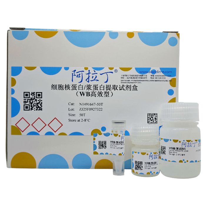
本产品可以从动物细胞或组织中方便高效地分离细胞核蛋白和细胞浆蛋白,其可逐步裂解细胞,将完整的细胞核 从细胞质中分离出来,然后从细胞核中提取出细胞核蛋白。
本产品含有强效去污剂,提取的核蛋白不仅包括核膜蛋白,也包括组蛋白和核内转录因子等可溶性蛋白。传统试 剂盒核质分离后,核蛋白提取时间通常在30~40 min之间,而本产品将核蛋白提取时间控制在10 min。此外,本产品 提取的核蛋白量显著高于其它试剂盒,且核质分离效果显著,尤其适用于Western Blot实验。
组分与储存:
组分货号
| 组分名称
| 物理外观 | 50T | 储存
| | N1491647A | WB核/浆试剂A | 液体 | 20 mL | 2-8℃ | | N1491647B | WB核/浆试剂B | 液体 | 0.5 mL | 2-8℃ | | N1491647C | WB核/浆试剂C | 液体 | 5 mL | 2-8℃ |
|
产品特点:
速度快—本产品优化了细胞核蛋白提取的操作方法,提取时间比传统试剂盒缩短10~20 min;
得率佳—本产品为Western Blot高效型试剂盒,优化了细胞核蛋白提取的试剂配方,蛋白提取量高于常规蛋白提取试剂盒,对于Western Blot应用效果极佳;
纯度高—核蛋白/浆蛋白分离效果显著,交叉污染率极低(效果优于传统试剂盒);
易操作—操作简单,无需梯度超速离心。
注意事项:
1. 本产品仅适用于动物细胞或组织,不可用于植物,酵母,大肠杆菌等样品胞核/胞浆蛋白的提取;
2. 试剂A和试剂C使用时请加入1×蛋白酶抑制剂 (货号P665818) 或1×蛋白酶和磷酸酶抑制剂 (货号P752090);
3. 本产品为WB高效型试剂盒,对于SDS不兼容性实验不适用;
4. 提取的蛋白样品可使用BCA蛋白定量试剂盒(货号R1491648/B665595)进行蛋白定量;
5. 为了您的安全和健康,请穿实验服并戴一次性手套操作;
6. 本产品仅限科研使用。
使用说明:
将试剂盒各组分置于冰上,其中试剂A和试剂 C使用时请添加 1×蛋白酶抑制剂 (货号P665818) 或1×蛋白酶和磷酸酶抑制剂 (货号P752090)。
动物细胞
1. 贴壁细胞:弃去培养皿中的培养基,加入适量PBS缓冲液,用细胞刮刀将细胞从培养皿表面刮下,将细胞重悬转移至离心管中。300×g离心5 min,吸弃上清。
悬浮细胞:将细胞转移至离心管中,300×g离心5 min,吸弃上清;再加入适量PBS缓冲液重悬细胞,300×g离心5 min,吸弃上清。
注意:不能用胰酶处理贴壁细胞。胰酶会使细胞变得很脆弱,影响后续核质分离。
2. 用适量PBS重悬细胞,取2×10⁶个细胞置于1.5 mL离心管中,300×g离心5 min,吸弃上清。按照表1所推荐的试剂体积进行细胞浆和细胞核蛋白提取,2×10⁶个细胞体积通常为20 μL,以下步骤都是以20 μL的细胞体积为例进行操作。
注意:不可仅凭经验收取细胞,一定要用细胞计数仪检测细胞活率和密度,再取2×10⁶个细胞。
表1. 不同细胞体积推荐的提取试剂体积
| 细胞体积/μL | 核/浆试剂A/μL | 核/浆试剂B/μL | 核/浆试剂C/μL | | 10 | 100 | 5 | 50 | | 20 | 200 | 10 | 100 | | 50 | 500 | 25 | 250 | | 100 | 1000 | 50 | 500 |
|
3. 向细胞沉淀中加入200 μL试剂A,充分混匀后,冰上静置10min。
4. 向上步所得细胞悬液中再加入10μL试剂B,最高速涡旋振荡5 sec,冰上静置1min。
5. 再进行最高速涡旋振荡5sec,冰上静置2 min。
注意:对于不同种类的细胞,此步骤的冰浴时间可适当延长 (3min) 或缩短 (1min),避免出现沉淀聚集现象即可。
6. 将孵育后的细胞离心10min (1,600×g, 4°C),小心吸去含有胞浆蛋白的上清液,如有需要可转移至新离心管中,置于冰上或分装后储存于-80℃备用。
注意:吸头切勿触及细胞沉淀,为降低胞浆组分对细胞核组分的污染,可以在沉淀上方保留极小体积的上清,换用10μL吸头尽可能吸尽上清。
7. 加入200 μL试剂A重悬细胞沉淀,充分混匀后,立即离心5 min (13,000×g, 4°C),弃尽上清。
注意:沉淀含有细胞核,可以在沉淀上方保留极小体积的上清,换用10μL吸头吸尽上清。
8. 向沉淀中加入100μL试剂C,充分混匀后,最高速涡旋振荡10sec,置于冰上,在10min内每隔2min重复涡旋一次。
9. 将上步孵育后的悬液离心10min (13,000×g, 4℃),将含有可溶性细胞核蛋白的上清液转移至新离心管中置于冰上或分装后储存于-80°C备用。
动物组织
1. 把20-80 mg动物组织尽可能剪成小的碎片置于2 mL离心管中 (可在组织中加入适量PBS缓冲液,使用注射器的活塞柄将其研碎),离心3min (1,600×g,4℃) 收集碎片沉淀。
注意:必须使用新鲜的组织样品提取胞核/胞浆蛋白。冻存的组织细胞会受损,导致核质分离效果变差;但是如果是刚刚取下的组织,不能立刻做实验,可以将组织于无菌环境下放入细胞培养基 (如DMEM) 中4℃暂时保存,并于48 h内完成实验 (需确保培养基未污染)。
2. 向组织碎片中加入试剂A,转移至1-2 mL玻璃匀浆器中,在冰上进行充分匀浆。按照表2所推荐的试剂体积进行细胞浆和细胞核蛋白提取。
注意:① 须使用玻璃匀浆器研磨组织,不可使用破碎仪破碎组织。
② 为充分转移组织小块,可用剪刀剪短移液器吸头末端,便于使用移液器吸起组织小块。
表2. 不同组织量推荐的提取试剂体积
组织重量/mg
| 核/浆试剂A/μL | 核/浆试剂B/μL | 核/浆试剂C/μL | | 20 | 200 | 10 | 100 | | 40 | 400 | 20 | 200 | | 60 | 600 | 60 | 600 | | 80 | 800 | 80 | 800 |
|
3. 把匀浆液转移至干净预冷的离心管中,冰上孵育15min。
4. 将试剂B加入匀浆液中,最高速涡旋振荡5sec,冰上静置1min。
5. 再进行最高速涡旋振荡5sec,冰上静置2min。
6. 将孵育后的匀浆液离心10min (1,600×g,4°C),小心吸去含有胞浆蛋白的上清液,如有需要可转移至新离心管中,置于冰上或分装后储存于-80°C备用。
注意:吸头切勿触及细胞沉淀,为降低胞浆组分对细胞核组分的污染,可以在沉淀上方保留极小体积的上清,换用10 μL吸头尽可能吸尽上清。
7. 加入与步骤2等体积的试剂A重悬细胞沉淀,充分混匀后,立即离心5min (13,000×g,4°C),弃尽上清。
注意:沉淀含有细胞核,可以在沉淀上方保留极小体积的上清,换用10μL吸头吸尽上清。
8. 向沉淀中加入试剂C,充分混匀后,最高速涡旋振荡10sec,置于冰上,在10min内每隔2min重复涡旋一次。
9. 将上步孵育后的悬液离心10min (13,000×g, 4°C),将含有可溶性细胞核蛋白的上清液转移至新离心管中置于冰上或分装后储存于-80°C备用。
This kit enables convenient and efficient separation of nuclear and cytoplasmic proteins from animal cells or tissues. It employs a stepwise cell lysis protocol to isolate intact nuclei from the cytoplasm, followed by extraction of nuclear proteins.
The kit contains potent detergents that extract not only nuclear membrane proteins but also soluble proteins such as histones and nuclear transcription factors. Compared to traditional kits requiring 30–40 minutes for nuclear protein extraction, this product reduces the extraction time to 10 minutes. It delivers higher nuclear protein yields and superior nuclear-cytoplasmic separation, making it particularly suitable for Western Blot applications.
N1491647
| Component
| 50T
| Storage
|
N1491647A
| WB nuclear/plasma reagent A | 20 mL
| 2-8℃
|
N1491647B
| WB nuclear/plasma reagent B
| 0.5 mL
| 2-8℃
|
N1491647C
| WB nuclear/plasma reagent C
| 5 mL
| 2-8℃ |
Key Features
1.Rapid: Optimized protocol reduces extraction time by 10–20 minutes compared to traditional kits.
2.High Yield: Formulated for Western Blot applications, delivering higher nuclear protein yields than conventional kits.
3.High Purity: Excellent separation of nuclear/cytoplasmic proteins with minimal cross-contamination (outperforms traditional kits).
4.Easy Operation: Simple protocol without ultracentrifugation gradients.
Protocol
Place all kit components on ice. Add 1× protease inhibitor (Cat. No. P665818) or 1× protease/phosphatase inhibitor (Cat. No. P752090) to Reagent A and Reagent C before use.
Animal Cells
1.Harvest Cells:
Adherent cells: Discard medium, wash with PBS, scrape cells, and transfer to a tube. Centrifuge at 300 ×g for 5 min.
Suspension cells: Centrifuge at 300 ×g for 5 min, wash with PBS, and repeat centrifugation.
2.Resuspend cells in PBS, transfer 2 × 10⁶ cells to a 1.5 mL tube, and centrifuge at 300 ×g for 5 min. Discard supernatant.
3.Add 200 μL Reagent A to the pellet, mix thoroughly, and incubate on ice for 10 min.
4.Add 10 μL Reagent B, vortex at maximum speed for 5 sec, and incubate on ice for 1 min.
5.Vortex again at maximum speed for 5 sec and incubate on ice for 2 min.
Note: Adjust ice incubation time (1–3 min) based on cell type to avoid aggregation.
6.Centrifuge at 1,600 ×g for 10 min (4°C). Carefully transfer the supernatant (cytoplasmic fraction) to a new tube. Store at -80°C if needed.
Note: Avoid touching the pellet. Retain a minimal volume of supernatant to reduce contamination.
7.Resuspend the pellet in 200 μL Reagent A, mix thoroughly, and centrifuge at 13,000 ×g for 5 min (4°C). Discard supernatant completely.
Note: The pellet contains nuclei. Use a 10 μL tip to remove residual supernatant.
8.Add 100 μL Reagent C to the pellet, vortex at maximum speed for 10 sec, and incubate on ice. Repeat vortexing every 2 min for 10 min.
9.Centrifuge at 13,000 ×g for 10 min (4°C). Transfer the supernatant (nuclear protein fraction) to a new tube and store at -80°C.
Animal Tissues
1.Mince 20–80 mg tissue into small fragments in a 2 mL tube (optional: add PBS and grind with a syringe plunger). Centrifuge at 1,600 ×g for 3 min (4°C) to collect fragments.
2.Add Reagent A (see Table 1 for volumes) to the fragments, transfer to a homogenizer, and homogenize on ice.
Note: Cut pipette tips to facilitate transfer of tissue fragments.
3.Transfer the homogenate to a pre-chilled tube and incubate on ice for 15 min.
4.Add Reagent B (Table 1), vortex at maximum speed for 5 sec, and incubate on ice for 1 min.
5.Vortex again for 5 sec and incubate on ice for 2 min.
6.Centrifuge at 1,600 ×g for 10 min (4°C). Transfer the supernatant (cytoplasmic fraction) to a new tube. Store at -80°C if needed.
7.Resuspend the pellet in the same volume of Reagent A as Step 2, mix, and centrifuge at 13,000 ×g for 5 min (4°C). Discard supernatant.
8.Add Reagent C (Table 1) to the pellet, vortex at maximum speed for 10 sec, and incubate on ice. Vortex every 2 min for 10 min.
9.Centrifuge at 13,000 ×g for 10 min (4°C). Transfer the supernatant (nuclear protein fraction) to a new tube and store at -80°C.
Table 1. Recommended Reagent Volumes for Tissue Extraction
组织重量/mg
| WB核/浆试剂A/μL
| WB核/浆试剂B/μL
| WB核/浆试剂C/μL
|
20
| 200
| 10
| 100
|
40
| 400
| 20
| 200
|
60
| 800
| 40
| 400
|
80
| 1000
| 50
| 500 |
Precautions
1.Add 1× protease inhibitor (Cat. No. P665818) or 1× protease/phosphatase inhibitor (Cat. No. P752090) to Reagent A and Reagent C before use.
2.This kit is optimized for Western Blot and is not compatible with SDS-sensitive applications.
3.Quantify extracted proteins using the BCA Protein Assay Kit (Cat. No. R1491648/B665595).
4.Wear a lab coat and disposable gloves for safety.
5.For research use only.

 ZJ26F0636540


 危险品化学品经营许可证(带存储)