
计算溶液所需的质量、体积或浓度。
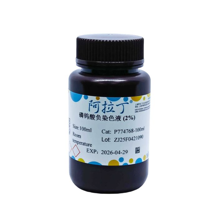
磷钨酸负染色液 (2%)
有货
BioReagent,生物染色剂,用于显微镜 级 ,适用于对基线干扰要求严格的色谱和分析工作流程。
避光,室温。常规运输 。请查阅批次 COA 获取详细规格。
SDS、COA、产品数据表及规格说明书均可下载。可通过批号查询获取批次 COA。
在色谱分析、有机合成和交叉偶联反应领域已被 0 篇同行评审文献引用。
负染色又称阴性染色,是由Hall发现的相对于普通染色(即正染色)而言的染色技术。其原理在于利用重金属盐包绕低电子密度的样品,增强样本四周的电子密度,造成细微结构之间的质量-厚度差异,增强散射吸收反差,使样品在黑暗的背景上呈现明亮的结构。负染色液有磷钨酸、钼酸铵、印度墨汁等,其中最常用的是1~3%磷钨酸。
磷钨酸负染色液(2%)适用于显示大分子、细菌、病毒、原生动物、噬菌体、细胞器、核酸大分子、蛋白质晶体及其他大分子材料等。染色后的样品图像呈现透明的亮光,而背景图像呈黑色。
使用说明:
一、滴染法
1. 样品低速离心 (2000g,10min) 或采用其他方法浓缩样品,制成悬浮液并且使其达到一定浓度和纯度。
2. 将样品悬浮液直接滴于带有支持膜的载网上,静置3~5min。
3. 用滤纸条从液滴边缘吸去多余液体,稍干燥。
4. 滴加负染色液,静置2~3min。
5. 吸去多余染色液,自然干燥,进行显微镜观察。
二、漂浮法
1. 样品低速离心 (2000g,10min) 或采用其他方法浓缩样品,制成悬浮液并且使其达到一定浓度和纯度。
2. 将带有支持膜的载网置于样品液滴上漂浮以沾取样品。
3. 载网置于负染色液上漂浮1~2min。
4. 吸去多余染色液,自然干燥,进行显微镜观察。
注意事项:
1、目的样本尽量新鲜。
2、样品应为均匀的悬浮液,其纯度和浓度应适宜,否则无法与染色剂之间产生特异和清晰的结合反应。
3、为了您的安全和健康,请穿实验服并戴一次性手套操作。
染色结果
| 样品 | 透明的亮光 |
| 背景 | 黑色 |
Negative staining, also known as negative contrast staining, was discovered by Hall. It is a staining technique in contrast to conventional staining (i.e., positive staining). Its principle relies on encasing low-electron-density specimens with heavy metal salts to raise the electron density surrounding the samples. This creates mass-thickness differences among fine structures, amplifies contrast via scattered and absorbed electrons, and renders bright structural features of the specimen against a dark background.Common negative staining solutions include phosphotungstic acid, ammonium molybdate, and India ink, among which 1–3% phosphotungstic acid is the most widely used.
2% phosphotungstic acid negative staining solution is suitable for visualizing macromolecules, bacteria, viruses, protozoa, bacteriophages, organelles, nucleic acid macromolecules, protein crystals, and other macromolecular materials. Stained specimens exhibit transparent bright outlines, while the background appears black in micrographs.
Instructions for Use
I. Drop Staining Method
1. Concentrate the sample via low-speed centrifugation (2000 g, 10 min) or alternative methods to prepare a suspension with appropriate concentration and purity.
2. Drop the sample suspension directly onto a copper grid coated with support film, and let it stand for 3–5 min.
3. Blot excess liquid from the edge of the droplet with filter paper and allow slight drying.
4. Add negative staining solution dropwise and incubate for 2–3 min.
5. Remove excess staining solution, air-dry naturally, and observe under an electron microscope.
II. Floating Staining Method
1. Concentrate the sample via low-speed centrifugation (2000 g, 10 min) or alternative methods to prepare a suspension with appropriate concentration and purity.
2. Float the support-film-coated copper grid on the sample droplet to adsorb the sample.
3. Float the grid on the negative staining solution for 1–2 min.
4. Remove excess staining solution, air-dry naturally, and observe under an electron microscope.
Precautions
1. The target sample should be as fresh as possible.
2. The sample must form a homogeneous suspension with suitable purity and concentration; otherwise, specific and distinct binding reactions with the stain cannot be achieved.
3. Wear a lab coat and disposable gloves during operation for personal safety and health protection.
Staining Results:
| Specimen | transparent bright areas |
| background | Black |
通过匹配包装上的批号来查找并下载产品的 COA,每批产品都进行了严格的验证,您可放心使用!
| 批号(Lot Number) | 证书类型 | 货号 |
|---|---|---|
 ZJ26F0736956 |
分析证书 | P774768 |
| ZJ25F0622938 |
分析证书 | P774768 |
| ZJ25F1028457 |
分析证书 | P774768 |
| ZJ25F0421090 |
分析证书 | P774768 |
我们的级别选择指南涵盖了目录中所有的变体的纯度、稳定剂状态和应用适用性。
查看 BioReagent 级别指南 → 查看 Biological Stain 级别指南 → 查看 for Microscopy 级别指南 →

 危险品化学品经营许可证(带存储)