
计算溶液所需的质量、体积或浓度。
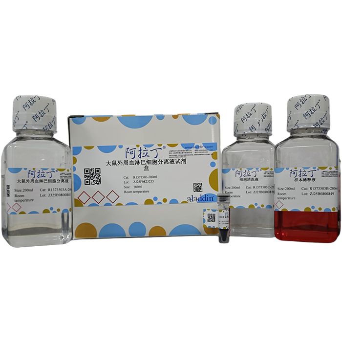
大鼠外周血淋巴细胞分离液试剂盒
有货
BioReagent,无菌,用于细胞培养 级 ,适用于对基线干扰要求严格的色谱和分析工作流程。
避光,室温。常规运输 。请查阅批次 COA 获取详细规格。
SDS、COA、产品数据表及规格说明书均可下载。可通过批号查询获取批次 COA。
在色谱分析、有机合成和交叉偶联反应领域已被 0 篇同行评审文献引用。
本产品是一种用于分离大鼠外周血中单个核细胞(主要为淋巴细胞)的无菌的密度梯度细胞分离液。其原理是根据大鼠外周血中各种细胞的密度不同,使用特定密度的细胞分离液(为1.084),经过梯度离心,可以使细胞在较短的时间内按照相应的密度梯度分布,从而将血液中的淋巴细胞分离出来。无菌条件下分离的淋巴细胞可用于后续培养。为了产品使用起来的方便与高效,本试剂盒配置了分离淋巴细胞过程中专用的样本稀释液、细胞清洗液和台盼蓝染色液。使用本试剂盒中的细胞清洗液清洗细胞可以极大的提高细胞回收率,减少在后续清洗过程中造成的细胞损失。
产品组分表:
| R1373503 | Component | 200 ml | Storage |
| R1373503A | 大鼠外周血淋巴细胞分离液 | 200 ml | RT.Store in the dark. |
| R1373503B | 样本稀释液 | 200 ml | RT.Store in the dark. |
| R1373503C | 细胞清洗液 | 200 ml | RT.Store in the dark. |
| R1373503D | 台盼蓝染色液(0.4%) | 1ml | RT.Store in the dark. |
产品适用范围
本产品适用于从大鼠的抗凝血液中分离单个核细胞(主要为淋巴细胞)。
保存条件
18-25℃,避光保存,有效期2年。本品易感染细菌,需在无菌条件下操作,启封后4℃避光保存。
操作步骤
1. 取新鲜的大鼠的抗凝血全血(EDTA、枸橼酸钠或肝素抗凝即可)或者去纤维蛋白血液,用等体积的本试剂盒中的样本稀释液稀释全血。
2. 在离心管中加入适量分离液(当稀释后血液体积小于 3ml 时,加入 3ml 分离液;大于等于 3ml加入等体积分离液。但二者的总体积不能超过离心管的三分之二,否则会影响分离效果),将稀释后的血液平铺到分离液液面上方,注意保持两液面界面清晰。(可以用巴氏德吸管吸取血液,然后将血液小心的平铺于分离液上。如果样品较多加样时间较长,在离心之前出现红细胞成团下沉属正常现象)
3. 室温,水平转子 400~1000g,离心 20~30min。(血液体积越大所需的离心力越大,离心时间越长,可根据具体情况摸索最佳分离条件)
4. 离心之后将出现明显的分层:最上层是稀释的血浆层,中间是透明的分离液层,血浆与分离液之间的白膜层即为目的细胞层,离心管底部是红细胞与粒细胞层。
5. 小心的吸取白膜层细胞到 15mL 洁净的离心管中,加入 5~10ml 左右本试剂盒中的细胞清洗液洗涤细胞。250-500g 离心 5-10min。
6. 洗涤 2-3 次,弃上清,用 PBS 重悬细胞备用。
分离过程示意图

注意事项
1. 开封前请将本产品颠倒混匀,本分离液为无菌产品,为延长分离液保存时间,请在无菌条件
下启封,启封后置于 4℃保存,防止微生物污染。
2. 室温下分离效果最好(20±2℃),低温可能会导致白膜层中红细胞污染严重,高温可能会导
致分离出的细胞数变少。
3. 血液样本最好为新鲜的抗凝血(2h 以内),为保持细胞的活性,避免冷冻和冷藏。血液放置超过 6h 会导致分离效果变差甚至不能达到分离目的。
4. 为保证本产品能够达到最佳的分离效果,请使用本试剂盒内配置的专用的细胞清洗液和样本稀释液进行操作。
5. 部分塑料制品(如聚苯乙烯)因其带有静电作用,可能会导致细胞挂壁,影响分离效果。
6. 吸取过多的分离液层可能会导致混杂的粒细胞数量增加;吸取过多的血浆层可能会导致细胞
7. 中血浆蛋白及血小板污染。
8. 如果要进一步对分离的细胞进行培养,需要在收集血液和分离的过程中,注意无菌操作。
This product is a sterile density‑gradient cell separation medium for isolating mononuclear cells (predominantly lymphocytes) from rat peripheral blood. Based on density differences among various cell populations in rat peripheral blood, this separation medium with a specific density of 1.084 enables cells to distribute according to their respective density gradients within a short period upon density‑gradient centrifugation, so as to isolate lymphocytes from blood. Lymphocytes isolated under sterile conditions can be used for subsequent cell culture. For convenient and efficient operation, this kit is equipped with dedicated sample dilution buffer, cell washing buffer and trypan blue staining solution for lymphocyte isolation. Washing cells with the cell washing buffer supplied in the kit can greatly improve cell recovery rate and reduce cell loss during subsequent washing steps.
Contents & Storage
|
Scope of Application
This product is intended for the isolation of mononuclear cells (predominantly lymphocytes) from anticoagulated rat blood.
Storage Conditions
Store at 18‑25 °C, protected from light, valid for 2 years from the date of manufacture. This product is susceptible to bacterial contamination; handle under aseptic conditions. After opening, store at 4 °C protected from light.
Operating Procedures
1. Take fresh anticoagulated rat whole blood (anticoagulants including EDTA, sodium citrate or heparin are acceptable) or defibrinated blood, and dilute the whole blood with an equal volume of Sample Dilution Buffer supplied in this kit.
2. Add an appropriate volume of separation medium into a centrifuge tube. Add 3 mL separation medium when the volume of diluted blood is less than 3 mL; add an equal volume of separation medium when the diluted blood volume is ≥ 3 mL. The total volume shall not exceed two‑thirds of the centrifuge‑tube capacity, otherwise the separation performance will be compromised. Layer the diluted blood carefully on top of the separation medium and maintain a distinct liquid‑liquid interface. Use a Pasteur pipette to slowly overlay the blood sample. If sample loading takes a long time due to large sample size, sedimentation of aggregated red blood cells prior to centrifugation is considered normal.
3. Centrifuge at room temperature in a horizontal rotor at 400‑1000 g for 20‑30 min. Larger blood volumes require higher centrifugal force and longer centrifugation duration; optimize the conditions empirically according to practical samples.
4. Distinct layers will form after centrifugation: the top layer is diluted plasma; the middle layer is transparent separation medium; the buffy coat located between plasma and separation medium contains target cells; red blood cells and granulocytes pellet at the bottom of the tube.
5. Carefully aspirate the buffy‑coat cells and transfer into a clean 15 mL centrifuge tube. Add approximately 5‑10 mL of Cell Washing Buffer from the kit for cell washing. Centrifuge at 250‑500 g for 5‑10 min.
6. Repeat the washing procedure 2‑3 times. Discard the supernatant and resuspend the cell pellet in PBS for subsequent use.
Schematic Diagram of Separation Process
 |
Precautions
1. Invert and mix thoroughly before opening. This separation medium is sterile. To extend shelf‑life, open the product under aseptic conditions. Store at 4 °C after opening to prevent microbial contamination.
2. Optimal separation performance is achieved at room temperature (20 ± 2 °C). Low temperature may cause severe red‑blood‑cell contamination in the buffy‑coat layer, while high temperature may reduce the yield of isolated cells.
3. Fresh anticoagulated blood samples (within 2 hours) are recommended. Avoid freezing or refrigeration to preserve cell viability. Blood samples stored longer than 6 hours will result in poor separation performance or even separation failure.
4. For optimal separation results, use the dedicated cell washing buffer and sample dilution buffer supplied within this kit.
5. Some plasticware (e.g. polystyrene) may cause cell adhesion due to static electricity, which compromises separation efficiency.
6. Excessive aspiration of separation medium may increase granulocyte contamination; excessive aspiration of plasma layer may introduce contamination from plasma proteins and platelets.
7. Strictly maintain aseptic conditions during blood collection and isolation if the harvested cells are intended for subsequent cell culture.
通过匹配包装上的批号来查找并下载产品的 COA,每批产品都进行了严格的验证,您可放心使用!
| 批号(Lot Number) | 证书类型 | 货号 |
|---|---|---|
 ZJ26F1039906 |
分析证书 | R1373503 |
| ZJ25F0825255 |
分析证书 | R1373503 |
我们的级别选择指南涵盖了目录中所有的变体的纯度、稳定剂状态和应用适用性。
查看 BioReagent 级别指南 → 查看 for Cell culture 级别指南 → 查看 Sterile 级别指南 →

 危险品化学品经营许可证(带存储)