Measured by the ability of the immobilized protein to support the adhesion of human monocyte-derived dendritic cells. When 5 x 10⁴ cells/well are added to human CD83/Fc Chimera coated plates (5 µg/mL, 100 µL/well), approximately 50-75% will adhere after 30 minutes at 37° C.
Function: May play a significant role in antigen presentation or the cellular interactions that follow lymphocyte activation.
Description: Human CD83 is a 40 - 50 kDa member of the Siglec (or sialic-acid-binding immunoglobulin-like lectin) family of transmembrane proteins. CD83 is synthesized as a type I transmembrane glycoprotein that contains a 125 amino acid (aa) extracellular region, a 22 aa transmembrane segment, and 39 aa cytoplasmic domain. It contains one V type Ig-like domain in the extracellular region with no inhibitory cytoplasmic motif(s). Although in vitro studies suggest CD83 may form membrane-bound covalent homodimers, in vivo this does not appear to be the case. In the extracellular region, mouse and human CD83 are 66% aa identical. Relative to human, mouse CD83 is 11 aa shorter in its extracellular domain and is expressed as a 30 - 35 kDa protein. Human CD83 is active in the mouse system. One alternate splice form has been reported. This leads to a small monomeric soluble form of 74 aa that includes aa’s # 20 - 52 and # 164 - 205 . In human, proteolytic cleavage and solubilization of CD83 has also been suggested, and this could lead to dimeric circulating CD83. CD83 is a primary marker for dendritic cells. It is also found on B cells, neutrophils, monocytes and macrophages. Except for dendritic cells, CD83 expression is often transient. CD83 binds to sialic acids on target cells. The function of CD83 is only now becoming clear. Membrane CD83 appears to promote T cell proliferation, particularly of CD8+ cytotoxic T cells. Soluble CD83, however, appears to be immunosuppressive and blocks T cell activation. On monocytes, CD83 is suggested to drive monocytes into a fibrocyte phenotype. A lack of membrane-expressed CD83 leads to an unusual IL-4/IL-10 producing CD4+ T cell phenotype.
规格
产品名称
Recombinant Human CD83 Protein
别名
重组人CD83蛋白 | 重组人 CD83 蛋白
英文别名
B-cell activation protein | BL11 | BL11CD83 antigen | CD83 antigen (activated B lymphocytes, immunoglobulin superfamily) | CD83 molecule | CD83 | Cell surface protein HB15 | cell-surface glycoprotein | HB15 | HB15hCD83 | B-cell activation protein | Cell s
Measured by the ability of the immobilized protein to support the adhesion of human monocyte-derived dendritic cells. When 5 x 10⁴ cells/well are added to human CD83/Fc Chimera coated plates (5 µg/mL, 100 µL/well), approximately 50-75% will adhere after 30 minutes at 37° C.
Lyophilized from PBS, pH 7.4 with 5%Trehalose, 5%Mannitol
复溶
Always centrifuge tubes before opening. Do not mix by vortex or pipetting. It is recommended to reconstitute to a concentration of 0.1-1mg/mL. Dissolve the lyophilized protein in distilled water. Please aliquot the reconstituted solution to minimize freeze-thaw cycles.
储存条件
-20°C储存,避免反复冻融
运输条件
超低温运输
稳定性与储存
建议在无菌条件下于 -20℃~ -80℃ 保存 1 年以上。避免反复冻融循环
图片
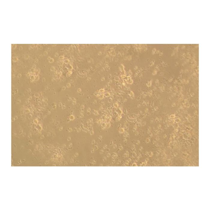
Recombinant Human CD83 Protein (rp144137) - Protein Bioactivity Measured by the ability of the immobilized protein to support the adhesion of human monocyte-derived dendritic cells. When 5 x 10⁴ cells/well are added to human CD83/Fc Protein coated plates (5 µg/mL, 100 µL/well), approximately 50-75% will adhere after 30 minutes at 37℃.
Recombinant Human CD83 Protein (rp144137) - SDS-PAGE 2 μg/lane of Recombinant Human CD83 Protein was resolved with SDS-PAGE under reducing (R) and non-reducing (N) conditions and visualized by Coomassie® Blue staining, showing the band at 16.2-27.5 kDa.


 ZJ23F0700592


 危险品化学品经营许可证(带存储)