Recombinant Human R-Spondin 1 protein stimulated Wnt signal pathway with Wnt-3a protein in HEK293T cells. After 6 hours, the stimulation when adding 300 ng/mL of R-Spondin-1 reached highest effect. Compared with only Wnt-3a stimulation, the Wnt signaling pathway was enhanced 3.1-fold after adding 300 ng/mL R-Spondin 1.
Activator of the canonical Wnt signaling pathway by acting as a ligand for LGR4-6 receptors. Upon binding to LGR4-6 (LGR4, LGR5 or LGR6), LGR4-6 associate with phosphorylated LRP6 and frizzled receptors that are activated by extracellular Wnt receptors, triggering the canonical Wnt signaling pathway to increase expression of target genes. Also regulates the canonical Wnt/beta-catenin-dependent pathway and non-canonical Wnt signaling by acting as an inhibitor of ZNRF3, an important regulator of the Wnt signaling pathway. Acts as a ligand for frizzled FZD8 and LRP6. May negatively regulate the TGF-beta pathway. Has a essential roles in ovary determination. Regulates Wnt signaling by antagonizing DKK1/KREM1-mediated internalization of LRP6 through an interaction with KREM1.
Post-translational: C-, and N-glycosylated. N-glycosylation at Asn-137, negatively influences its secretion and enhancing effect on Wnt/beta-catenin signaling. C-mannosylation at Trp-156 by DPY19L3 is required for its secretion and regulates the enhancing activity of Wnt signaling
生物活性
Recombinant Human R-Spondin 1 protein stimulated Wnt signal pathway with Wnt-3a protein in HEK293T cells. After 6 hours, the stimulation when adding 300 ng/mL of R-Spondin-1 reached highest effect. Compared with only Wnt-3a stimulation, the Wnt signaling pathway was enhanced 3.1-fold after adding 300 ng/mL R-Spondin 1.
Lyophilized from 10mM PBS, 5% Trehalose, 5% Mannitol, pH7.4.
复溶
Reconstitute in sterile water to a concentration of 0.1-0.5 mg/ml.
储存条件
-20°C储存,避免反复冻融
运输条件
超低温运输
稳定性与储存
Store at -20~-80℃ for more than 1 year. Upon delivery aliquot. Avoid freeze/thaw cycle.
图片
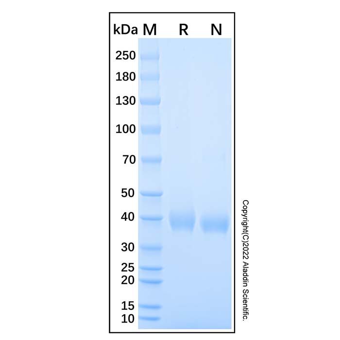
Recombinant Human RSPO1 Protein (rp151196) - SDS-PAGE 2 μg/lane of Recombinant Human RSPO1 Protein was resolved with SDS-PAGE under reducing (R) and non-reducing (N) conditions and visualized by Coomassie® Blue staining, showing the band at 35 - 42 kDa.

 ZJ25F0723562


 危险品化学品经营许可证(带存储)