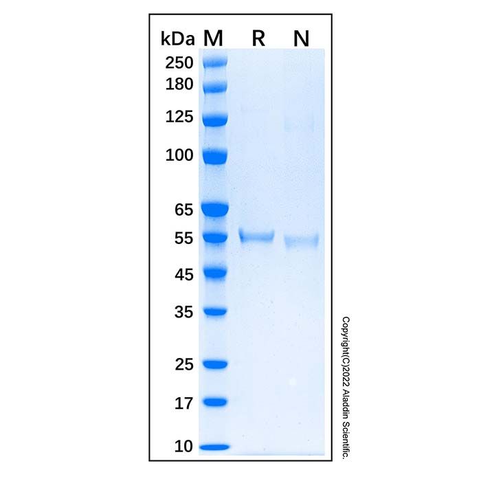
Purity:>90%, by SDS-PAGE visualized with Coomassie® Blue Staining. Description: p53 is well known for its key role as a tumor suppressor protein. It is 393 amino acids (aa) in length with a predicted molecular weight of 44 kDa. It belongs to the p53 family that also includes p63 and p73. Structurally, p53 is characterized by an N-terminal transactivation domain, central DNA-binding and oligomerization domains, and a C-terminal regulatory domain. It is thought to exist as a homotetramer, and it exhibits approximately 72% and 76% aa identity with its mouse and rat orthologs, respectively. Mutations in the p53 gene are one of the most frequent genomic events accompanying oncogenic transformation. p53 responds to signals such as DNA damage or cell stress primarily through its actions as a transcription factor. Among its gene targets are a range factors that promote DNA repair mechanisms or apoptosis, including cell cycle regulatory proteins and members the Bcl-2 family. Because of its critical role in genomic homeostasis, p53 activities are tightly regulated by a network of protein-protein interactions, microRNAs, and a range of post-translational modifications, including phosphorylation, acetylation, methylation, and ubiquitination. A widely studied regulator is Murine Double Minute 2 (MDM2). MDM2 is known to suppress p53 activity through direct binding or through its actions as a Ubiquitin ligase (E3) that catalyzes p53 ubiquitination and proteasome-mediated degradation.
We recommend that this vial be briefly centrifuged prior to opening to bring the contents to the bottom. Reconstitute at 0.5 mg/mL in sterile distilled water. Stock solutions should be apportioned into working aliquots and stored at ≤ -20 °C. Further dilutions should be made in appropriate buffered solutions.
储存条件
-20°C储存,避免反复冻融
运输条件
超低温运输
稳定性与储存
在 -20°C 下可保存 1 年。避免冷冻/解冻循环。
图片
Recombinant Human p53 Protein (rp170134) - SDS-PAGE 2 μg/lane of Recombinant Human p53 Protein was resolved with SDS-PAGE under reducing (R) and non-reducing (N) conditions and visualized by Coomassie® Blue staining, showing a band at 55.4 kDa under reducing conditions and 52.5 & 122.4 kDa under non-reducing conditions.

 ZJ24F0303510


 危险品化学品经营许可证(带存储)