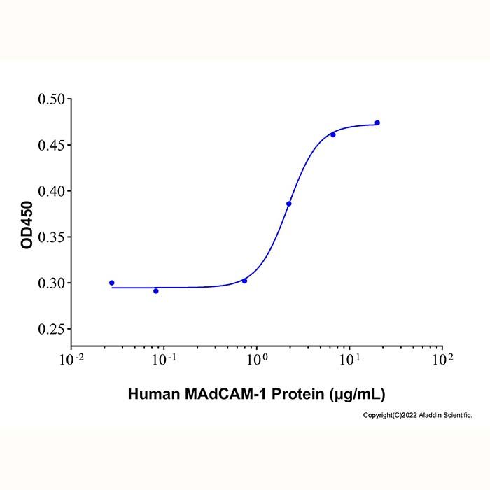
Measured by the ability of the immobilized protein to support the adhesion of Jurkat human T-lymphocyte leukemia cells in the presence of 1.0 mM MnCl2, The ED50 for this effect is 2.19 μg/mL.
Protein Function: Mucosal addressin cell adhesion molecule-1 (MAdCAM-1) is an approximately 60 kDa type 1 transmembrane glycoprotein. It is an endothelial cell adhesion molecule that belongs to the immunoglobulin (Ig) superfamily of proteins. Human MAdCAM-1 is synthesized as a 382 amino acid (aa) precursor that contains an 18 aa signal sequence, a 299 aa extracellular domain (ECD), a 21 aa transmembrane segment, and a 44 aa cytoplasmic tail. Within the ECD, there is one potential site for N-linked glycosylation. The ECD comprises two Ig-like domains of 90 aa and 119 aa, respectively, each possessing invariant cysteine residues that stabilize the Ig loop. There is also a Ser-Thr-Pro-rich (71%) mucin-like 48 aa domain that is (aa 206 ‑ 317) formed by six tandem repeats of an eight aa sequence having the general consensus DTTSPEP/SP. This mucin domain contains 19 potential sites for O-linked glycosylation. A splicing variant in which a single Ala residue is substituted for aa 223 ‑ 334 in isoform 1 produces a second isoform. Human mature MAdCAM-1 shares only 44% aa sequence identity with mature mouse MAdCAM-1. The integrin alpha (4) beta (7), which is expressed on lymphocytes, functions as the MAdCAM-1 receptor. The Ig domains of MAdCAM-1 are critical to alpha (4) beta (7) binding, and the mucin domain has activity in L‑Selectin binding. MAdCAM-1 expression is up-regulated by TNF-alpha and IL‑1 beta. MAdCAM-1 is expressed on the surface of high endothelial venules (HEV) in the gut and in Peyer’s patches, on endothelial cells of the mesenteric lymph nodes, lamina propria of the small and large intestine, and the mammary gland during lactation, and on brain endothelial cells. MAdCAM‑1 has also been reported to be expressed in the liver portal region in autoimmune hepatitis, and in bone marrow following allogenic (genetically non-identical) hematopoietic stem cell transplantation, where it recruits donor T cells, which may lead to graft versus host disease. MAdCAM‑1 functions as a homing receptor and plays a central role in leukocyte migration into HEVs and Peyer’s patch. In addition to its normal role in lymphocyte trafficking to mucosal tissue, MAdCAM‑1 expression is also dramatically increased in chronic inflammatory and disease states, including inflammatory bowel disease (Crohn’s disease and ulcerative colitis), sclerosing cholangitis, and diabetes, and may play an important role in these conditions.
Measured by the ability of the immobilized protein to support the adhesion of Jurkat human T-lymphocyte leukemia cells in the presence of 1.0 mM MnCl2, The ED50 for this effect is 2.19 μg/mL.
48.2-77.1 kDa, under reducing conditions; 48.2-77.1 kDa, under non-reducing conditions
分子类型
蛋白质
产品线
生物产品
储存与运输
物理形态
冻干(Lyophilized)
储存缓冲液
Lyophilized from PBS, pH 7.4
复溶
We recommend that this vial be briefly centrifuged prior to opening to bring the contents to the bottom. Reconstitute at 0.5 mg/mL in sterile distilled water. Stock solutions should be apportioned into working aliquots and stored at ≤ -20 °C. Further dilutions should be made in appropriate buffered solutions.
储存条件
-20°C储存,避免反复冻融
运输条件
超低温运输
稳定性与储存
在 -20°C 下可保存 1 年。避免冷冻/解冻循环。
图片
Recombinant Human MAdCAM-1 Protein (rp181270) - Protein Bioactivity Measured by the ability of the immobilized protein to support the adhesion of Jurkat human T-lymphocyte leukemia cells in the presence of 1.0 mM MnCl₂, The ED
₅₀
for this effect is 2.19 μg/mL.
Recombinant Human MAdCAM-1 Protein (rp181270) - SDS-PAGE 3 μg/lane of Recombinant Human MAdCAM-1 Protein was resolved with SDS-PAGE under reducing (R) and non-reducing (N) conditions and visualized by Coomassie® Blue staining, showing a band at 48.2-77.1 kDa.

 ZJ24F0708066


 危险品化学品经营许可证(带存储)