Stem cell factor (SCF), also known as c-kit ligand (KL), mast cell growth factor (MGF), and steel factor (SLF), is a widely expressed 28‑40 kDa type I transmembrane glycoprotein. It promotes the survival, differentiation, and mobilization of multiple cell types including myeloid, erythroid, megakaryocytic, lymphoid, germ cell, and melanocyte progenitors. SCF is a primary growth and activation factor for mast cells and eosinophils. Mature human SCF consists of a 189 amino acid (aa) extracellular domain (ECD), a 23 aa transmembrane segment, and a 36 aa cytoplasmic tail. The ECD shows both N‑linked and O-linked glycosylation. Proteolytic cleavage at two alternate sites in the extracellular juxtamembrane region releases a 25 kDa soluble molecule which is comparable to the only form produced by Steel-dickie mutant mice. An alternately spliced isoform of human SCF lacks 28 aa that encompasses the primary proteolytic recognition site. Within the ECD of the long isoform (corresponding to this recombinant protein), human SCF shares 79%‑87% aa sequence identity with canine, feline, mouse, and rat SCF. Rat SCF is active on mouse and human cells, but human SCF is only weakly active on mouse cells. Noncovalent dimers of transmembrane or soluble SCF interact with the receptor tyrosine kinase SCF R/c‑kit to trigger receptor dimerization and signaling. SCF assists in the recovery of cardiac function following myocardial infarction by increasing the number of cardiomyocytes and vascular channels. SCF is a versatile factor in the differentiation of many specific cell types like spermatogonial stem cells and megakaryocyte progenitors. Apart from differentiation, SCF also can maintain stemness in cells. This is the case for human bone marrow mesenchymal cells, which require SCF and hepatocyte growth factor for maintenance. Hematopoietic stem cells similarly require SCF from surrounding cells in their niche to maintain their stemness and their progenitors. SCF has also improved protocols for continuous generation of cells in culture systems, like granulocytes and macrophages. For treatment of graft versus host disease, SCF is used in combination with other cytokines to generate myeloid-derived suppressor cells from human umbilical cord blood. SCF is also used to generate T cells for cell-based therapies, drug screening, and disease modeling. In regenerative studies, SCF is applied in wound healing hydrogel as a means of increasing its adhesion strength and tissue regeneration.
Post-translational: A soluble form (sKITLG) is produced by proteolytic processing of isoform 1 in the extracellular domain. Found in two differentially glycosylated forms, LMW-SCF and HMW-SCF. LMW-SCF is fully N-glycosylated at Asn-145, partially N-glycosylated at Asn-90, O-glycosylated at Ser-167, Thr-168 and Thr-180, and not glycosylated at Asn-97 or Asn-118. HMW-SCF is N-glycosylated at Asn-118, Asn-90 and Asn-145, O-glycosylated at Ser-167, Thr-168 and Thr-180, and not glycosylated at Asn-97. A soluble form exists as a cleavage product of the extracellular domain.
生物活性
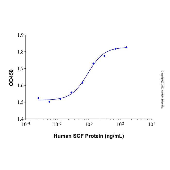
Measured in a cell proliferation assay using TF-1 cells. The ED50 for this effect is typically 0.86 ng/mL.
20.4-31.6 kDa, under reducing conditions; 18.1-28.3 kDa, under non-reducing conditions.
分子类型
蛋白质
产品线
生物产品
储存与运输
物理形态
冻干(Lyophilized)
储存缓冲液
Lyophilized from PBS, pH 7.4
浓度
见COA
复溶
We recommend that this vial be briefly centrifuged prior to opening to bring the contents to the bottom. Reconstitute at 1.0 mg/mL in sterile distilled water. Stock solutions should be apportioned into working aliquots and stored at ≤ -20 °C. Further dilutions should be made in appropriate buffered solutions.
储存条件
-20°C储存,避免反复冻融
运输条件
超低温运输
稳定性与储存
Store at -20°C for 1 year. Avoid freeze/thaw cycle.
图片
Recombinant Human SCF Protein (rp215100) - Bioactivity Measured in a cell proliferation assay using TF-1 cells. The ED50 for this effect is typically 0.86 ng/mL.
Recombinant Human SCF Protein (rp215100) - SDS-PAGE 3 μg/lane of Recombinant Human SCF Protein was resolved with SDS-PAGE under reducing (R) and non-reducing (N) conditions and visualized by Coomassie® Blue staining, showing a band at 20.4-31.6 kDa under reducing conditions and 18.1-28.3 kDa under non-reducing conditions.

 ZJ25F0521176


 危险品化学品经营许可证(带存储)