FAK (Focal Adhesion Kinase) is a non-receptor protein tyrosine kinase involved in signal transduction from integrin-enriched focal adhesion sites that mediate cell contact with the extracellular matrix. FAK-enhanced signals have been shown to mediate the survival of anchorage-dependent cells and are critical for efficient cell migration in response to growth factor receptor and integrin stimulation. Elevated expression of FAK in human tumors has been correlated with increased malignancy and invasiveness. Elevated FAK expression in anaplastic astrocytoma and glioblastoma tumor biopsy samples has been demonstrated.
Post-translational: Phosphorylated on tyrosine residues upon activation, e.g. upon integrin signaling. Tyr-397 is the major autophosphorylation site, but other kinases can also phosphorylate this residue. Phosphorylation at Tyr-397 promotes interaction with SRC and SRC family members, leading to phosphorylation at Tyr-576, Tyr-577 and at additional tyrosine residues. FGR promotes phosphorylation at Tyr-397 and Tyr-576. FER promotes phosphorylation at Tyr-577, Tyr-861 and Tyr-925, even when cells are not adherent. Tyr-397, Tyr-576 and Ser-722 are phosphorylated only when cells are adherent. Phosphorylation at Tyr-397 is important for interaction with BMX, PIK3R1 and SHC1. Phosphorylation at Tyr-925 is important for interaction with GRB2. Dephosphorylated by PTPN11; PTPN11 is recruited to PTK2 via EPHA2 (tyrosine phosphorylated). Microtubule-induced dephosphorylation at Tyr-397 is crucial for the induction of focal adhesion disassembly; this dephosphorylation could be catalyzed by PTPN11 and regulated by ZFYVE21. Phosphorylation on tyrosine residues is enhanced by NTN1 (By similarity). Sumoylated; this enhances autophosphorylation.
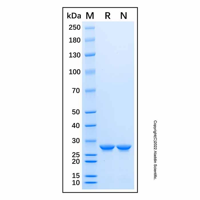
27.5 kDa, under reducing conditions; 27.5 kDa, under non-reducing conditions.
分子类型
蛋白质
产品线
生物产品
储存与运输
物理形态
液体
储存缓冲液
PBS, 5% Glycerol, pH 7.4
浓度
见COA
储存条件
-20°C储存,避免反复冻融
运输条件
超低温运输
稳定性与储存
-20℃ 存储12个月;收货后建议分装,避免反复冻融。
图片
Recombinant Human FAK Protein (rp217136) - SDS-PAGE 3 μg/lane of Recombinant Human FAK Protein was resolved with SDS-PAGE under reducing (R) and non-reducing (N) conditions and visualized by Coomassie® Blue staining, showing a band at 27.5 kDa.

 ZJ25F0622900


 危险品化学品经营许可证(带存储)