Tyrosine protein kinase ABL1 (Abelson murine leukemia viral oncogene homolog 1) is a ubiquitously expressed, magnesium-dependent, cytosolic member of the ABL subfamily of non-receptor protein tyrosine kinases. Human nonreceptor tyrosine kinases share a conserved domain structure that contributes to activity regulation and substrate specificity in many key processes linked to cell growth and survival. In addition, ABL1 contains domains unique to the ABL1 paralog that enable its function in DNA binding and DNA-damage response. ABL1 is composed of an N-terminal glycine myristoyl group that blocks the surface pocket of the kinase domain, a cap that stabilizes an inactive conformation, SH3 and SH2 domains that impose a locked inactive state, a protein kinase region, three nuclear localization signals, a DNA-binding region, and C-terminal nuclear export signal motifs, and actin F-binding domain. Activity is regulated through disruption of autoinhibitory interactions and several phosphorylation events that increase or decrease activity or promote function through stabilization. The SH2 domain contributes to both catalytic activity and target site specificity as has been shown with domain swapping. In chronic myelogenous, acute myeloid, and acute lymphoblastic leukemias, ABL1 is fused to BCR gene, resulting in deletion of the regulatory domains and production of a constitutively active tyrosine kinase. Several mutations in ABL1 lead to misregulation of activity in a variety of cancers. ABL1 kinase inhibition is used for therapeutic treatment in leukemic cancers and requires further development of drugs to address ABL1-mutation conferred resistance.
Post-translational: Acetylated at Lys-711 by EP300 which promotes the cytoplasmic translocation. Phosphorylation at Tyr-70 by members of the SRC family of kinases disrupts SH3 domain-based autoinhibitory interactions and intermolecular associations, such as that with ABI1, and also enhances kinase activity. Phosphorylation at Tyr-226 and Tyr-393 correlate with increased activity. DNA damage-induced activation of ABL1 requires the function of ATM and Ser-446 phosphorylation (By similarity).Phosphorylation at Ser-569 has been attributed to a CDC2-associated kinase and is coupled to cell division (By similarity).Phosphorylation at Ser-618 and Ser-619 by PAK2 increases binding to CRK and reduces binding to ABI1. Phosphorylation on Thr-735 is required for binding 14-3-3 proteins for cytoplasmic translocation. Phosphorylated by PRKDC (By similarity). Polyubiquitinated. Polyubiquitination of ABL1 leads to degradation.
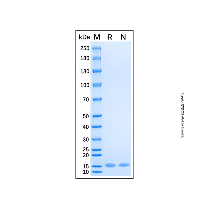
15.4 kDa, under reducing conditions; 15.4 kDa, under non-reducing conditions.
分子类型
蛋白质
产品线
生物产品
储存与运输
物理形态
冻干(Lyophilized)
储存缓冲液
Lyophilized from 20mM Tris, 300mM NaCl, pH 8.0.
浓度
expressed in E. coli; 见COA
复溶
We recommend that this vial be briefly centrifuged prior to opening to bring the contents to the bottom. Reconstitute at 1.0 mg/mL in sterile distilled water. Stock solutions should be apportioned into working aliquots and stored at ≤ -20 °C. Further dilutions should be made in appropriate buffered solutions.
储存条件
-20°C储存,避免反复冻融
运输条件
超低温运输
稳定性与储存
-20℃保存 12个月;复溶后建议分装,避免反复冻融。
图片
Recombinant Human ABL1 Protein (rp217149) - SDS-PAGE
2 μg/lane of Recombinant Human ABL1 Protein was resolved with SDS-PAGE under reducing (R) and non-reducing (N) conditions and visualized by Coomassie® Blue staining, showing a band at 15.4 kDa.

 ZJ26F0435021


 危险品化学品经营许可证(带存储)