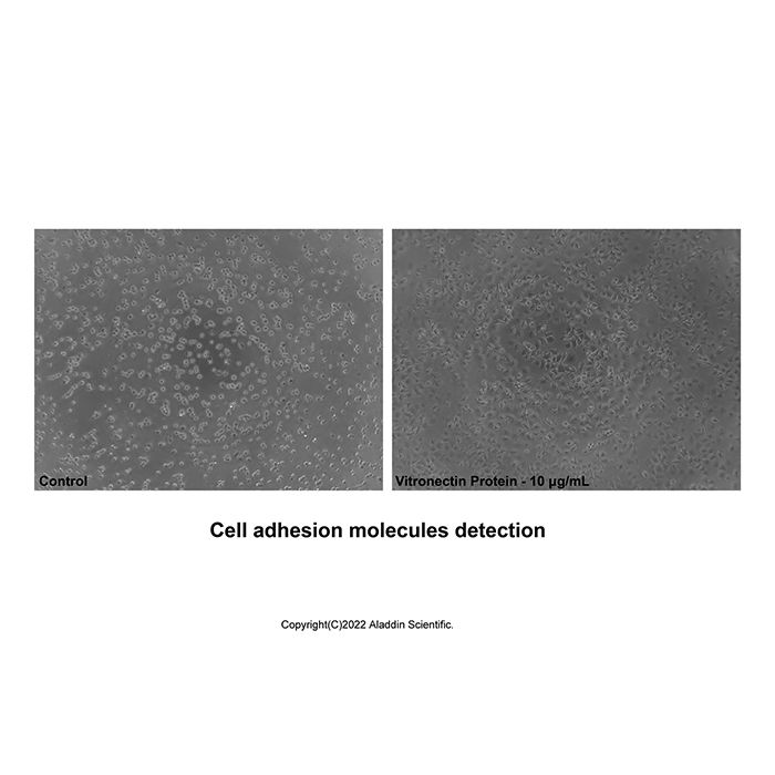
Measured by the ability of the immobilized protein to support the adhesion of NIH3T3 mouse embryonic fibroblast cells. The ED50 for this effect is 2.0-10.0 μg/mL.
Vitronectin is a large glycoprotein found in blood and the extracellular matrix (ECM). The gene for Vitronectin encodes a 19 amino acid (aa) signal peptide and a 459 aa protein. The amino terminal 130 aa residues of Vitronectin contain multiple binding sites for a variety of structures. Included is a site for binding to plasminogen activator inhibitor-1 (PAI‑1) and urokinase receptor, an (RGD) sequence that binds alpha v beta 3, alpha v beta 5, alpha v beta 1, alpha IIb beta 3, alpha v beta 6, and alpha v beta 8 integrins, a stretch of acidic amino acids that includes two sulfated tyrosine residues that bind thrombin-anti-thrombin III complexes, and a collagen binding site. The major part of the Vitronectin molecule (aa 132-459) contains six hemopexin-like repeats. The carboxyl-terminal end of Vitronectin also has multiple sites and functions. It contains a stretch of basic amino acids that binds the acidic amino acids of the amino-terminal region, thereby stabilizing Vitronectin’s three dimensional structure. The carboxyl-terminal end also contains a plasminogen binding site, a heparin binding site that binds complement factor C7, C8 or C9, a glycosaminoglycan binding site, and a second PAI-1 binding site (aa 348-370). Vitronectin also contains an endogenous cleavage site, plus cleavage sites for elastase, thrombin and plasmin. Vitronectin has also been shown to bind IGF-2 and TGF-beta. The apparent molecular weight of human Vitronectin is 75 kDa, with ~30% of its molecular mass being attributed to glycosylation at 3 different sites. In blood and plasma, Vitronectin is found predominantly as a single chain monomer. It can also be found as a dimer after endogenous cleavage. The dimer is composed of a 65 kDa and 10 kDa component held together by a disulfide bond. Binding of thrombin-anti-thrombin II complex or complement leads to an unfolding of Vitronectin. Unfolding of Vitronectin generates disulfide-linked multimers that are found in platelet secretions and extracellular matrix. Vitronectin is produced at high levels by the liver and many tumors. As might be expected by its structure, Vitronectin is involved in a number of biological activities including cell adhesion, cell spreading and migration, cell proliferation, extracellular anchoring, fibrinolysis, hemostasis, and complement mediated immune defense.
Post-translational: Sulfated on tyrosine residues.N- and O-glycosylated.Phosphorylation on Thr-69 and Thr-76 favors cell adhesion and spreading.It has been suggested that the active SMB domain may be permitted considerable disulfide bond heterogeneity or variability, thus two alternate disulfide patterns based on 3D structures are described with 1 disulfide bond conserved in both.Phosphorylation sites are present in the extracellular medium.
生物活性
Measured by the ability of the immobilized protein to support the adhesion of NIH3T3 mouse embryonic fibroblast cells. The ED50 for this effect is 2.0-10.0 μg/mL.
62.8-68.7 & 70.4-80.2 kDa, under reducing conditions; 65.7-75.2 kDa, under non-reducing conditions.
分子类型
蛋白质
产品线
生物产品
储存与运输
物理形态
冻干(Lyophilized)
储存缓冲液
Lyophilized from PBS, pH 7.4
浓度
见COA
复溶
We recommend that this vial be briefly centrifuged prior to opening to bring the contents to the bottom. Reconstitute at 1.0 mg/mL in sterile distilled water. Stock solutions should be apportioned into working aliquots and stored at ≤ -20 °C. Further dilutions should be made in appropriate buffered solutions.
储存条件
-20°C储存,避免反复冻融
运输条件
超低温运输
稳定性与储存
-20℃保存 12个月;复溶后建议分装,避免反复冻融。
图片
Recombinant Human Vitronectin Protein (rp220404) - Bioactivity Measured by the ability of the immobilized protein to support the adhesion of NIH3T3 mouse embryonic fibroblast cells. The ED50 for this effect is 2.0-10.0 μg/mL.
Recombinant Human Vitronectin Protein (rp220404) - SDS-PAGE 3 μg/lane of Recombinant Human Vitronectin Protein was resolved with SDS-PAGE under reducing (R) and non-reducing (N) conditions and visualized by Coomassie® Blue staining, showing a band at 62.8-68.7 & 70.4-80.2 kDa under reducing conditions and 65.7-75.2 kDa under non-reducing conditions. Vitronectin exists in two forms: a single chain 75 kDa form (V75) and a clipped form composed of two chains (65 kDa and 10 kDa) (V65+V10) which are held together by a disulfide bond(PMID: 12447888).

 ZJ25F0825757


 危险品化学品经营许可证(带存储)