基于人类免疫缺陷病毒-1(HIV-1)的慢病毒载体是一种用于基因转移研究的载体。慢病毒载体可以将外源基因或外源的shRNA有效地整合到宿主染色体上,从而达到持久性表达目的序列的效果。在感染能力方面,慢病毒载体可有效地感染神经元细胞、肝细胞、心肌细胞、肿瘤细胞、内皮细胞、干细胞等多种类型的细胞。对于一些较难转染的细胞,如原代细胞、干细胞、不分化的细胞等,使用慢病毒载体能大大提高目的基因或目的shRNA的转导效率,且目的基因或目的shRNA整合到宿主细胞基因组的几率大大增加,能够比较方便快捷地实现目的基因或目的shRNA的长期、稳定表达。在体外实验及体内实验的研究中,慢病毒己经成为表达外源基因或外源shRNA的常用载体形式之一,并且正在获得越来越广泛的应用。
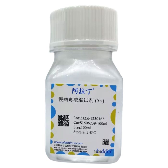
Lentivirus Concentration Solution (5x) 慢病毒浓缩试剂是针对慢病毒富集而优化的聚合物材料,它提供了一种简单、快速、高效的慢病毒颗粒浓缩方法。只需将慢病毒上清液与慢病毒浓缩试剂按比例混合,短时间孵育,采用标准离心机进行离心即可得到慢病毒颗粒沉淀。超滤、超速离心法等病毒浓缩法,操作时间长,步骤繁琐,且通常会有细胞碎片和血清蛋白等的污染,病毒纯度低。使用本产品进行病毒浓缩后,病毒滴度可提高10-100倍,病毒回收率最高可达约90%,病毒损失少,并且病毒在浓缩过程中能保持病毒生物活性。整个实验过程可在 4 小时内快速完成,无需超速离心即可获得出色的回收效率。产品特点:
⁕ 简单快速——操作简单,无需超速离心,确保活性,实验耗时不超过4小时
⁕ 浓缩效果优——病毒滴度可浓缩10-100倍以上(Transduction Units/mL)
⁕ 回收效率高——慢病毒回收效率高达 90% 以上
⁕ 应用范围广——适用于所有类型的慢病毒颗粒
实验流程概要:

图一. 慢病毒浓缩实验流程图
实验步骤:
1.将含有慢病毒颗粒的培养基转移至无菌容器中,800xg离心10分钟以除去细胞碎片。
⁕ 为保证病毒活性,浓缩前的病毒上清液应尽量选择新鲜收集的病毒原液。
2. 使用0.45μm过滤器过滤上清液至无菌容器中。
⁕ 如果使用滤膜过滤,推荐使用低蛋白结合力的纤维素醋酸酯或聚醚砜(PES)膜,不推荐使用硝酸纤维素膜(NC)。
3. 向每4体积含慢病毒的上清液中加入1体积慢病毒浓缩试剂(5x),使慢病毒浓缩试剂(5x)稀释为工作浓度(1x)。
4. 将上述混合物于4℃ 摇床中慢速孵育3小时或过夜。
⁕ 慢病毒颗粒在 4℃ 可稳定长达4天。
5. 将混合物4℃ 4000 x g 离心25分钟。离心后,慢病毒颗粒可能在容器底部显示为米色或白色沉淀。
6. 小心弃去上清液,4℃ 4000 x g 离心5分钟,再次弃尽残余液体,应尽量避免吸走沉淀物,该沉淀物即为慢病毒颗粒。
7. 用初始体积(原上清液体积)1/10 -1/100 预冷的慢病毒保存液重悬病毒颗粒,使用移液器小心吹打,重悬慢病毒颗粒。
⁕ 重悬病毒沉淀时,吹打操作要轻柔,可选择慢病毒稳定储存液(L1505998)、完全培养基或FBS重悬慢病毒。
8. 小体积分装慢病毒,储存于 -80℃ 冰箱或液氮中,留取少量病毒测定滴度。
⁕ 应尽量避免慢病毒反复冻融,否则会降低病毒滴度。
适用范围:
适用于任何慢病毒颗粒的浓缩方法。
注意事项:
1. 为了您的健康安全,请规范操作,穿戴实验服与手套开展实验。
2. 所有慢病毒操作均需在BSL2级生物安全柜中进行。
3. 本产品仅供科研使用,请勿用于临床诊断及治疗。
实验案例分析:
1.使用10cm细胞培养皿培养Lentivirus Concentration Solution (5x),待细胞密度为75%左右开始包装慢病毒。
2.使用Lentiviral Packaging Kit(L1455942)进行慢病毒包装(阳性对照质粒含GFP绿色荧光蛋白基因),具体包装步骤按照说明书进行。
3.准备目的细胞:将需感染的目的细胞293T以(1-3)x10⁴的密度接种于96孔板内,次日即可进行慢病毒感染。
4.待病毒包装48h后,收集慢病毒上清液约9mL,800xg离心10分钟以除去细胞碎片,使用0.45μm过滤器过滤上清液至无菌容器中。
5.取1mL病毒上清液作为浓缩前对照1*,剩余8mL病毒上清液中加入2mL本产品进行慢病毒浓缩,具体慢病毒浓缩步骤按照说明书进行,保留1mL浓缩后应弃去的上清液作为对照2#,使用800μL 慢病毒稳定储存液(L1505998)轻轻重悬慢病毒颗粒,即可得到浓缩10倍的慢病毒。
6.慢病毒感染目的细胞:将96孔板细胞弃去上清,每孔加入新鲜培养基100μL,Polybrene/聚凝胺( P1501912 )0.2μL,浓缩后慢病毒100μL或对照1*(浓缩前慢病毒原液)100μL或对照2#(浓缩后应弃去的慢病毒上清液)100μL。
7.病毒感染16小时后更换新鲜完全培养基,继续培养细胞48h后,使用荧光显微镜观察绿色荧光强度,实验结果如图2所示。
此外,我们在多种常见细胞株上进行了慢病毒浓缩效果验证,实验结果如图3所示。

图2. 慢病毒感染目的细胞293T观察GFP表达情况。(左)浓缩10x慢病毒,(中)对照1*(浓缩前慢病毒原液),(右)对照2#(浓缩后应弃去的慢病毒上清液)


图3. 采用 Lentiviral Packaging Kit(L1455942)高滴度慢病毒包装试剂盒进行慢病毒包装,收集慢病毒,采用慢病毒浓缩试剂 (S1506239) 进行10倍浓缩,感染目的细胞,检测荧光蛋白表达水平。(左)浓缩10x慢病毒,(中)对照1,浓缩前慢病毒原液,(右)对照2,浓缩后应弃去的慢病毒上清液。
Lentiviral vectors based on Human Immunodeficiency Virus-1 (HIV-1) are widely used tools for gene transfer research. These vectors can efficiently integrate exogenous genes or exogenous shRNA (short hairpin RNA) into the host chromosome, thereby achieving long-term stable expression of the target sequence. In terms of infectivity, lentiviral vectors can effectively transduce various cell types, including neuronal cells, hepatocytes, cardiomyocytes, tumor cells, endothelial cells, and stem cells. For cells that are difficult to transfect—such as primary cells, stem cells, and undifferentiated cells—using lentiviral vectors significantly improves the transduction efficiency of target genes or target shRNA. Additionally, it greatly increases the probability of target genes or target shRNA integrating into the host cell genome, enabling convenient and rapid long-term, stable expression of the target sequence. In both in vitro and in vivo research, lentiviruses have become one of the most common vector systems for expressing exogenous genes or exogenous shRNA, and their applications are expanding increasingly.
Lentivirus Concentration Solution (5×) is a polymer-based reagent optimized for lentivirus enrichment. It provides a simple, rapid, and efficient method for concentrating lentiviral particles. The protocol involves mixing lentiviral supernatant with the Lentivirus Concentration Solution at the recommended ratio, incubating for a short period, and then centrifuging using a standard centrifuge to obtain precipitated lentiviral particles. In contrast, other viral concentration methods such as ultrafiltration and ultracentrifugation are time-consuming and cumbersome to operate. They also often suffer from contamination by cell debris and serum proteins, resulting in low viral purity. When using this product for viral concentration, the viral titer can be increased by 10–100 fold, with a maximum viral recovery rate of approximately 90%. This method minimizes viral loss while preserving the biological activity of lentiviruses during the concentration process. The entire experimental procedure can be completed quickly within 4 hours, and excellent recovery efficiency is achieved without the need for ultracentrifugation.
Product Features:
⁕ Simple & Rapid – Easy to operate, no ultracentrifugation required, ensures viral activity, and the experiment takes no more than 4 hours.
⁕ Excellent Concentration Effect – Viral titer can be concentrated by 10–100 folds or more (Transduction Units/mL).
⁕ High Recovery Efficiency – Lentivirus recovery efficiency is as high as 90% or more.
⁕ Wide Application Range – Suitable for all types of lentiviral particles.
Summary of Experimental Procedure:

Figure 1. Flowchart of Lentivirus Concentration Experiment
Experimental Steps:
1. Transfer the culture medium containing lentiviral particles to a sterile container, and centrifuge at 800×g for 10 minutes to remove cell debris.
⁕ To ensure viral activity, freshly collected viral lentiviral supernatant is recommended for concentration.
2. Filter the supernatant through a 0.45 μm filter into a sterile container.
⁕ For membrane filtration, low protein-binding cellulose acetate or polyethersulfone (PES) membranes are recommended; nitrocellulose (NC) membranes are not recommended.
3. Add 1 volume of Lentivirus Concentration Solution (5×) to every 4 volumes of lentivirus-containing supernatant to dilute the Lentivirus Concentration Solution (5×) to the working concentration (1×).
4. Incubate the mixture slowly in a shaker at 4°C for 3 hours or overnight.
⁕ Lentiviral particles remain stable at 4°C for up to 4 days.
5. Centrifuge the mixture at 4000×g for 25 minutes at 4°C. After centrifugation, lentiviral particles may appear as a beige or white precipitate at the bottom of the container.
6. Carefully discard the supernatant, then centrifuge at 4000×g for 5 minutes at 4°C. Discard any remaining liquid completely, taking care to avoid aspirating the precipitate, which contains the lentiviral particles.
7. Resuspend the viral particles in pre-chilled lentivirus preservation solution at 1/10 to 1/100 of the original volume (volume of the initial supernatant). Gently pipette to resuspend the lentiviral particles.
⁕ When resuspending the viral precipitate, pipette gently. Lentivirus can be resuspended in Lentivirus Stable Storage Solution (L1505998), complete medium, or FBS.
8. Aliquot the lentivirus into small volumes and store in a -80°C freezer or liquid nitrogen. Retain a small amount of virus for titer determination.
⁕ Avoid repeated freeze-thaw cycles of lentivirus, as this will reduce viral titer.
Scope of Application:
Suitable for the concentration method of any lentiviral particles.
Precautions:
1. For your health and safety, please operate in a standardized manner and wear a lab coat and gloves when conducting experiments.
2. All lentivirus operations must be performed in a BSL-2 (Biosafety Level 2) biological safety cabinet.
3. This product is for scientific research use only and shall not be used for clinical diagnosis or treatment.
Experimental Case Analysis:
1. Culture cells in a 10 cm cell culture dish; when the cell confluency reaches approximately 75%, start lentivirus packaging.
2. Use the Lentiviral Packaging Kit (L1455942) for lentivirus packaging (the positive control plasmid contains the GFP green fluorescent protein gene). Follow the instructions for specific packaging steps.
3. Prepare target cells: Seed the target cells (293T cells to be infected) into a 96-well plate at a density of (1–3)×10⁴ cells/well, and lentiviral infection can be performed the next day.
4. After 48 hours of virus packaging, collect approximately 9 mL of lentiviral supernatant. Centrifuge at 800×g for 10 minutes to remove cell debris, then filter the supernatant through a 0.45 μm filter into a sterile container.
5. Take 1 mL of the viral supernatant as *Control 1 (pre-concentration control)**. Add 2 mL of this product to the remaining 8 mL of viral supernatant for lentivirus concentration. Follow the instructions for specific lentivirus concentration steps. Retain 1 mL of the supernatant that should be discarded after concentration as Control 2# (post-concentration discarded supernatant control). Gently resuspend the lentiviral particles in 800 μL of Lentivirus Stable Storage Solution (L1505998) to obtain lentivirus concentrated 10-fold.
6. Lentiviral infection of target cells: Aspirate the supernatant from the cells in the 96-well plate. To each well, add 100 μL of fresh medium, 0.2 μL of Polybrene (P1501912), and 100 μL of either concentrated lentivirus, Control 1* (pre-concentration lentiviral stock), or Control 2# (post-concentration discarded lentiviral supernatant).
7. Replace with fresh complete medium 16 hours after viral infection. After continuing to culture the cells for 48 hours, observe the green fluorescent intensity using a fluorescence microscope. The experimental results are shown in Figure 2.
In addition, we verified the lentivirus concentration effect in a variety of common cell lines, and the experimental results are shown in Figure 3.

Figure 2. Observation of GFP expression in target 293T cells infected with lentivirus. (Left) 10× concentrated lentivirus; (Middle) Control 1* (pre-concentration lentiviral stock); (Right) Control 2# (post-concentration lentiviral supernatant to be discarded)


Figure 3. Lentivirus was packaged using the Lentiviral Packaging Kit (L1455942) (a high-titer lentivirus packaging kit). After collecting the lentivirus, it was concentrated 10-fold with the Lentivirus Concentration Solution (5×) (S1506239). The concentrated lentivirus was then used to infect target cells, and the expression level of fluorescent protein was detected. (Left) 10× concentrated lentivirus; (Middle) Control 1: Pre-concentration lentiviral stock; (Right) Control 2: Post-concentration lentiviral supernatant to be discarded

 ZJ25F1230163


 危险品化学品经营许可证(带存储)