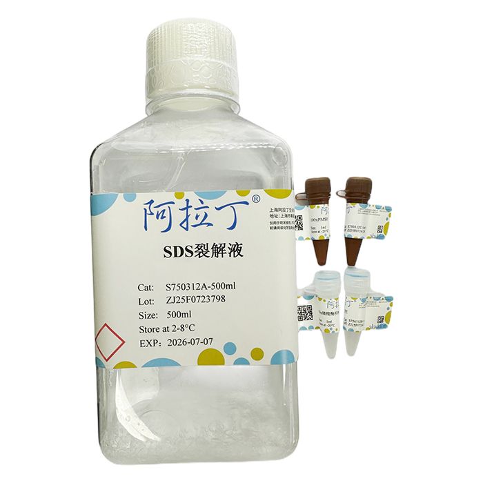
SDS裂解液(SDS Lysis Buffer)是一种比较强烈的细胞组织裂解液,通常适用于难溶性蛋白质的提取,同时由于SDS裂解液中由于含有较高浓度的SDS,因此有可能引起提取蛋白质的变性。因此裂解得到的蛋白样品可以用于常规的Westem、ChIP(染色质免疫共沉淀,chromatin immunoprecipitation)等,但不适用于蛋白活性方面的检测,同时由于SDS等干扰物质的存在,蛋白定量时不能用Bradford法测定由本裂解液裂解得到样品的蛋白浓度,但是可以用BCA蛋白浓度测定试剂盒测定蛋白浓度。
| S750312 | Component | 100mL
| 500mL
| Storage | | S750312A | SDS裂解液 | 100mL | 500mL | 2-8℃ | | S750312B | 50x磷酸酶抑制剂 | 2x1mL | 10x1mL | -20℃. Avoid freeze/thaw cycle. | | S750312C | 100xPMSF | 1mL | 5x1mL | -20℃. Avoid freeze/thaw cycle. |
|
注意事项:
1、为取得最佳的使用效果,可以适当分装后-20℃保存使用,避免过多的反复冻融;
2、裂解样品的所有步骤都需在冰上或 2~8℃进行;
3、为了您的自身安全,使用试剂前,请做好防护,如穿实验服,带手套等。
使用说明:
1、试剂准备
融解SDS裂解液,混匀。取适当量的裂解液,在使用前数分钟内将蛋白酶抑制剂复合物或/和磷酸酶抑制剂复合物按照1:50 加入SDS裂解液中,或者使用100mM的PM SF,并使PM SF的最终浓度为1 mM。
2、细胞/组织的裂解
对于贴壁细胞:去除培养液,用PBS、生理盐水或无血清培养液洗一遍。按照6孔板每孔加入150-250uL裂解液的比例加入裂解液。用枪吹打数下,使裂解液和细胞充分接触。通常裂解液接触细胞1-2秒后,细胞就会被裂解。如果用于ChIP,初步裂解后需在冰浴上继续裂解 10 分钟:对于悬浮细胞:离心收集细胞,用手指把细胞用力弹散。按照6孔板每孔细胞加入150-250mL 裂解液的比例加入裂解液。再用手指轻弹以充分裂解细胞。如果细胞量较多,必需分装成 50-100 万细胞/管,然后再裂解。充分裂解后应没有明显的细胞沉淀。如果用于ChIP,初步裂解后需在冰浴上继续裂解 10 分钟。对于组织样品:用组织剪将组织剪成碎片后,按照每20毫克组织加入150-250 uL裂解液的比例加入裂解液,并使用用玻璃匀浆器匀浆,直至充分裂解。如果用于ChIP,初步裂解后需在冰浴上继续裂解10分钟。
3、蛋白样品收集
充分裂解后,10000-14000g离心3-5分钟,取上清,即可进行后续的PAGE、Western和ChIP等操作。
注:裂解液用量说明:对于培养细胞来说,通常6孔板每孔细胞加入150uL裂解液已经足够但如果细胞密度非常高可以适当加大裂解液的用量到200uL或250uL。如果用于ChIP,建议6孔板每孔细胞至少加入200uL裂解液。对于组织样品,通常每20mq组织中需要加入150-250uL裂解液,但是对于蛋白含量较高的组织可以增加到300-400μL。
SDS Lysis Buffer is a relatively strong cell tissue lysis buffer, usually suitable for the extraction of insoluble proteins. At the same time, due to the high concentration of SDS in SDS lysis buffer, it may cause denaturation of extracted proteins. Therefore, the protein samples obtained from the lysis can be used for conventional Westem, ChIP (chromatin immunoprecipitation), etc., but are not suitable for detecting protein activity. At the same time, due to the presence of interfering substances such as SDS, the protein concentration of the sample obtained from the lysis of this lysate cannot be determined by the Bradford method during protein quantification, but can be determined by the BCA protein concentration determination kit.
| S750312 | Component | 100mL
| 500mL
| Storage | | S750312A | SDS cracking solution
| 100mL | 500mL | 2-8℃ | | S750312B | 50x phosphatase inhibitor | 2x1mL | 10x1mL | -20℃. Avoid freeze/thaw cycle. | | S750312C | 100xPMSF | 1mL | 5x1mL | -20℃. Avoid freeze/thaw cycle. |
|
Precautions:
1. To achieve the best usage effect, it can be appropriately packaged and stored at -20 ℃ to avoid excessive repeated freezing and thawing;
2. All steps of sample cracking must be carried out on ice or at 2-8 ℃;
3. For your own safety, please take protective measures such as wearing lab coats and gloves before using the reagents.
Instructions for Use:
1. Reagent preparation
Dissolve SDS lysate and mix well. Take an appropriate amount of lysis buffer and add protease inhibitor complex and/or phosphatase inhibitor complex to SDS lysis buffer at a ratio of 1:50 within a few minutes before use, or use 100mM PM SF and make the final concentration of PM SF 1 mM.
2. Cell/tissue lysis
For adherent cells: Remove the culture medium and wash once with PBS, physiological saline, or serum-free culture medium. Add lysis buffer in a ratio of 150-250uL per well to a 6-well plate. Blow with a gun several times to ensure full contact between the lysate and the cells. Usually, after 1-2 seconds of contact with the cell lysate, the cell will be lysed. If used for ChIP, it needs to be further lysed on an ice bath for 10 minutes after initial lysis. For suspended cells, collect the cells by centrifugation and use your fingers to forcefully disperse them. Add lysis buffer at a ratio of 150-250mL per well of cells in a 6-well plate. Gently flick with your fingers to fully lyse the cells. If there are a large number of cells, they must be divided into 500000 to 1 million cells/tube and then lysed. After sufficient lysis, there should be no obvious cell precipitation. If used for ChIP, it needs to be further cracked on an ice bath for 10 minutes after initial cracking. For tissue samples: Cut the tissue into fragments using a tissue cutter, add 150-250 uL of lysis buffer per 20 milligrams of tissue, and homogenize using a glass homogenizer until fully lysed. If used for ChIP, it needs to be further cracked on an ice bath for 10 minutes after initial cracking.
3. Collection of protein samples
After sufficient lysis, centrifuge at 10000-14000g for 3-5 minutes, take the supernatant, and proceed with subsequent page, Western language, and ChIP operations.
Note: Explanation of lysis buffer dosage: For cultured cells, adding 150uL lysis buffer to each well of a 6-well plate is usually sufficient. However, if the cell density is very high, the amount of lysis buffer can be appropriately increased to 200uL or 250uL. If used for ChIP, it is recommended to add at least 200uL lysis buffer to each well of cells in a 6-well plate. For tissue samples, typically 150-250uL of lysis buffer needs to be added every 20mq of tissue, but for tissues with high protein content, it can be increased to 300-400 μL.

 ZJ25F1230254


 危险品化学品经营许可证(带存储)