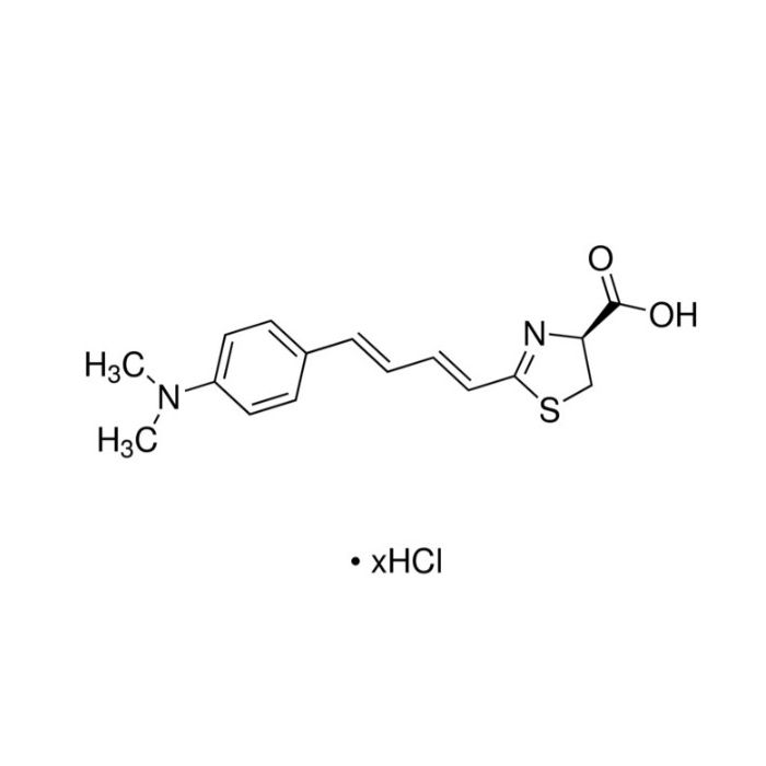
AkaLumine盐酸盐是一种发光峰值在 670-680 nm处的荧光素类似物,能够穿透血脑屏障并产生在体内组织中更容易观察到的红光。产生的Akaluc蛋白既是一种更加有效的底物催化剂,而且也在细胞中更加大量地表达。
AkaLumine盐酸盐有以下优势及特点:
1.相较于传统产品 AkaLumine ,AkaLumine盐酸盐的溶解度高 50倍以上。
2.非常适合用于活体成像实验,因为它的峰值范围在近红外(NIR)窗内,不易受水和血红蛋白吸收峰影响。
3.信号更强,更清晰:在小鼠大脑中,AkaLuc和 AkaLumine盐酸盐的这种组合,被称为AkaBLI,它们产生的生物发光信号比天然的荧光素酶-荧光素反应产生的信号强1000倍。在体内的其他部位,比如在小鼠肺部,仅一两个发光细胞就足以清晰可见,使得AkaLumine盐酸盐可用于监控移植细胞。
使用方法
1.制备储备液:可用去离子水或二甲基亚砜(DMSO)溶解本产品,配制浓度不超过 50 mg/mL 的 AkaLumine 盐酸盐储备溶液;建议储备液浓度为:40 mg/mL,如称取 40 mg 的 AkaLumine 盐酸盐粉末直接溶于 1 mL 的去离子水或 DMSO 中,混匀后立即使用或低温保存。
2.工作液稀释:根据实验条件,可选用生理盐水、PBS 或者其他类型生理缓冲液(pH6.6 - 7.2)来稀释本产品;如选择去离子水作为储备液,稀释至工作液后,可用 0.22 μm 的滤膜过滤除菌后使用。
注:⼀旦配成溶液,请分装保存,避免反复冻融造成产品失效;对于检测灵敏度要求特别高的实验,建议使用新鲜配制的本产品;稀释后DMSO的比例需小于0.5%(如储备液体系中含有DMSO)。
3.体外实验:在细胞表达的荧光素酶检测体系中,推荐的工作液浓度为0.004 mg/mL。
体外实验在加样完成后即可进行发光检测或拍照。
4.体内实验:在动物活体实验中,推荐按2 mg/kg 的AkaLumine盐酸盐用量/体重的比例进行注射相应的量。如用生理盐水将AkaLumine盐酸盐储备液稀释至 0.4 mg/mL 的工作液,20 g重的小鼠通过尾静脉注射或腹腔注射100 μL工作液。
体内实验推荐拍摄时间:尾静脉注射入体内 0-5 min 、腹腔注射入体内 10-20 min。
注:上述工作浓度及检测时间只做推荐参考;本产品用于动物活体成像分析、体外生物发光检测的最佳工作浓度、检测时间请自行参考相关文献,或根据实验目的、所培养的特定细胞和组织等,进行预实验摸索和优化。
AkaLumine hydrochloride is a luciferin analogue with an emission peak at 670–680 nm. It can cross the blood‑brain barrier and generate red light that is readily detectable in living tissues. The AkaLuc protein acts not only as a more efficient substrate catalyst but also exhibits higher expression levels in cells.
AkaLumine hydrochloride has the following advantages and features:
1. Compared with native AkaLumine, AkaLumine hydrochloride possesses over 50‑fold higher solubility.
2. It is highly suitable for in vivo imaging. Its emission maximum falls within the near‑infrared (NIR) window, minimizing interference from absorption by water and hemoglobin.
3. Brighter and better‑resolved signals: The pairing of AkaLuc with AkaLumine hydrochloride, termed AkaBLI, yields a bioluminescent signal in the mouse brain that is approximately 1000‑fold stronger than that from the native luciferase‑luciferin reaction. In other in vivo regions such as the mouse lung, only one or a few luminescent cells can be clearly visualized. This property enables AkaLumine hydrochloride to be used for monitoring transplanted cells.
Protocol
1. Preparation of stock solution: Dissolve the product in deionized water or dimethyl sulfoxide (DMSO) to prepare AkaLumine hydrochloride stock solution with a concentration no higher than 50 mg/mL. The recommended stock concentration is 40 mg/mL. For example, weigh 40 mg of AkaLumine hydrochloride powder and dissolve directly in 1 mL of deionized water or DMSO. Mix well, and use immediately or store at low temperature.
2. Dilution for working solution: Dilute the stock with normal saline, PBS, or other physiological buffers (pH 6.6–7.2) according to experimental requirements. If deionized water is used for the stock solution, filter the diluted working solution through a 0.22 μm filter for sterilization prior to use.
Notes: Aliquot the solution after preparation to avoid product inactivation caused by repeated freeze-thaw cycles. Freshly prepared solution is recommended for assays requiring ultra-high detection sensitivity. If the stock solution is prepared in DMSO, the final DMSO proportion after dilution should be less than 0.5%.
3. In vitro assay: The recommended working concentration is 0.004 mg/mL for luciferase reporter assays with expressing cells.
Luminescence measurement or imaging can be performed immediately after sample addition for in vitro experiments.
4. In vivo assay: For animal in vivo studies, the recommended dosage of AkaLumine hydrochloride is 2 mg/kg based on body weight. As an example, dilute the AkaLumine hydrochloride stock with normal saline to a working concentration of 0.4 mg/mL. A 20 g mouse can be administered 100 μL of the working solution via tail vein injection or intraperitoneal injection.
Recommended imaging time window for in vivo experiments: 0–5 min after tail vein injection; 10–20 min after intraperitoneal injection.
Notes: The above working concentrations and imaging time windows are for reference only. Optimize the optimal working concentration and acquisition time for in vivo bioluminescence imaging and in vitro bioluminescence assays by referring to relevant literature or conducting preliminary tests according to your experimental goals, specific cell lines and tissues.

 A2516345


 危险品化学品经营许可证(带存储)