
计算溶液所需的质量、体积或浓度。
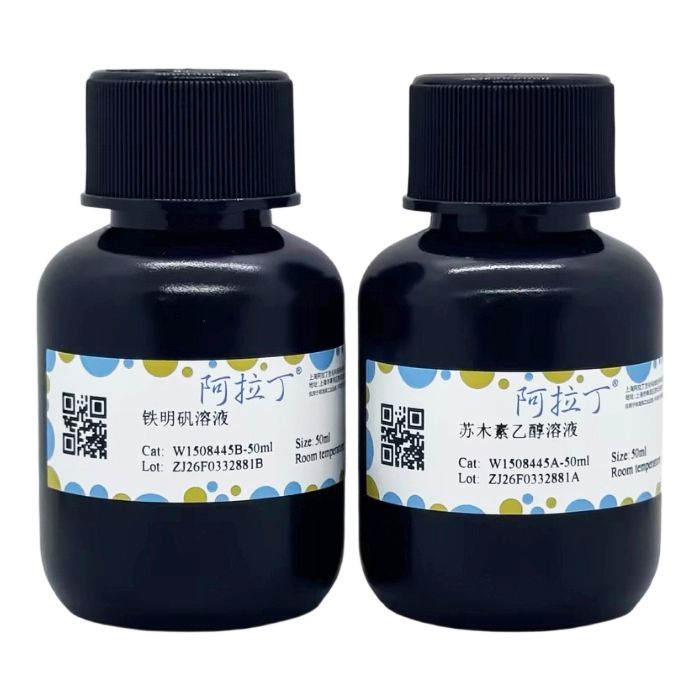
Weil铁明矾苏木素染色液
有货
BioReagent,生物染色剂,用于显微镜 级 ,适用于对基线干扰要求严格的色谱和分析工作流程。
避光,室温。常规运输 。请查阅批次 COA 获取详细规格。
SDS、COA、产品数据表及规格说明书均可下载。可通过批号查询获取批次 COA。
在色谱分析、有机合成和交叉偶联反应领域已被 0 篇同行评审文献引用。
髓鞘 (Myelin Sheath)是包裹在神经细胞轴突外面的一层膜,即髓鞘由髓鞘细胞和细胞膜组成,是神经膜细胞的质膜沿着轴索的轴心螺旋缠绕形成的多层脂双层结构, 髓鞘上有郎飞氏结,可使神经冲动跳跃传递,髓鞘染色在病理诊断中有一定意义,髓鞘的病理变化分为早期、中期和晚期。在早期着色较深;病变中期阶段的髓鞘变性形成脂滴,可用脂质染色加以显示,后期彻底溃变并被吞噬细胞清除,故不再有髓鞘的阳性结果。
很多疾病都可以引起髓鞘的变化,Weil髓鞘染色可以显示病理情况下髓鞘是否完整、变性、坏死程度及修复情况,对神经组织的病理诊断和研究均有意义, 例如神经纤维受损时,髓鞘可出现膨胀、曲折成球形、断裂或脱鞘完全消失等改变。
Weil铁明矾苏木素染色液又称Weil苏木素染色液,主要由苏木素、铁明矾、乙醇等组成,临用前混合。用于髓鞘的石蜡切片染色该试剂仅用于科研领域,不适用于临床诊断或其他用途。
自备材料:
1、蒸馏水、梯度乙醇、二甲苯或脱蜡透明液、中性树胶
2、Weil分化液、2%明矾溶液、氨水蓝化液
3、恒温箱或水浴锅、显微镜
操作步骤(仅供参考):
1、临用前配制Weil铁明矾苏木素染色液:取A、B试剂等量混合即成,不宜提前配制。
2、石蜡切片15-20μm,二甲苯或脱蜡透明液脱蜡至水洗,蒸馏水冲洗。
3、入配制好的Weil铁明矾苏木素染色液,置于58℃温箱或水浴锅染色20-30min;若置于室温,染色1h,蒸馏水冲洗。
4、用明矾溶液分化2-3min,并镜下控制区分正常髓鞘与灰质或变性区域,蒸馏水冲洗。
5、Weil分化液分化2-10min (如分化较好此步可省略),蒸馏水冲洗。
5、滴加Weil蓝化液处理5min,充分水洗。
6、常规脱水,二甲苯或脱蜡透明液透明,中性树胶封固。
染色结果:
髓鞘: 黑色
背景: 无色
注意事项:
1、此试剂盒简便快速,明矾分化这一步很关键,需在镜下观察分化程度。
2、固定液以10%的福尔马林为佳。
3、置于58℃温箱或水浴锅染色时,应注意防止染色液挥发。
4、为了您的安全和健康,请穿实验服并戴一次性手套操作。
5、试剂开封后请尽快使用,以防影响后续实验效果。
The myelin sheath is a membrane wrapping around the axon of nerve cells. Specifically, it is composed of myelinating cells and cell membranes, forming a multi-layered lipid bilayer structure through the spiral wrapping of the plasma membrane of neurolemmocytes around the axis of the axon. The myelin sheath is interspersed with nodes of Ranvier, which enable the saltatory conduction of nerve impulses. Myelin staining holds certain significance in pathological diagnosis. Pathological changes of the myelin sheath are classified into early, intermediate, and late stages. In the early stage, the myelin sheath shows deep staining. During the intermediate stage of pathological changes, myelin degeneration leads to the formation of lipid droplets, which can be visualized by lipid staining. In the late stage, the myelin sheath undergoes complete degeneration and is cleared by phagocytes, resulting in the absence of positive myelin staining.
Numerous diseases can induce changes in the myelin sheath. Weil myelin staining can demonstrate the integrity, degeneration degree, necrosis extent, and repair status of the myelin sheath under pathological conditions, which is of great significance for the pathological diagnosis and research of nerve tissues. For instance, when nerve fibers are damaged, the myelin sheath may exhibit changes such as swelling, spherical tortuosity, fragmentation, or complete demyelination.
Weil’s ferric alum hematoxylin staining solution, also known as Weil’s hematoxylin staining solution, mainly consists of hematoxylin, ferric alum, ethanol, etc., and should be mixed immediately before use. It is used for the staining of paraffin sections of myelin sheaths. This reagent is intended for research purposes only and not for clinical diagnosis or any other applications.
Materials to Be Prepared by the User
1. Distilled water, graded ethanol, xylene or dewaxing clearing solution, neutral gum
2. Weil’s differentiating solution, 2% alum solution, ammonia bluing solution
3. Incubator or water bath, microscope
Operating Procedures (for reference only)
1. Prepare Weil’s ferric alum hematoxylin staining solution before use: Mix equal volumes of Reagent A and Reagent B. Do not prepare the solution in advance.
2. Cut paraffin sections with a thickness of 15-20 μm. Dewax the sections with xylene or dewaxing clearing solution and rinse with distilled water.
3. Immerse the sections in the prepared Weil’s ferric alum hematoxylin staining solution. Incubate in a 58℃ incubator or water bath for 20-30 minutes; alternatively, stain at room temperature for 1 hour, followed by rinsing with distilled water.
4. Differentiate the sections with alum solution for 2-3 minutes. Monitor the differentiation process under a microscope to distinguish normal myelin sheaths from gray matter or degenerated areas, then rinse with distilled water.
5. Differentiate with Weil’s differentiating solution for 2-10 minutes (this step can be omitted if differentiation is satisfactory), followed by rinsing with distilled water.
6. Add Weil’s bluing solution dropwise to treat the sections for 5 minutes, then rinse thoroughly with distilled water.
7. Dehydrate the sections using the standard procedure, clear with xylene or dewaxing clearing solution, and mount with neutral gum.
Staining Results
Myelin sheath: Black
Background: Colorless
Precautions
1. This kit is simple and fast to use. The alum differentiation step is critical, and the differentiation degree must be observed under a microscope.
2. 10% formalin is the preferred fixative.
3. When staining in a 58℃ incubator or water bath, take precautions to prevent the volatilization of the staining solution.
4. For your safety and health, wear a lab coat and disposable gloves during operation.
5. Please use the reagent as soon as possible after opening to avoid affecting subsequent experimental results.
| W1508445 | Component | 2×50 mL | Storage |
| W1508445A | 苏木素乙醇溶液 | 50 mL | RT. Store in the dark. |
| W1508445B | 铁明矾溶液 | 50 mL | RT. |
通过匹配包装上的批号来查找并下载产品的 COA,每批产品都进行了严格的验证,您可放心使用!
| 批号(Lot Number) | 证书类型 | 货号 |
|---|---|---|
 ZJ26F0332881 ZJ26F0332881 |
分析证书 | W1508445 |
我们的级别选择指南涵盖了目录中所有的变体的纯度、稳定剂状态和应用适用性。
查看 BioReagent 级别指南 → 查看 Biological Stain 级别指南 → 查看 for Microscopy 级别指南 →

 危险品化学品经营许可证(带存储)