
计算溶液所需的质量、体积或浓度。
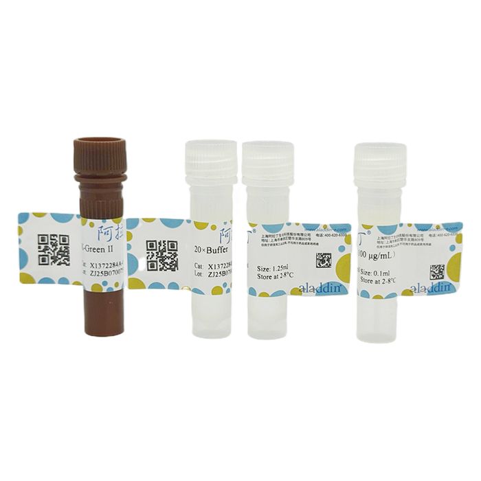
X-Green II 双链DNA定量试剂盒升级版
有货
BioReagent,分子生物学级 ,适用于对基线干扰要求严格的色谱和分析工作流程。
2-8°C储存,避光。低温运输 。请查阅批次 COA 获取详细规格。
SDS、COA、产品数据表及规格说明书均可下载。可通过批号查询获取批次 COA。
在色谱分析、有机合成和交叉偶联反应领域已被 0 篇同行评审文献引用。
X-Green II是荧光检测dsDNA并进行定量的一种产品,这种方法非常灵敏。常用于分子生物学技术:cDNA文库的构建、亚克隆的DNA片段纯化及应用,比如进行DNA定量、产物扩增和引物的进一步检测。常规的DNA含量的检测方法是在260 nm处测其吸光值。这种方法的主要缺点是核苷酸、单链核酸和蛋白质对信号的影响很大,并且还会受到核酸制备过程中污染物的干扰,无法区分DNA和RNA,而且这种方法不灵敏(5 μg/mL dsDNA溶液A260=0.1)。X-Green II定量方法简单、方便,成为生物制品残留DNA检测的标准。
X-Green II只有与dsDNA结合后才发出荧光,并且所发荧光强度与DNA浓度成正比X-Green II双链DNA定量试剂盒升级版可以检测出25 pg/mL-1000 ng/mL范围内的dsDNA,且线性关系较好(R2>0.99)。
产品组分表:
|
储存条件
4℃避光保存,有效期见外包装。对于长期储存,20×Buffer 和 dsDNA 标准液可以储存在≤-20℃。
产品参数
Ex/Em:480/520 nm(结合 dsDNA)
使用方法
试剂制备
X-Green II是以1 mL的浓缩液形式保存在有机溶剂中。实验时,配制2×X-Green II工作液:将组分A用1×Buffer(组分B 稀释20倍)按1:200的比例稀释。对于终体积为2 mL的检测体系,如需准备足够20个样品测定的工作液,可在 19.9 mL 1×Buffer 中加入100 μL组分A;对于终体积为200 μL的检测体系,如需准备足够20个样品测定的工作液,可在1.99 mL 1×Buffer中加 入10 μL组分A。由于试剂容易吸附到玻璃表面,要在塑料容器中配制。组分A试剂见光易降解,因此要注意避光保存。溶液最好在配制好的数小时内使用,以保证最佳的实验结果。
实验方法
1. 用无菌水将组分 B 稀释成 1×Buffer。如制备 50 mL 1×Buffer,需要将 2.5 mL 组分 B 加入到 47.5 mL 无菌水中。
2. 稀释DNA标准品,制作标准曲线。用1×Buffer将组分C由100 μg/ mL稀释至2 μg/ mL。如取40 μL组分C,加入1.96 mL无菌
水。对于低浓度的标准曲线,可以把2 μg/mL的DNA标准品稀释40倍,制备50 ng/mL的DNA储液,然后再进一步稀释。具体 稀释方法可参照表1、表2。
表 1. 宽线度标准曲线制备
|
表2. 低浓度标准曲线制备
|
3. 对于每个未知样品,将1-10 μL样品与99-90 μL的1×Buffer混匀,加入微孔板孔中检测。
4. 用1×Buffer按照1:200稀释组分A,制备2×X-Green II工作液。对于每个标准品和每个未知样品,都需要100 μL的体积。确定要测试的样品和未知样品的总数,并将该数值乘以100 μL,即为所需稀释的X-Green II试剂的总体积。X-Green II对光敏感,融化和稀释时要注意避光操作。
5. 添加100 μL稀释的X-Green II工作液到每个标准和样品孔。吹吸三次混匀。
6. 用锡箔纸覆盖微板,并在室温下孵育5 min。
7. 读取数据,取平均值生成标准曲线,并确定未知样品DNA的浓度。
注意事项
1. 使用前请将产品瞬时离心至管底,再进行后续实验。
2. 荧光染料均存在淬灭问题,请尽量注意避光,以减缓荧光淬灭。
3. X-Green II工作液最好现配现用,以保证最佳结果。
4. 为了您的安全和健康,请穿实验服并戴一次性手套操作。
X-Green II is a product for fluorescent detection and quantification of dsDNA. This method is highly sensitive and widely applied in molecular biology workflows, including cDNA library construction, purification of DNA fragments for subcloning and related applications such as DNA quantification, product amplification and further primer detection.
The conventional method for DNA quantitation measures absorbance at 260 nm. Its major drawbacks include strong signal interference from nucleotides, single-stranded nucleic acids and proteins, as well as contaminants carried over during nucleic acid preparation. It cannot distinguish DNA from RNA and suffers from low sensitivity (A₂₆₀ = 0.1 for 5 μg/mL dsDNA solution). Simple and convenient, the X-Green II quantitation method has become a standard test for residual DNA in biological products.
X-Green II emits fluorescence only upon binding to dsDNA, and the fluorescence intensity is proportional to DNA concentration. The upgraded X-Green II Double-stranded DNA Quantification Kit detects dsDNA within the range of 25 pg/mL to 1000 ng/mL with good linearity (R²>0.99).
|
Storage Conditions
Store at 4 °C protected from light. See outer packaging for shelf life. For long-term storage, 20×Buffer and dsDNA Standard can be stored at ≤ −20 °C.
Product Specifications
Ex/Em: 480/520 nm (when bound to dsDNA)
Protocol
Reagent Preparation
X-Green II is supplied as a 1 mL concentrate in organic solvent. Prepare 2× X-Green II Working Solution for assay: dilute Component A at a 1:200 ratio with 1×Buffer (20-fold dilution of Component B).
1) For a final assay volume of 2 mL: to prepare working solution sufficient for 20 samples, add 100 μL Component A into 19.9 mL 1×Buffer.
2) For a final assay volume of 200 μL: to prepare working solution sufficient for 20 samples, add 10 μL Component A into 1.99 mL 1×Buffer.
The reagent readily adsorbs to glass surfaces; prepare the solution in plastic containers. Component A is light-sensitive and prone to degradation, so keep it protected from light. Prepare the solution and use it within several hours to obtain optimal assay results.
Assay Procedure
1.Dilute Component B with sterile water to make 1×Buffer. For preparation of 50 mL 1×Buffer, add 2.5 mL Component B to 47.5 mL sterile water.
2.Dilute the DNA standard to generate the standard curve. Dilute Component C from 100 μg/mL to 2 μg/mL using 1×Buffer. Example: take 40 μL Component C and add to 1.96 mL sterile water.
For low-range standard curves, further dilute the 2 μg/mL DNA stock 40-fold to prepare a 50 ng/mL DNA stock solution, followed by serial dilution. Refer to Table 1 and Table 2 for detailed dilution schemes.
Table 1. Preparation of Wide-Range Standard Curve
|
Table 2. Preparation of Low-Concentration Standard Curve
|
3.For each unknown sample, mix 1–10 μL sample with 99–90 μL 1×Buffer, then transfer into a microplate well for detection.
4.Prepare 2× X-Green II Working Solution by diluting Component A 200-fold with 1×Buffer. 100 μL working solution is required for each standard and unknown sample. Calculate total reagent volume by multiplying 100 μL by the total number of standards and unknown samples. X-Green II is photosensitive; perform thawing and dilution under light protection.
5.Add 100 μL diluted X-Green II Working Solution to each standard and sample well. Mix by pipetting up and down three times.
6.Cover the microplate with aluminum foil and incubate at room temperature for 5 min.
7.Read fluorescence signals. Take average values to plot the standard curve and calculate DNA concentration of unknown samples.
Notes
1.Briefly centrifuge the product to collect liquid at the bottom of the tube before use, then proceed with experiments.
2.Fluorescent dyes are subject to photobleaching. Minimize light exposure to slow down fluorescence quenching.
3.Prepare X-Green II Working Solution freshly before use for optimal results.
4.For personal safety and health, wear a lab coat and disposable gloves during operation.
通过匹配包装上的批号来查找并下载产品的 COA,每批产品都进行了严格的验证,您可放心使用!
| 批号(Lot Number) | 证书类型 | 货号 |
|---|---|---|
 ZJ25F0724920 |
分析证书 | X1372284 |
我们的级别选择指南涵盖了目录中所有的变体的纯度、稳定剂状态和应用适用性。
查看 BioReagent 级别指南 → 查看 Suitable for molecular biology 级别指南 →

 危险品化学品经营许可证(带存储)