Calcein AM,中文名钙黄绿素乙酰氧基甲酯,是一种可渗透进入细胞、常用于测定真核细胞活力或线粒体通透性转换孔 (Mitochondrial Permeability Transition Pore, MPTP)的绿色荧光探针。Calcein AM近年来被广泛应用到细胞活性分析实验中。
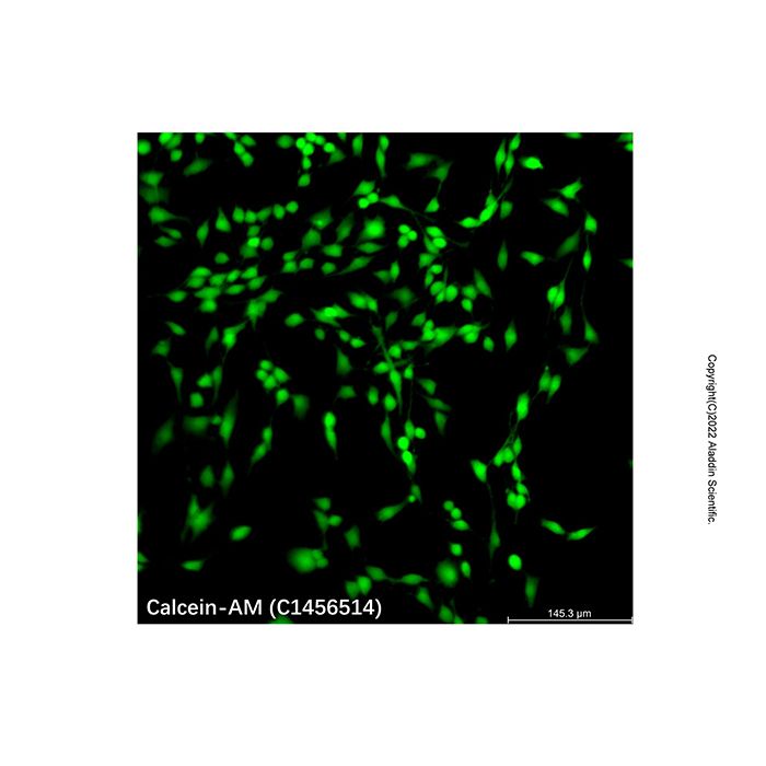
Calcein AM 是在Calcein (钙黄绿素)的基础上增加了乙酰氧基甲酯(AM)基团,加强了疏水性,因此能够轻易穿透活细胞膜。Calcein AM本身并无荧光,进入细胞后被细胞中内源性酯酶水解生成具有强负电荷的不能通透细胞膜的极性分子钙黄绿素(Calcein),从而被滞留在细胞内,而Calcein可发出强绿色荧光(Ex/Em = 494/517 nm)。与其它同类探针相比,由于Calcein AM的细胞毒性非常低,几乎不会影响细胞功能如细胞增殖或淋巴细胞的趋化等,而且对pH值敏感性低,所以Calcein AM是目前染活细胞的最理想荧光探针之一。
由于死细胞缺乏酯酶,Calcein AM仅用于对活细胞的活力测试和短期标记,而核酸红色荧光染料碘化丙啶(Propidium Iodide, PI) 由于不能穿透活细胞的细胞膜而只能染色细胞膜完整性被破坏的死细胞,所以Calcein AM常常与碘化丙啶(PI)联合使用,对活细胞和死细胞同时进行双重荧光染色。
使用方法
1. 工作液的配制
用合适的缓冲液,如无血清培养液、HBSS或PBS按照1:1000稀释本产品,即得到染色工作液。
注:Calcein AM的最终浓度建议根据细胞类型和实验实际情况进行适当调整。
2. 染色
a. 对于贴壁细胞:
(a) 将细胞接种于培养皿、多孔细胞培养板或者细胞爬片上,按实验设计对细胞进行一定处理。轻轻吸除孔板中的培养液,PBS洗涤约10秒,吸除PBS。
(b) 加入适当体积的Calcein AM染色工作液,轻轻晃动使染料均匀覆盖所有细胞。
注:对于在六孔板中培养、汇合度超过80%的贴壁细胞,建议按1 mL/well 加入染色工作液,也可根据具体的实验体系进行优化。
(c) 37ºC孵育细胞10-45min,不同的细胞最佳孵育时间不同。可以考虑以10min作为初始孵育时间,根据具体的实验情况进行适当优化得到更加理想的检测效果。
(d) 吸除染色液,用PBS清洗2-3次,然后加入无血清细胞培养液后即可在荧光显微镜下观察,在 Ex/Em = 494/517 nm 下观察绿色荧光,也可以在染色结束后进行流式细胞仪分析检测或使用荧光酶标仪检测细胞的荧光。
b. 对于悬浮细胞:
(a) 细胞按实验设计进行一定处理后,计数。取适当细胞,500×g室温离心5分钟,轻轻吸除培养基后加适量PBS重悬,500×g室温离心5分钟,去除PBS。
(b) 加适量Calcein AM染色工作液,使细胞密度约为 10⁶ 个/mL 。
(c) 37ºC孵育细胞10-45min,不同的细胞最佳孵育时间不同。可以考虑以10min作为初始孵育时间,根据具体的实验情况进行适当优化得到更加理想的检测效果。
(d) 直接滴加于载玻片上,加盖盖玻片,置于显微镜下观察,在 Ex/Em = 494/517 nm 下观察绿色荧光,也可以在染色结束后进行流式细胞仪分析检测或使用荧光酶标仪检测细胞的荧光。
注:若背景影响严重,可以离心去除染液后使用PBS清洗1-2遍,再置于显微镜下观察。
注意事项
1. 为了您的安全和健康,请穿实验服并戴一次性手套。
2. 荧光染料存在淬灭的问题,建议染色后尽量当天完成检测。
Calcein-AM is a cell-permeant green fluorescent probe commonly used for determining eukaryotic cell viability or the Mitochondrial Permeability Transition Pore (MPTP). In recent years, Calcein-AM has been widely applied in cell viability analysis experiments.
Calcein-AM is derived from Calcein by the addition of acetoxymethyl ester (AM) groups, which enhance its hydrophobicity, thereby allowing it to easily penetrate live cell membranes. Calcein-AM itself is non-fluorescent. Once inside the cell, it is hydrolyzed by endogenous esterases to generate calcein, a strongly negatively charged, polar molecule that cannot permeate the cell membrane and is therefore retained within the cell. Calcein emits intense green fluorescence (Ex/Em=494/517 nm). Compared to other similar probes, Calcein-AM is one of the most ideal fluorescent probes for staining live cells due to its very low cytotoxicity—it hardly affects cellular functions such as cell proliferation or lymphocyte chemotaxis—and its low sensitivity to pH changes.
Since dead cells lack esterase activity, Calcein-AM is used specifically for viability testing and short-term labeling of live cells. The red fluorescent nucleic acid dye Propidium Iodide (PI) can only stain dead cells with compromised membrane integrity, as it cannot penetrate the membranes of live cells. Therefore, Calcein-AM is often used in combination with Propidium Iodide (PI) for simultaneous dual-fluorescence staining of live and dead cells.
Procedure
1. Preparation of Working Solution
Dilute this product 1:1000 in an appropriate buffer, such as serum-free culture medium, HBSS, or PBS, to prepare the staining working solution.
Note: The optimal final concentration of Calcein-AM is suggested to be adjusted appropriately based on cell type and specific experimental conditions.
2. Staining Procedure
a. For adherent cells:
(a) Seed cells in a culture dish, multi-well plate, or on glass coverslips. Treat the cells as required by the experimental design. Gently aspirate the culture medium from the wells and wash with PBS for approximately 10 seconds. Aspirate the PBS.
(b) Add an appropriate volume of Calcein-AM working solution. Gently swirl to ensure even coverage of all cells.
Note: For adherent cells cultured in a 6-well plate with a confluence exceeding 80%, it is recommended to add the staining working solution at a volume of 1 mL per well. This volume can be optimized based on the specific experimental system.
(c) Incubate cells at 37°C for 10-45 minutes. The optimal incubation time varies for different cell types. Consider starting with 10 minutes and optimize for ideal results based on specific experimental conditions.
(d) Aspirate the staining solution and wash the cells 2-3 times with PBS. Then, add serum-free culture medium before observing under a fluorescence microscope. Observe green fluorescence at Ex/Em=494/517 nm. Alternatively, proceed with analysis by flow cytometry or measure fluorescence intensity using a fluorescence microplate reader after staining is complete.
b. For suspension cells:
(a) Treat cells as required by the experimental design and perform a cell count. Take an appropriate number of cells and centrifuge at 500 x g for 5 minutes at room temperature. Gently aspirate the medium, resuspend the pellet in an adequate volume of PBS, and centrifuge again at 500 x g for 5 minutes at room temperature to remove the PBS.
(b) Add an appropriate volume of Calcein-AM working solution to resuspend the cells at a density of approximately 1x10^6 cells/mL.
(c) Incubate cells at 37°C for 10-45 minutes. The optimal incubation time varies for different cell types. Consider starting with 10 minutes and optimize for ideal results based on specific experimental conditions.
(d) Place a drop of the cell suspension directly onto a glass slide, cover with a coverslip, and observe under a microscope. Observe green fluorescence at Ex/Em=494/517 nm. Alternatively, proceed with analysis by flow cytometry or measure fluorescence intensity using a fluorescence microplate reader after staining is complete.
Note: If background interference is significant, centrifuge to remove the staining solution and wash 1-2 times with PBS before microscopic observation.
Precautions
1. For your safety and health, wear a lab coat and disposable gloves.
2. Fluorescent dyes are susceptible to quenching. It is recommended to complete detection on the same day after staining.

 ZJ26F0939280


 危险品化学品经营许可证(带存储)