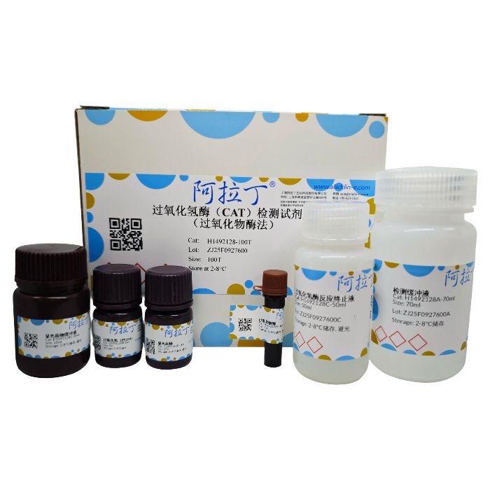
该试剂盒可通过显色反应来检测细胞、组织或其它样品中过氧化氢酶(Catalase)活性。在过氧化氢相对比较充足的情况下,过氧化氢酶可以催化过氧化氢产生水和氧气。残余的过氧化氢在过氧化物酶的催化下可以氧化生色底物,产生红色的产物 (N-(4-antipyryl)-3-chloro- 5-sulfonate-p-benzoquinonemonoimine),最大吸收波长为 520nm。用过氧化氢标准品,制作标准曲线,这样就可以计算出样品中的过氧化氢酶在单位时间单位体积内催化了多少量的过氧化氢转变为水和氧气,从而可以计算出样品中过氧化氢酶的酶活力。该试剂盒检测灵敏度高,可检测出1U/mL的过氧化氢酶。
过氧化氢酶分布非常广泛。在肝脏、肾脏和红细胞中,过氧化氢酶的水平非常高,是清除能导致氧化损伤的过氧化氢的主要场所。
本试剂盒可检测全血、红细胞裂解产物、血清、组织匀浆产物、细胞裂解产物等生物样品中的过氧化氢酶的活性。
一个试剂盒共可以进行100次检测。
H1492128
| Component
| 100T
| Storage
|
H1492128A
| 检测缓冲液 | 70 mL | 2-8℃ |
H1492128B
| 过氧化氢(约1M) | 5 mL
| 2-8℃
|
H1492128C
| 过氧化氢酶反应终止液 | 50 mL
| 2-8℃. Store in the dark.
|
H1492128D
| 显色底物 | 1 瓶 | 2-8℃. Store in the dark.
|
H1492128E
| 显色底物缓冲液 | 20 mL
| 2-8℃. Store in the dark.
|
H1492128F
| 过氧化物酶 | 1 管 | 2-8℃. Store in the dark. |
检测仪器:酶标仪
使用说明
1、试剂盒的准备
1.1 过氧化物酶溶液:将1管过氧化物酶溶解于1.45mL检测缓冲液中,分装后-20°C保存。
1.2 显色底物溶液:将20mL显色底物缓冲液加入显色底物中,使其溶解,分装后-20°C保存。
1.3 配制250mM过氧化氢溶液。本试剂盒提供的过氧化氢浓度约为1M。由于过氧化氢不是非常稳定,使用前,需使用分光光度计准确测量其浓度。把浓度约为1M的过氧化氢,用本试剂盒提供的检测缓冲液稀释100倍,使过氧化氢的浓度约为10mM。将10µL1M过氧化氢溶液加入至990µL检测缓冲液中,置于光径为1cm的紫外比色皿中,用紫外分光光度计测定A240。
过氧化氢浓度(mM)=22.94 × A240
从而计算出本试剂盒提供的过氧化氢的实际浓度。然后再根据实际的过氧化氢浓度配制 250mM 过氧化氢溶液。
1.4 配制5mM过氧化氢溶液。根据测定得到的实际过氧化氢浓度配制5mM过氧化氢溶液。
1.5 配制显色工作液。在冰浴上溶解显色底物溶液,适当分装后再使用,尽量避免反复冻融。其它试剂放置在冰浴上备用。取适当量的过氧化物酶溶液,按照1:1000的比例用显色底物溶液稀释,配制成显色工作液。例如取5μL过氧化物酶溶液,加入5mL显色底物溶液中,混匀即得到5mL显色工作液。
2、样品的准备
用含有去垢剂(如Western及IP细胞裂解液)或低渗缓冲液的裂解液处理样本时,可用检测缓冲液(试剂盒中自带)稀释样本。如果用等渗缓冲液处理样本,为保持过氧化物酶体的完整性,用含有0.1%TritonX-100的检测缓冲液(在检测缓冲液中加入0.1%TritonX-100)。 如果样本的蛋白浓度非常低(低于0.025mg/mL),将BSA加入缓冲液中,使其终浓度为0.5mg/ml,可稳定酶。
当分析组织提取物时,过氧化氢酶的量依赖于组织来源。在肝脏及肾脏中活性最高,在结缔 组织中活性最低。组织的细胞提取物,不需要稀释,可直接用于分析。血液裂解液显示相当 高的活性,因此需要稀释。具体的稀释倍数可以参考下表。
2.1 细胞样品的准备。可用Western及IP细胞裂解液裂解细胞(参考说明书)。
2.2 组织样品的准备。可用Western及IP细胞裂解液裂解组织(参考说明书)。
2.3 全血样品的准备。收集全血至一抗凝管内,颠倒混匀。取100μL全血冻融一次,用检测缓冲液稀释1000倍后进行后续检测。
2.4 红细胞裂解液的准备。用抗凝管收集血液,颠倒混匀。取至少500μL全血4°C2500g 离心5min。弃上清,沉淀用冰冷的生理盐水(0.9%氯化钠)洗涤3次。用约5倍细胞体积的冰冷的去离子水,例如Milli-Q纯水,重悬细胞沉淀,冰浴10min。使用前用检测缓冲液稀释400倍后进行后续检测。
表1:
| 样品名称 | 蛋白浓度
| 检测时的样品使用量
| 反应时间
| A520(空白对照-样品)/反应
|
空白对照
| -
| -
| -
| 1.20-1.32 (A520 初始值)
|
人红细胞裂解液
| 0.2mg/mL
| 2-6μL
| 2min
| 0.21-0.71
|
鼠肝裂解液
| 0.3mg/mL
| 5-10μL
| 1min
| 0.36-0.78
|
鼠肾裂解液
| 0.3mg/mL
| 5-10μL
| 3min
| 0.28-0.57
|
鼠脾裂解液
| 0.3mg/mL
| 5-10μL
| 3min
| 0.11-0.21
|
鼠脑裂解液
| 0.7mg/mL
| 10-20μL
| 3min
| 0.014-0.028
|
HepG2 裂解液
| 2.0mg/mL
| 2-4μL
| 3min
| 0.21-0.43
|
Jurkat 裂解液
| 2.0mg/mL
| 2-4μL
| 3min
| 0.28-0.64
|
过氧化氢酶标准
| 10000倍稀释
| 2-4μL
| 1min
| 0.36-0.71 |
说明:表中的数据仅供参考,实际测定值由于检测体系的不同等可能与参考值之间存在误差。 对于相同的样品,同样的实验体系,测得的A520值和表1相比可能会有较大差异。如果实 验体系不同,例如测得蛋白浓度的方法不同,使用的检测波长不同等,测得的A520值和提 供的参考数值相比可能会有更大的差异。对于表中没有列出的样品,可以参考表中的类似样 品进行检测,然后再根据检测结果适当调整蛋白浓度和样品使用量,或反应时间。 通常对于完全未知的样品,可以对样品进行1、10、20和50倍稀释,然后取10μL,反应 1min。样品稀释后的浓度,最好可使反应体系中的过氧化氢在反应1-5min后减少30-50% 左右,这时的检测结果更加精确。
3、标准曲线测定
3.1 取0、12.5、25、50或75μL配制好的5mM过氧化氢溶液至1.5mL或0.5mL塑料离心管 中,分别加入检测缓冲液至最后体积为100μL,混匀。
3.2 各取4μL,加入到96孔板中的孔内。加入200μL显色工作液。25°C至少孵育15min 后测定A520,但孵育时间不宜超过45min。(注:本步骤可以和样品测定步骤中的最后一步同时进行)
注意:过氧化氢标准品需当天新鲜制备。
4、样品测定
表2:
| 空白对照 (blank)
| 样品 (sample)
|
样品体积
| 0 μL
| X μL
|
检测缓冲液
| 40 μL
| 40-X μL
|
250mM 过氧化氢溶液
| 10 μL
| 10 μL |
4.1 参考表2,取XμL (0-40μL)样品至1.5mL离心管中,加入检测缓冲液至体积为40μL (即加入40-XμL检测缓冲液),吹打混匀。
4.2 再加入10μL 250mM过氧化氢溶液,迅速吹打混匀。参考表1,25°C反应1~5min。
4.3 最后加入450μL过氧化氢酶反应终止液,颠倒混匀或Vortex混匀以终止反应。需在终止反应后15min内完成下面的步骤4.4和步骤4.5。
4.4 在新的离心管内加入40μL检测缓冲液,再加入10μL已终止的上述反应体系4.3制备,混匀。
4.5 从上一步骤的50μL体系中取10μL加入到96孔板中的一个孔内。加入200μL显色工作液。 4.6 25°C至少孵育15min后测定A520,但孵育时间不宜超过45min。
5、样品中过氧化氢酶酶活力的计算:
5.1 计算出标准曲线。A520=k[过氧化氢μM]+b,由标准曲线计算出k和b的值。
5.2 计算出样品中残余的过氧化氢μM。残余过氧化氢μM=(A520-b)/k
5.3 过氧化氢酶酶活力单位的定义:1个酶活力单位(1unit)在25°C,pH7.0的条件下,在 1min 内可以催化分解1μM过氧化氢。
5.4 对于细胞或组织样品的过氧化氢酶活力计算:
[样品过氧化氢酶酶活力]=[消耗过氧化氢μM]×[稀释倍数]/([反应分钟数] ×[样品体积] ×[蛋白浓度])
[样品过氧化氢酶酶活力]的单位为units/mg蛋白
[消耗过氧化氢μM]=[空白对照残余过氧化氢μM]-[样品残余过氧化氢μM]
[稀释倍数]=250
[反应分钟数]即为实际的反应分钟数
[样品体积]为表2中的XμL,以mL来表示即为X/1000mL。
[蛋白浓度]为取XμL样品时,样品中的蛋白浓度,单位为mg/mL。
5.5 对于血浆等液体样品的过氧化氢酶活力计算:
[样品过氧化氢酶酶活力]=[消耗过氧化氢μM] ×[稀释倍数]/([反应分钟数] ×[样品体积])
[样品过氧化氢酶酶活力]的单位为units/ml样品。
注意事项:
1、待测的过氧化氢酶样品,无论是纯的过氧化氢酶还是细胞或组织裂解产物,在4°C通常可以保存1周,-70°C可以长期保存,但-20°C保存后过氧化氢酶的活力会显著下降。
2、在检测过程中加入过氧化氢酶反应终止液后15min内需开始显色反应。
3、过氧化氢不太稳定,精确的过氧化氢浓度需参考本说明书中的方法进行测定。
4、如需对样品中的过氧化氢酶活力进行精确定量,需自备蛋白浓度测定试剂盒,例如,BCA 蛋白定量试剂盒。
5、为了您的自身安全,使用试剂前,请做好防护,如穿实验服,带手套等。
This kit enables the colorimetric detection of catalase activity in cells, tissues, and other biological samples. Under conditions of sufficient hydrogen peroxide (H₂O₂) supply, catalase catalyzes the decomposition of H₂O₂ into water and oxygen. The residual H₂O₂ oxidizes a chromogenic substrate in the presence of peroxidase to produce a red product (N-(4-antipyryl)-3-chloro-5-sulfonate-p-benzoquinonemonoimine) with a maximum absorbance at 520 nm. By generating a standard curve using H₂O₂ standards, the amount of H₂O₂ decomposed per unit time and volume can be quantified, allowing calculation of catalase activity in the sample. The kit exhibits high sensitivity, capable of detecting catalase levels as low as 1 U/mL.
Catalase is widely distributed in living organisms. It is highly abundant in the liver, kidneys, and red blood cells, where it plays a major role in clearing H₂O₂ to prevent oxidative damage.
This kit is suitable for measuring catalase activity in whole blood, erythrocyte lysates, serum, tissue homogenates, cell lysates, and other biological samples.
One kit provides sufficient reagents for 100 assays.
H1492128
| Component
| 100T
| Storage
|
H1492128A
| Assay Buffer
| 70 mL | 2-8℃ |
H1492128B
| H₂O₂ (~1 M)
| 5 mL
| 2-8℃
|
H1492128C
| Stop Solution
| 50 mL
| 2-8℃. Store in the dark.
|
H1492128D
|
Chromogenic Substrate
| 1 bottle
| 2-8℃. Store in the dark.
|
H1492128E
| Substrate Buffer
| 20 mL
| 2-8℃. Store in the dark.
|
H1492128F
| Peroxidase
| 1 tube
| 2-8℃. Store in the dark. |
Protocol
1. Reagent Preparation
1.1 Peroxidase Solution: Dissolve one tube of peroxidase in 1.45 mL Assay Buffer. Aliquot and store at -20°C.
1.2 Chromogenic Substrate Solution: Add 20 mL Substrate Buffer to the bottle of chromogenic substrate and mix to dissolve. Aliquot and store at -20°C.
1.3 Preparation of 250 mM H₂O₂: The provided H₂O₂ has a nominal concentration of ~1 M. Due to instability, accurately determine its actual concentration spectrophotometrically before use. Dilute 10 µL of the ~1 M H₂O₂ solution with 990 µL Assay Buffer (~10 mM). Measure A₂₄₀ using a UV spectrophotometer (1 cm pathlength).
H₂O₂ concentration (mM) = 22.94 × A₂₄₀
Calculate the actual concentration of the stock H₂O₂, then prepare a 250 mM H₂O₂ solution using Assay Buffer.
1.4 Preparation of 5 mM H₂O₂: Dilute the accurately measured H₂O₂ stock to 5 mM using Assay Buffer.
1.5 Chromogenic Working Solution: Thaw the Chromogenic Substrate Solution on ice and aliquot to avoid repeated freeze-thaw cycles. Keep other reagents on ice. Dilute Peroxidase Solution 1:1000 in Chromogenic Substrate Solution to prepare the working solution (e.g., add 5 µL Peroxidase Solution to 5 mL Chromogenic Substrate Solution). Mix well.
2. Sample Preparation
When using lysis buffers containing detergents (e.g., Western & IP Lysis Buffer) or hypotonic buffers, dilute samples with the provided Assay Buffer. For samples in isotonic buffers (to maintain peroxisome integrity), use Assay Buffer containing 0.1% Triton X-100. For samples with very low protein concentration (<0.025 mg/mL), add BSA to the buffer to a final concentration of 0.5 mg/mL to stabilize the enzyme.
Tissue extracts exhibit variable catalase activity (highest in liver and kidney, lowest in connective tissue). Cell lysates can typically be used directly. Blood lysates show high activity and require dilution. Refer to the table below for guidance.
2.1 Cell Samples: Lyse cells using Western & IP Lysis Buffer (refer to product instructions).
2.2 Tissue Samples: Lyse tissues using Western & IP Lysis Buffer (refer to product instructions).
2.3 Whole Blood: Collect blood into an anticoagulant tube and mix by inversion. Freeze-thaw 100 µL of whole blood once, then dilute 1000-fold with Assay Buffer before assay.
2.4 Erythrocyte Lysate: Collect blood into an anticoagulant tube and mix. Centrifuge at least 500 µL at 2500 g for 5 min at 4°C. Discard supernatant and wash the pellet three times with ice-cold saline (0.9% NaCl). Resuspend the pellet in ~5 volumes of ice-cold deionized water (e.g., Milli-Q) and incubate on ice for 10 min. Dilute 400-fold with Assay Buffer before use.
Table 1: Sample Usage Guide (Reference Only)
Sample Type
| Protein Conc. |
Sample Volume
| Reaction Time
| ΔA₅₂₀ (Blank - Sample)
|
Blank Control
| -
| -
| -
| 1.20–1.32 (Initial)
|
Human Erythrocyte Lysate
| 0.2 mg/mL
| 2–6 µL
| 2 min
| 0.21–0.71
|
Mouse Liver Lysate
| 0.3 mg/mL
| 5–10 µL
| 1 min
| 0.36–0.78
|
Mouse Kidney Lysate
| 0.3 mg/mL
| 5–10 µL
| 3 min
| 0.28–0.57
|
Mouse Spleen Lysate
| 0.3 mg/mL
| 5–10 µL
| 3 min
| 0.11–0.21
|
Mouse Brain Lysate
| 0.7 mg/mL
| 10–20 µL
| 3 min
| 0.014–0.028
|
HepG2 Lysate
| 2.0 mg/mL
| 2–4 µL
| 3 min
| 0.21–0.43
|
Jurkat Lysate
| 2.0 mg/mL
| 2–4 µL
| 3 min
| 0.28–0.64
|
Catalase Standard
| 10000x diluted
| 2–4 µL
| 1 min
| 0.36–0.71 |
*Note: The values in this table are for reference only. Actual readings may vary significantly due to differences in experimental systems. For uncharacterized samples, try 1, 10, 20, and 50-fold dilutions, use 10 µL, and react for 1 min. Optimal dilution should result in 30–50% consumption of H₂O₂ in 1–5 min for accurate results.*
3. Standard Curve
3.1 Add 0, 12.5, 25, 50, or 75 µL of the prepared 5 mM H₂O₂ solution to 1.5 mL or 0.5 mL microcentrifuge tubes. Adjust the volume to 100 µL with Assay Buffer and mix.
3.2 Transfer 4 µL of each standard to a 96-well plate. Add 200 µL Chromogenic Working Solution. Incubate at 25°C for at least 15 min (not exceeding 45 min) and measure A₅₂₀.
Note: H₂O₂ standards must be prepared fresh daily. This step can be performed concurrently with step 4.6.
4. Sample Assay
Table 2: Reaction Setup
Component
| Blank
| Sample
|
Sample Volume
| 0 µL
| X µL
|
Assay Buffer
| 40 µL
| (40 - X) µL
|
250 mM H₂O₂
| 10 µL
| 10 µL
|
4.1 Pipette X µL (0–40 µL) of sample into a 1.5 mL tube. Add Assay Buffer to a total volume of 40 µL (i.e., add (40 - X) µL Buffer). Mix gently.
4.2 Add 10 µL of 250 mM H₂O₂ solution and mix quickly. Incubate at 25°C for 1–5 min (refer to Table 1).
4.3 Add 450 µL Stop Solution to terminate the reaction. Mix thoroughly by inversion or vortexing. Proceed to steps 4.4 and 4.5 within 15 min.
4.4 In a new tube, add 40 µL Assay Buffer and 10 µL of the terminated reaction mixture from step 4.3. Mix.
4.5 Transfer 10 µL from the 50 µL mixture in step 4.4 to a well of a 96-well plate. Add 200 µL Chromogenic Working Solution.
4.6 Incubate at 25°C for at least 15 min (not exceeding 45 min) and measure A₅₂₀.
5. Calculation of Catalase Activity
5.1 Generate standard curve: A₅₂₀ = k × [H₂O₂ (µM)] + b. Determine k and b.
5.2 Calculate residual H₂O₂ in sample: [Residual H₂O₂ (µM)] = (A₅₂₀ - b) / k
5.3 Unit Definition: One unit (U) of catalase activity is defined as the amount of enzyme that decomposes 1 µM of H₂O₂ per minute at 25°C and pH 7.0.
5.4 For cell or tissue samples:
[Catalase Activity] = [ΔH₂O₂ (µM)] × [Dilution Factor] / ([Reaction Time (min)] × [Sample Volume (mL)] × [Protein Concentration (mg/mL)])
Units: U/mg protein
[ΔH₂O₂ (µM)] = [Residual H₂O₂ in Blank (µM)] - [Residual H₂O₂ in Sample (µM)]
[Dilution Factor] = 250
[Reaction Time] = actual reaction time in minutes
[Sample Volume] = X µL from Table 2, expressed in mL (X/1000 mL)
[Protein Concentration] = protein concentration of the sample (mg/mL) when X µL was used.
5.5 For liquid samples (e.g., plasma):
[Catalase Activity] = [ΔH₂O₂ (µM)] × [Dilution Factor] / ([Reaction Time (min)] × [Sample Volume (mL)])
Units: U/mL sample
Precautions
1. Catalase samples (pure enzyme or lysates) can be stored at 4°C for ~1 week or at -70°C for long-term storage. Avoid -20°C, as it significantly reduces activity.
2. The chromogenic reaction (step 4.5) must be initiated within 15 min after adding Stop Solution.
3. H₂O₂ is unstable. Determine its concentration accurately as described before use.
4. For precise quantification of catalase activity, a protein assay kit (e.g., BCA Protein Assay Kit) is required to determine sample protein concentration.
5. Wear appropriate personal protective equipment (lab coat, gloves) during reagent handling.
6. For research use only.

 ZJ26F0737119


 危险品化学品经营许可证(带存储)