
计算溶液所需的质量、体积或浓度。
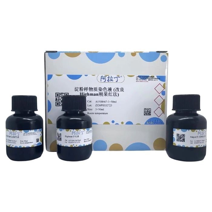
淀粉样物质染色液 (改良Highman刚果红法)
有货
BioReagent,生物染色剂,用于显微镜 级 ,适用于对基线干扰要求严格的色谱和分析工作流程。
避光,室温。常规运输 。请查阅批次 COA 获取详细规格。
SDS、COA、产品数据表及规格说明书均可下载。可通过批号查询获取批次 COA。
在色谱分析、有机合成和交叉偶联反应领域已被 0 篇同行评审文献引用。
淀粉样物质是一种无固定形状的细胞外嗜酸性物质,可存在于不同的组织、器官,导致的疾病称为淀粉样变,淀粉样物质主要是由蛋白质构成,该蛋白大部分排列成反向的β-折叠层结构,在电子显微镜下淀粉样物质呈原纤维排列,病例材料中为大量细胞外的、不分支的细丝,大多随机排列。用于识别淀粉样物质的组织学方法有甲紫染色、刚果红染色、偏振光显微镜观察等,目前研究发现传统的甲紫染色法灵敏度低、特异性差,经典的而且有效的方法是刚果红染色,1922年Bennhold发现了刚果红可以用于活体内淀粉样物质的鉴别,并应用到组织切片,后来经过Highman改良,染色效果更好。
改良Highman刚果红染色又称甲醇刚果红染色,主要由刚果红染色液和Mayer苏木素染色液等组成,该染色法性能稳定,并且已经被科研和临床领域广泛应用,推荐该法作为淀粉样物质染色的主要方法之一。该试剂仅用于科研领域,不适用于临床诊断或其他用途。
|
自备材料:
10%中性福尔马林、蒸馏水、系列乙醇、二甲苯或环保浸蜡脱蜡透明液、中性树胶
操作步骤(仅供参考):
1、常规固定,常采用10%的中性福尔马林固定液,常规脱水包埋。
2、切片厚度4μm,常规二甲苯或脱蜡透明液脱蜡至水。
3、入改良Highman染色液浸染10-30min,弃余液。
4、Highman分化液分化1-5s,立即入水终止分化,水洗2次后镜下控制至恰当程度。
5、自来水冲洗5min。
6、入Mayer苏木素染色液浅染细胞核1-2min或更短时间。
7、自来水冲洗10min。
8、逐级常规乙醇脱水,二甲苯或脱蜡透明液透明,中性树胶封固。
染色结果:
淀粉样物质: 红色
细胞核: 蓝色
注: 在偏光显微镜下,淀粉样物质呈黄绿色的双折光。

改良Highman染色液 (淡染效果,蓝色为核)
注意事项:
1、切片脱蜡应尽量干净,否则影响染色效果。
2、Highman分化液应密闭保存,一旦开启尽快用完。
3、改良Highman染色液染色时尽量采用浸染,如果滴染,应置于湿盒防止溶液挥发。
4、Highman分化液分化步骤很重要,分化时间较短,胶原纤维也被染成红色;分化过度,淀粉样物质也被脱色;如果脱色过度,可以将切片清洗后重新用刚果红染色液浸染。
5、脱水应迅速,避免脱色。
6、由于组织特异性或环境变化等因素,有时会出现红色不明显的情况,应增加染色时间。
7、为了您的安全和健康,请穿实验服并戴一次性手套操作。
8、试剂开封后请尽快使用,以防影响后续实验效果。
Amyloid is an amorphous extracellular eosinophilic substance that can be present in various tissues and organs, and the diseases caused by it are called amyloidosis. Amyloid is mainly composed of proteins, most of which are arranged in an antiparallel β-pleated sheet structure. Under an electron microscope, amyloid presents a fibrillary arrangement, appearing as a large number of unbranched extracellular filaments in pathological specimens, mostly arranged randomly. Histological methods for identifying amyloid include methyl violet staining, Congo red staining, and polarized light microscopy. Current research has shown that the traditional methyl violet staining method has low sensitivity and poor specificity, while Congo red staining is a classic and effective approach. In 1922, Bennhold discovered that Congo red could be used for the identification of amyloid in vivo and applied this technique to tissue sections. Later, the method was modified by Highman, resulting in improved staining efficacy.
The modified Highman Congo red staining, also known as methanol Congo red staining, mainly consists of Congo red staining solution and Mayer's hematoxylin staining solution. This staining method has stable performance and has been widely used in scientific research and clinical fields. It is recommended as one of the primary methods for amyloid staining. This reagent is for research use only and is not suitable for clinical diagnosis or any other purposes.
|
Materials to be Prepared by User
10% neutral formalin, distilled water, graded ethanol, xylene or environment-friendly paraffin removal and clearing solution, neutral balsam
Procedure (for reference only)
1. Perform routine fixation, usually with 10% neutral formalin fixative, followed by routine dehydration and embedding.
2. Cut sections with a thickness of 4 μm, and perform routine dewaxing to water using xylene or dewaxing and clearing solution.
3. Immerse the sections in Modified Highman Staining Solution for 10-30 min, then discard the residual solution.
4. Differentiate with Highman Differentiating Solution for 1-5 s, immediately transfer the sections to water to terminate differentiation, wash twice with water, and check under a microscope to ensure appropriate differentiation.
5. Rinse with tap water for 5 min.
6. Immerse the sections in Mayer's Hematoxylin Staining Solution for light staining of cell nuclei for 1-2 min or a shorter duration.
7. Rinse with tap water for 10 min.
8. Conduct routine dehydration with graded ethanol, clear the sections with xylene or dewaxing and clearing solution, and mount them with neutral balsam.
Staining Results
Amyloid: Red
Cell Nucleus: Blue
Note: Under a polarized light microscope, amyloid exhibits yellowish-green birefringence.
Modified Highman Staining Solution (Light Staining Effect, Nuclei Stained Blue)
Precautions
1. Ensure thorough dewaxing of sections; otherwise, the staining effect will be affected.
2. Store Highman Differentiating Solution in a sealed container and use it as soon as possible after opening.
3. Prefer immersion staining when using Modified Highman Staining Solution; if drop staining is adopted, place the sections in a moist chamber to prevent solution evaporation.
4. The differentiation step with Highman Differentiating Solution is critical. A short differentiation time will result in collagen fibers being stained red; over-differentiation will cause amyloid to decolorize. If over-decolorization occurs, rinse the sections and re-stain with Congo red staining solution.
5. Perform dehydration rapidly to avoid decolorization.
6. Due to factors such as tissue specificity or environmental changes, the red color may sometimes appear faint, and the staining time should be appropriately extended in such cases.
7. For your safety and health, wear a lab coat and disposable gloves during operation.
8. Use the reagent as soon as possible after opening to avoid affecting the results of subsequent experiments.
通过匹配包装上的批号来查找并下载产品的 COA,每批产品都进行了严格的验证,您可放心使用!
| 批号(Lot Number) | 证书类型 | 货号 |
|---|---|---|
 ZJ26F0332725 ZJ26F0332725 |
分析证书 | A1508467 |
我们的级别选择指南涵盖了目录中所有的变体的纯度、稳定剂状态和应用适用性。
查看 BioReagent 级别指南 → 查看 Biological Stain 级别指南 → 查看 for Microscopy 级别指南 →

 危险品化学品经营许可证(带存储)