
 E2527686
计算溶液所需的质量、体积或浓度。
为什么选择此级别
提供 ≥98% 纯度,适用于对基线干扰要求严格的色谱和分析工作流程。
储存与运输
避光,-20°C储存,干燥。超低温运输 。请查阅批次 COA 获取详细规格。
质量文档
SDS、COA、产品数据表及规格说明书均可下载。可通过批号查询获取批次 COA。
文献证明
在色谱分析、有机合成和交叉偶联反应领域已被 1 篇同行评审文献引用。
概述
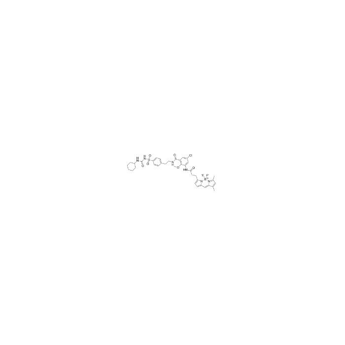
ER-Tracker 染料是与 Glibenclamide 偶联的 BODIPY 列染料的衍生物,对内质网高选择性结合,在低浓度下对细胞无毒性,该类染料属于环境敏感型的探针,与甲醛处理后仍能保留部分荧光,具有荧光寿命高,消光系数好等特点。Glibenclamide 是一种 ATP 依赖性 K+ 通道阻滞剂 (Kir6, KATP) 和 CFTR Cl-通道阻滞剂,在内质网中发生结合。ER-Tracker 不适合对固定后细胞进行染色。
ER-Tracker dye is a derivative of BODIPY series dyes coupled with Glibenclamide , highly selective binding to the endoplasmic reticulum, non-toxic to cells at low concentrations, this type of dye is an environmentally sensitive probe, and formaldehyde treatment can still retain part of the fluorescence, with high fluorescence life, good extinction coefficient and other characteristics. Glibenclamide is an atp-dependent K + channel blocker (Kir6, KATP) and CFTR Cl-channel blocker that binds in the endoplasmic reticulum. ER-Tracker is not suitable for staining cells after fixation.
In Vitro
1. Preparation of ER-Tracker working solution 1.1 Preparation of the stock solution Dissolve 100 ug ER-Tracker in 128 μL DMSO to obtain 1 mM of stock solution. Note: It is recommended to store the stock solution at -20℃or -80℃away from light and avoid repetitive freeze-thaw cycles. 1.2 Preparation of ER-Tracker working solution Dilute the stock solution in serum-free cell culture medium or PBS to obtain 100 nM-1 μM of working solution. Note: Please adjust the concentration of ER-Tracker working solution according to the actual situation. 2. Cell staining 2.1 Suspension cells (6-well plate) a. Centrifuge at 1000 g at 4℃for 3-5 minutes and then discard the supernatant. Wash twice with PBS, 5 minutes each time.The cell density is 1×10 6 /mL. b. Add 1 mL of working solution, and then incubate at room temperature for 5-30 minutes. c. Centrifuge at 400 g at 4℃for 3-4 minutes and then discard the supernatant. d. Wash twice with PBS, 5 minutes each time. e. Resuspend cells with serum-free cell culture medium or PBS. Observation by fluorescence microscopy or flow cytometry. 2.2 Adherent cells a. Culture adherent cells on sterile coverslips. b. Remove the coverslip from the medium and aspirate excess medium. c. Add 100 μL of working solution, gently shake it to completely cover the cells,and then incubate at room temperature for 5-30 minutes. d. Wash twice with medium, 5 minutes each time. Observation by fluorescence microscopy or flow cytometry. Note: If detection by flow cytometry, cells need to be resuspended before staining. MCE has not independently confirmed the accuracy of these methods. They are for reference only.
Form:Solid
规格
规格或纯度
≥98%
英文名称
ER-Tracker Green
生化机理
ER-Tracker 染料是 BODIPY 系列染料的衍生物,与格利本酰胺偶联。
储存条件
避光,-20°C储存,干燥
运输条件
超低温运输
纯度
≥98%
名称和识别符
分子类型
小分子
Smiles
O=C(NS(=O)(C1=CC=C(CCNC(C2=C(OC)C(NC(CCC3=CC=C4C=C5C(C)=CC(C)=[N]5[B+3]([F-])([N-]43)[F-])=O)=CC(Cl)=C2)=O)C=C1)=O)NC6CCCCC6
分子量
783.09
技术文档
高级数据
化学和物理性质
溶解性
DMSO : 50 mg/mL (63.85 mM; Need ultrasonic)
敏感性
对湿度敏感
质检证书(CoA,COO,BSE/TSE 和分析图谱)
技术文档和文章
此产品的引用文献
溶液计算器
常见问题
本产品的纯度是多少?
本产品的化学纯度(含量)为 ≥98%。批次的具体数值以分析证书(COA)为准。
本产品应如何储存?
请于?20 °C、避光、干燥条件下保存。请与干燥剂一同密封保存,本品对湿气敏感。
本产品如何运输?
本产品采用保温箱配冰袋运输。收货后请立即拆包,并按上述储存条件保存。
本产品的 CAS 号、分子式和分子量是多少?
本产品的 CAS 号为 730931-46-1,分子式为 C37H42BClF2N6O6S,分子量为 783.09 g/mol。
如何获取批次专用COA?
每个订单都会随附分析证书(COA),您也可以从账户订单历史中下载。如需订购前的COA,请联系您的客户代表,或使用PDP上的"申请COA"按钮——我们将提供代表性批次的COA。
产品仅提供给经过验证的企业、机构和合格的专业人员用于研发。不适用于人类、动物、诊断或治疗。
Copyright © 2023至今 上海阿拉丁生化科技股份有限公司 SSE:688179 保留所有权利。


 危险品化学品经营许可证(带存储)