
Classification of Chemokines, Receptor Signaling, and Regulatory Mechanisms of Immune Cell Migration
Classification of Chemokines, Receptor Signaling, and Regulatory Mechanisms of Immune Cell Migration
Chemokines are a class of small secreted proteins that regulate directional cell migration. They primarily establish spatial signals through chemokine receptors, cell-surface glycosaminoglycans, and cell-adhesion systems, thereby guiding neutrophils, monocytes, T cells, NK cells, and other cells into specific tissues. Chemokine networks participate in immune surveillance, inflammatory-cell recruitment, hematopoietic-cell homing, tissue repair, and formation of the tumor microenvironment, and their effects are jointly influenced by ligand concentration, receptor expression, spatial gradients, protein-processing status, and the responding cell type.
Keywords: chemokines; CXC chemokines; CC chemokines; CXCR; CCR; cell migration; chemotactic gradient; immune-cell recruitment; inflammatory response
1 Basic Characteristics of Chemokines
1.1 Basic Functions of Chemokines
(1) Directional Cell Migration
Chemokines can form concentration gradients in tissue fluid, on the vascular endothelial surface, or within the extracellular matrix. After cells expressing the corresponding receptors sense the gradient, they establish front-rear polarity and migrate toward increasing chemokine concentrations, accompanied by integrin activation, cell adhesion, and actin remodeling.
(2) Inflammatory and Homeostatic Regulation
Inflammatory chemokines are usually rapidly expressed after infection or tissue injury and are responsible for recruiting inflammatory cells, whereas homeostatic chemokines are constitutively expressed in specific tissues and participate in hematopoietic-cell homing, lymphocyte positioning, tissue residency, and immune surveillance.
(3) Ligand-Receptor Networks
Chemokine networks exhibit extensive cross-reactivity. The same chemokine may bind multiple receptors, and the same receptor may respond to multiple ligands. For example, CXCR2 recognizes multiple ELR-positive CXC chemokines, whereas CCL5 can act on CCR1, CCR3, and CCR5. Therefore, increased chemokine expression should be interpreted together with receptor expression and functional assays.
1.2 Structural Classification of Chemokines
(1) CXC Chemokines
In CXC chemokines, the two conserved cysteines near the amino terminus are separated by one amino acid. Some members contain an ELR motif composed of glutamic acid, leucine, and arginine before the first cysteine. ELR-positive members mainly participate in neutrophil recruitment, whereas ELR-negative members are more involved in lymphocyte migration, vascular regulation, and tissue positioning.
(2) CC Chemokines
In CC chemokines, the two conserved cysteines near the amino terminus are adjacent to each other. These chemokines mainly regulate the migration of monocytes, T cells, eosinophils, and dendritic cells through CCR-family receptors.
(3) CX3C and XC Chemokines
In CX3C chemokines, the two amino-terminal conserved cysteines are separated by three amino acids, with CX3CL1 as the representative member. XC chemokines retain only the second and fourth of the four conserved cysteines found in other typical chemokine classes and therefore lack the first and third conserved cysteines; representative members include XCL1 and XCL2.
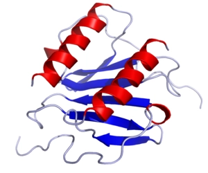
Figure 1 Three-dimensional structure of chemokines
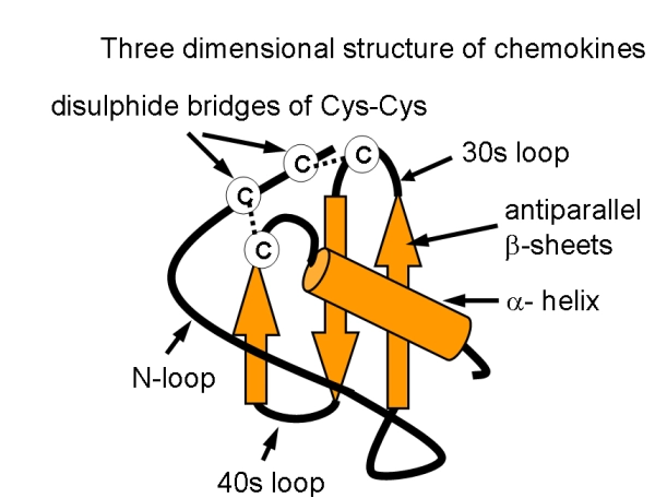
Figure 2 Conserved structural elements and disulfide bonding pattern of chemokines
Chemokine Category | Representative Members | Major Receptors or Functional Axes | Major Functions |
ELR-positive CXC chemokines | CXCL1, CXCL2, CXCL3, CXCL5, CXCL6, CXCL7, and CXCL8 | CXCR1 and CXCR2 | Neutrophil recruitment, degranulation, reactive oxygen species generation, and acute inflammation |
ELR-negative and specialized CXC chemokines | CXCL4, CXCL9, CXCL10, and CXCL11 | CXCR3-associated or glycosaminoglycan-dependent mechanisms | Recruitment of activated T cells and NK cells, vascular regulation, and inflammatory modulation |
Tissue-positioning-associated CXC chemokines | CXCL12, CXCL16, and CXCL17 | CXCR4, ACKR3, CXCR6, or receptors not yet clearly identified | Hematopoietic-cell homing, tissue residency, mucosal immunity, and cell adhesion |
CC chemokines | CCL1, CCL3, CCL5, CCL8, CCL15, and CCL17 | CCR1, CCR2, CCR3, CCR4, CCR5, CCR8, and others | Recruitment of monocytes and lymphocytes, chronic inflammation, and allergic responses |
CX3C chemokines | CX3CL1 | CX3CR1 | Cell adhesion and chemotaxis |
XC chemokines | XCL1 and XCL2 | XCR1 | Recruitment of cross-presenting dendritic cells |
1.3 Chemokine Receptors
(1) Classical Chemokine Receptors
Classical chemokine receptors are seven-transmembrane G protein-coupled receptors and are divided into CXCR, CCR, CX3CR, and XCR families according to their ligand types. Different immune cells have distinct receptor-expression profiles. For example, CXCR2 mainly participates in neutrophil recruitment, CXCR3 is commonly expressed on activated T cells and NK cells, CXCR4 participates in hematopoietic-cell homing, and CCR4 is associated with the migration of certain Th2 cells and regulatory T cells.
(2) Atypical Chemokine Receptors
Atypical chemokine receptors such as ACKR1, ACKR2, ACKR3, and ACKR4 generally do not generate classical Gi protein-dependent chemotactic signaling. Instead, they regulate chemokine distribution within tissues through ligand uptake, transport, presentation, or clearance. ACKR3 can bind CXCL12 and regulate the signaling range of the CXCL12-CXCR4 axis.
(3) Species Differences
Chemokine networks exhibit marked species differences. Human CXCL8 is an important ligand for CXCR1 and CXCR2, whereas mice lack a direct homolog that fully corresponds to human CXCL8. Mouse studies commonly use CXCL1, CXCL2, or CXCL5 to analyze similar neutrophil-recruitment processes.
2 Major Functions of CXC Chemokines
2.1 ELR-Positive CXC Chemokines and Neutrophil Recruitment
(1) CXCL1, CXCL2, and CXCL3
CXCL1, CXCL2, and CXCL3 are also known as GROα, GROβ, and GROγ, respectively. They mainly promote the migration of neutrophils and certain myeloid cells through CXCR2 and participate in inflammatory-cell recruitment during infection, tissue injury, and within the tumor microenvironment.
(2) CXCL5 and CXCL6
CXCL5 mainly promotes neutrophil migration through CXCR2. CXCL6 can act on both CXCR1 and CXCR2, and its receptor-activating capacity may also be affected by amino-terminal proteolytic processing.
(3) CXCL7 and Its Processed Forms
CXCL7 is generated by processing the platelet precursor protein PPBP. PBP, CTAP-III, and NAP-2 represent different processed forms, among which NAP-2 has strong CXCR2 agonistic and neutrophil-chemotactic activity. Therefore, different CXCL7 protein forms should not be regarded as completely equivalent.
(4) CXCL8
CXCL8, also known as IL-8, promotes neutrophil migration, integrin activation, degranulation, and reactive oxygen species generation through CXCR1 and CXCR2. Animal-derived CXCL8 should preferentially be used with cells from the same species, and receptor-activating activity should be confirmed through functional assays.
2.2 CXCL4 and Platelet-Associated Inflammatory Regulation
(1) Source and Release
CXCL4, also known as platelet factor 4, is mainly stored in platelet α-granules and released after platelet activation. It participates in vascular injury, leukocyte recruitment, and regulation of thromboinflammation.
(2) Glycosaminoglycan Binding
CXCL4 has strong heparin- and glycosaminoglycan-binding capacity. Heparin and other negatively charged substances may alter its aggregation state, spatial distribution, and effective concentration. Therefore, the formation of protein complexes should be considered in experimental systems containing heparin.
(3) Regulation of Vascular and Myeloid Cells
CXCL4 can regulate endothelial-cell, monocyte, and macrophage functions. Its mechanism of action is strongly dependent on cell type and experimental conditions and cannot be determined solely from expression of a single receptor.
2.3 CXCL9, CXCL10, CXCL11, and CXCR3 Signaling
(1) Recruitment of CXCR3-Positive Cells
CXCL9, CXCL10, and CXCL11 are commonly induced by interferon signaling and mainly promote migration of activated T cells and NK cells into inflamed tissues through CXCR3, participating in antiviral immunity, Th1-type inflammation, and tumor immunity.
(2) Functional Differences Among Ligands
Although the three ligands share CXCR3, they are not identical in receptor binding, receptor internalization, β-arrestin recruitment, and signaling duration. Therefore, they should not be used interchangeably as completely equivalent stimuli.
2.4 CXCL12, CXCL16, and CXCL17
(1) CXCL12-CXCR4/ACKR3 Axis
CXCL12, also known as SDF-1, mainly acts on CXCR4 and can also bind ACKR3. It participates in hematopoietic stem-cell homing to the bone marrow, immune-cell migration, tissue repair, and tumor-cell dissemination.
(2) CXCL16-CXCR6 Axis
CXCL16 exists in both membrane-bound and soluble forms. Membrane-bound CXCL16 can participate in cell adhesion and lipid uptake, whereas soluble CXCL16 mainly promotes migration of activated T cells and NK cells through CXCR6. Soluble recombinant protein cannot completely substitute for the functions of membrane-bound CXCL16.
(3) CXCL17 and Its Receptor
CXCL17 is mainly expressed in certain mucosal tissues and can affect migration of monocytes and other myeloid cells. Its functional receptor has not yet been conclusively identified, so migration, dose-response, and functional-blockade results should serve as the primary evidence in related studies.
3 Major Functions of CC Chemokines
3.1 CCL1-CCR8 Signaling
(1) Receptor and Responding Cells
CCL1 mainly acts on CCR8 and can recruit certain CCR8-positive T cells, regulatory T cells, and myeloid cells. Mouse CCL1 is also known as TCA-3.
(2) Immunoregulatory Effects
The CCL1-CCR8 axis participates in Th2-type immunity, positioning of tissue-resident immune cells, and regulation of the tumor immune microenvironment. When CCL1 is used in migration assays, CCR8 surface expression and receptor-blocking results should be evaluated together.
3.2 CCL3 and CCL5
(1) CCL3-CCR1/CCR5 Axis
CCL3, also known as MIP-1α, mainly acts on CCR1 and CCR5 and can promote migration of monocytes, macrophages, T cells, and other inflammatory cells. It can also cooperate with other inflammatory mediators to amplify local cellular infiltration.
(2) Multireceptor Activity of CCL5
CCL5, also known as RANTES, can act on CCR1, CCR3, and CCR5 and participates in the migration of T cells, monocytes, and eosinophils. CCL5 also has strong glycosaminoglycan-binding and aggregation capacities, and its immobilized state and aggregation form may influence receptor activation.
3.3 CCL8, CCL15, and CCL17
(1) Multireceptor Characteristics of CCL8
CCL8, also known as MCP-2, can interact with receptors including CCR1, CCR2, CCR3, and CCR5. When studying CCL8 function, receptor expression and individual or combined receptor-blocking experiments should be used to analyze the contribution of different receptors.
(2) Processing Dependence of CCL15
CCL15, also known as MIP-5, is mainly associated with CCR1 and CCR3. Amino-terminal processing can alter its receptor activation and chemotactic capacity, so the effective concentrations of different protein forms may vary.
(3) CCL17-CCR4 Axis
CCL17, also known as TARC, is an important ligand for CCR4 and can recruit Th2 cells, certain regulatory T cells, and skin-homing T cells. It is commonly used in research on allergic inflammation, skin inflammation, and tumor immunity.
4 Chemokine Receptors and Cell-Migration Signaling
4.1 G Protein-Dependent Signaling
(1) Gi Protein Activation
Classical chemokine receptors are mainly coupled to Gi-family G proteins. After ligand binding, Gαi inhibits adenylyl cyclase, whereas Gβγ activates signaling molecules such as PLCβ and PI3K.
(2) PLCβ-Ca²⁺ Signaling
PLCβ promotes the breakdown of phosphatidylinositol bisphosphate to generate IP3 and DAG. IP3 promotes intracellular Ca²⁺ release, whereas DAG participates in protein kinase C activation. Ca²⁺ flux can be used to evaluate early receptor activation but cannot independently demonstrate directional cell migration.
(3) PI3K and Small GTPases
PI3K signaling becomes enriched at the leading edge of the cell and regulates actin polymerization, pseudopod formation, and contraction of the cell rear through small GTPases including Rac, Cdc42, and Rho, enabling migration along a chemokine gradient.
(4) Integrin Activation
Chemokine receptors can increase integrin affinity through inside-out signaling, allowing leukocytes to adhere firmly to the vascular endothelial surface and subsequently complete crawling and transendothelial migration.
4.2 Glycosaminoglycans and Chemotactic Gradients
(1) Immobilized Gradients
Chemokines can bind glycosaminoglycans such as heparan sulfate and chondroitin sulfate on endothelial-cell surfaces and in the extracellular matrix, thereby restricting free diffusion and forming immobilized gradients.
(2) Protein Aggregation State
Glycosaminoglycan binding may alter the monomeric, dimeric, and higher-order oligomeric states of chemokines. Therefore, total chemokine concentration is not equivalent to the free concentration available for direct receptor activation.
(3) Differences Among Experimental Models
Transwell assays mainly simulate soluble gradients, whereas endothelial-cell, three-dimensional matrix, and microfluidic models more closely resemble immobilized gradients in tissues. Migration results obtained from different models cannot be considered directly equivalent.
4.3 Receptor Desensitization and Internalization
(1) Receptor Desensitization
Sustained or high-concentration chemokine stimulation can induce receptor phosphorylation, β-arrestin recruitment, and receptor internalization, thereby reducing cellular responsiveness to subsequent stimulation.
(2) Bell-Shaped Dose-Response Curves
Chemokine concentrations that are too low cannot generate effective stimulation, whereas excessively high concentrations may cause receptor desensitization or disrupt the spatial gradient. Therefore, chemotaxis experiments commonly exhibit bell-shaped dose-response curves.
(3) Cross-Desensitization
Cross-desensitization may occur among different chemokine receptors. When multiple chemokines are used together, the resulting effects may be synergistic, competitive, or mutually inhibitory.
5 Experimental Detection and Interpretation of Chemokines
5.1 Cell-Migration Models
(1) Transwell Migration Assay
The Transwell assay places a chemokine in the lower chamber and measures the number of cells migrating through a porous membrane. It is suitable for concentration screening and comparison of multiple groups. Pore size, cell-seeding density, incubation time, and serum concentration should be controlled.
(2) Microfluidic Migration Assay
Microfluidic systems can establish stable and controllable chemotactic gradients and continuously record cell trajectories. Analysis of migration speed, directionality, and forward-migration index can distinguish directional chemotaxis from increased random motility.
(3) Three-Dimensional Matrix Models
Three-dimensional matrix models can be used to observe cell migration in a spatial environment, but the results are jointly affected by matrix pore size, stiffness, degradability, and chemokine adsorption.
(4) Distinguishing Chemotaxis From Chemokinesis
Chemotaxis is directional migration along a concentration gradient, whereas chemokinesis is an increase in random motility. Equal-concentration controls in the upper and lower chambers should be included to exclude effects caused by changes in cell survival, adhesion, or random movement.
5.2 Validation of Receptor Dependence
(1) Receptor-Expression Detection
Chemokine receptor expression can be analyzed by flow cytometry, immunofluorescence, Western blotting, or transcript detection. Among these methods, measurement of cell-surface receptor expression generally more closely reflects the actual response capacity of cells.
(2) Receptor Blockade and Genetic Intervention
Blocking antibodies, small-molecule antagonists, siRNA, or CRISPR knockout can be used to validate receptor dependence. For chemokines that bind multiple receptors, individual and combined receptor blockade should be performed.
(3) Validation of Ligands Sharing the Same Receptor
The common pathway used by CXCL1, CXCL2, CXCL3, and CXCL5 can be validated through CXCR2 blockade. When studying CXCL6 or CXCL8, the involvement of CXCR1 should also be evaluated according to the cell type.
5.3 Conditions for Using Recombinant Chemokines
(1) Species Matching
Human-, mouse-, porcine-, and rat-derived chemokines should preferably be used with cells from the same species. Before cross-species use, activity should be confirmed through migration, Ca²⁺-flux, or receptor-internalization assays.
(2) Mature Protein Forms
Amino-terminal processing of chemokines such as CXCL7 and CCL15 alters receptor-activating capacity. Therefore, product sequences and mature protein forms should be considered in experiments.
(3) Concentration and Time Gradients
Chemokine experiments should include both concentration and time gradients. A single high-concentration treatment can readily cause receptor desensitization and cannot provide a complete dose-response relationship.
(4) Endotoxin and Protein Stability
Neutrophils, monocytes, and macrophages are highly sensitive to endotoxin. The endotoxin level of recombinant proteins should be considered, and repeated freeze-thaw cycles or improper storage that may cause protein aggregation and reduced activity should be avoided.
Experimental Method | Main Detection Content | Key Indicators | Interpretation Considerations |
Transwell migration assay | Endpoint cell migration | Number of migrated cells, migration rate, and chemotactic index | Include no-gradient and equal-concentration controls |
Microfluidic chemotaxis assay | Dynamic migration in a stable gradient | Directionality, speed, and trajectory length | Distinguish chemotaxis from increased random motility |
Ca²⁺-flux assay | Early receptor activation | Fluorescence peak and duration | Increased Ca²⁺ alone does not prove migration |
Receptor-internalization assay | Receptor activation and desensitization | Changes in cell-surface receptor levels | Degree of internalization does not necessarily equal chemotactic strength |
Downstream-signaling assay | PI3K, AKT, ERK, p38, and related signals | Phosphorylation levels and temporal changes | Include receptor blockade and time-course analysis |
Receptor knockdown or knockout | Receptor dependence | Degree of reduction in migration and signaling | Exclude compensation by other receptors |
Flow-based adhesion assay | Leukocyte adhesion and transendothelial migration | Rolling, arrest, firm adhesion, and transmigration counts | Results are affected by shear stress and endothelial status |
6 Products
6.1 CXC Chemokine Products Related to Neutrophil Recruitment
Catalog # | Name | Grade & Purity | Major Receptor or Functional Axis | Main Research Application |
Recombinant Mouse CXCL1/KC Protein | Carrier-free, PBS Only, ≥95% (SDS-PAGE) | CXCR2 | Mouse neutrophil migration, acute inflammation, and CXCR2-function research | |
Recombinant Mouse CXCL1/KC Protein | Animal Free, carrier-free, Bioactive, ActiBioPure™, azide-free, high performance, PBS Only, ≥97% (SDS-PAGE and HPLC) | CXCR2 | Mouse neutrophil chemotaxis, inflammatory-cell recruitment, and CXCR2-signaling research | |
Recombinant Mouse CXCL1 Protein | ≥90% (SDS-PAGE) | CXCR2 | Mouse CXCL1 dose-response, cell-migration, and receptor-blocking research | |
Recombinant Human CXCL1/GRO alpha Protein | Carrier-free, PBS Only, ≥90% (SDS-PAGE), see COA | CXCR2 | Human neutrophil migration, acute inflammation, and recruitment of tumor-associated myeloid cells | |
Recombinant Mouse CXCL2 Protein | Carrier-free, PBS Only, ≥90% (SDS-PAGE) | CXCR2 | Mouse infection, tissue injury, and neutrophil-recruitment research | |
Recombinant Human CXCL2/GRO beta Protein | Carrier-free, PBS Only, ≥90% (SDS-PAGE) | CXCR2 | Human neutrophil chemotaxis and comparative functional research on CXCL1 and CXCL2 | |
Recombinant Human GRO Gamma Protein | Animal Free, carrier-free, Bioactive, ActiBioPure™, azide-free, high performance, PBS Only, ≥97% (SDS-PAGE and HPLC) | CXCR2 | Human CXCL3/GROγ-mediated neutrophil migration and CXCR2-signaling research | |
Recombinant Human CXCL3/GRO gamma Protein | Carrier-free, PBS Only, ≥95% (SDS-PAGE), see COA | CXCR2 | Comparative analysis of CXCL1, CXCL2, and CXCL3 chemotactic activity and receptor responses | |
Recombinant Human CXCL5 Protein | Animal Free, carrier-free, Bioactive, ActiBioPure™, azide-free, high performance, PBS Only, ≥97% (SDS-PAGE and HPLC) | CXCR2 | CXCL5-CXCR2 axis, neutrophil recruitment, and inflammatory-microenvironment research | |
Recombinant Human CXCL5/ENA-70 Protein | Carrier-free, PBS Only, ≥95% (SDS-PAGE), see COA | CXCR2 | Human CXCL5 chemotactic activity and CXCR2-dependence research | |
CXCL6 | Moligand™ | CXCR1 and CXCR2 | Neutrophil migration, CXCR1 and CXCR2 receptor selectivity, and signaling research | |
Recombinant Mouse CXCL7 Protein | ≥90% (SDS-PAGE) | CXCR2-associated axis | Mouse CXCL7 function, platelet-associated inflammation, and neutrophil-migration research | |
Recombinant Human CXCL7/PBP Protein | Animal Free, carrier-free, Bioactive, ActiBioPure™, azide-free, high performance, PBS Only, ≥97% (SDS-PAGE and HPLC) | CXCL7 processing and CXCR2-associated axis | CXCL7 precursor processing, platelet-derived chemokines, and comparison of mature forms | |
Recombinant Human CXCL7/NAP-2 Protein | Carrier-free, PBS Only, ≥90% (SDS-PAGE), see COA | CXCR2 | Neutrophil migration, degranulation, and platelet-derived inflammatory-signaling research | |
Recombinant Porcine IL-8/CXCL8 Protein | Carrier-free, PBS Only, ≥95% (SDS-PAGE) | CXCR1 and CXCR2 | Porcine neutrophil migration, infection-associated inflammation, and CXCL8-receptor research | |
Recombinant Porcine IL-8/CXCL8 Protein | Carrier-free, His tag, SUMO tag, PBS Only, ≥90% (SDS-PAGE) | CXCR1 and CXCR2 | Porcine-cell chemotaxis and CXCL8-signaling research |
6.2 CXCL4, CXCR3 Ligands, and Tissue-Positioning-Related CXC Chemokine Products
Catalog # | Name | Grade & Purity | Major Receptor or Functional Axis | Main Research Application |
Recombinant Human CXCL4/PF4 Protein | Carrier-free, Bioactive, ActiBioPure™, His tag, ≥95% (SDS-PAGE), see COA | Glycosaminoglycan binding and cell-type-dependent signaling | Human platelet-associated inflammation, monocyte regulation, vascular injury, and CXCL4-complex research | |
Recombinant Rat PF4 Protein | ≥90% (SDS-PAGE) | CXCL4-associated signaling | Rat platelet activation, vascular injury, and thromboinflammation research | |
Recombinant Mouse PF4 Protein | ≥90% (SDS-PAGE) | CXCL4-associated signaling | Mouse platelet-associated inflammation, monocyte regulation, and CXCL4-function research | |
Recombinant Human CXCL9 Protein | Animal Free, carrier-free, Bioactive, ActiBioPure™, azide-free, high performance, ≥97% (SDS-PAGE and HPLC) | CXCR3 | Migration of human activated T cells and NK cells, Th1-type immunity, and tumor-immunity research | |
Recombinant Human CXCL9/MIG Protein | Carrier-free, Bioactive, ActiBioPure™, high performance, PBS Only, ≥95% (SDS-PAGE) | CXCR3 | CXCR3 activation, effector-lymphocyte recruitment, and interferon-associated inflammation research | |
Recombinant Mouse CXCL9/MIG Protein | Carrier-free, Bioactive, ActiBioPure™, high performance, His tag, MBP tag, ≥95% (SDS-PAGE), expressed in E. coli, see COA | CXCR3 | Mouse T-cell migration, infection-associated inflammation, and tumor-immunity model research | |
Recombinant Human CXCL10/IP-10 Protein | Carrier-free, Bioactive, ActiBioPure™, PBS Only, ≥90% (SDS-PAGE) | CXCR3 | Human T-cell and NK-cell chemotaxis, antiviral immunity, and tumor-immunity research | |
Recombinant Mouse CXCL10/IP-10 Protein | Animal Free, carrier-free, Bioactive, ActiBioPure™, azide-free, high performance, PBS Only, ≥97% (SDS-PAGE and HPLC) | CXCR3 | Mouse interferon responses, lymphocyte infiltration, and inflammatory-model research | |
CXCL11 | Moligand™ | CXCR3 | CXCR3 ligand-biased signaling, receptor internalization, and T-cell migration research | |
Recombinant Human/Rhesus Macaque/Feline CXCL12/SDF-1 alpha Protein | Carrier-free, ≥95% (SDS-PAGE), expressed in E. coli, see COA | CXCR4 and ACKR3 | Hematopoietic-cell homing, CXCR4 signaling, cell migration, and cross-species functional research | |
Recombinant Mouse CXCL16 Protein | Animal Free, carrier-free, Bioactive, ActiBioPure™, azide-free, high performance, PBS Only, ≥98% (SDS-PAGE and HPLC) | CXCR6 | Migration of mouse CXCR6-positive T cells and NK cells and tissue-inflammation research | |
Recombinant Human CXCL16 Protein | Animal Free, carrier-free, Bioactive, ActiBioPure™, high performance, His tag, ≥95% (SDS-PAGE) | CXCR6 | Human CXCL16-CXCR6 axis, lymphocyte migration, and vascular-inflammation research | |
Recombinant Mouse CXCL17/VCC-1 Protein | Carrier-free, His tag, PBS Only, ≥90% (SDS-PAGE), see COA | Functional receptor not yet conclusively identified | Mouse mucosal immunity, monocyte migration, and CXCL17-receptor exploration research |
6.3 CC Chemokine-Related Products
Catalog # | Name | Grade & Purity | Major Receptor or Functional Axis | Main Research Application |
Recombinant Human CCL1 Protein | Animal Free, carrier-free, Bioactive, ActiBioPure™, azide-free, high performance, PBS Only, ≥97% (SDS-PAGE and HPLC) | CCR8 | Human CCL1-CCR8 axis, regulatory T-cell migration, and tumor-immunity research | |
Recombinant Mouse CCL1/TCA-3 Protein | Animal Free, carrier-free, recombinant, ActiBioPure™, high performance, His tag, Fc tag, ≥95% (SDS-PAGE) | CCR8 | Migration of mouse CCR8-positive cells, Th2-type immunity, and tissue immune-regulation research | |
Recombinant Human CCL3/MIP-1 alpha Protein | Carrier-free, Bioactive, high performance, ≥90% (SDS-PAGE) | CCR1 and CCR5 | Migration of monocytes, T cells, and other inflammatory cells and CCR1/CCR5-function research | |
Recombinant Human CCL5/RANTES Protein | ≥95% (SDS-PAGE) | CCR1, CCR3, and CCR5 | Migration of T cells, monocytes, and eosinophils and chronic-inflammation research | |
Recombinant Human CCL8/MCP-2 Protein | Animal Free, carrier-free, Bioactive, ActiBioPure™, azide-free, high performance, PBS Only, ≥96% (SDS-PAGE and HPLC) | CCR1, CCR2, CCR3, and CCR5 | Migration of monocytes, T cells, and eosinophils and receptor-selectivity research | |
Recombinant Human CCL8/MCP-2 Protein | Carrier-free, Bioactive, ActiBioPure™, high performance, ≥95% (SDS-PAGE) | CCR1, CCR2, CCR3, and CCR5 | Human CCL8 dose-response, receptor blockade, and inflammatory-cell recruitment research | |
Recombinant Human CCL15 Protein | Animal Free, carrier-free, Bioactive, ActiBioPure™, azide-free, high performance, PBS Only, ≥97% (SDS-PAGE and HPLC) | CCR1 and CCR3 | Monocyte and myeloid-cell migration, CCL15 processing, and receptor-function research | |
Recombinant Human CCL15/MIP-5 Protein | Carrier-free, Bioactive, ActiBioPure™, high performance, ≥95% (SDS-PAGE), see COA | CCR1 and CCR3 | CCL15-CCR1 axis, chronic inflammation, and myeloid-cell recruitment research | |
Recombinant Human TARC/CCL17 Protein | Animal Free, carrier-free, Bioactive, ActiBioPure™, azide-free, high performance, PBS Only, ≥97% (SDS-PAGE and HPLC) | CCR4 | Migration of Th2 cells and certain regulatory T cells, allergic inflammation, and tumor-immunity research | |
Recombinant Human CCL17/TARC Protein | Carrier-free, ≥95% (SDS-PAGE) | CCR4 | CCL17-CCR4 axis, skin inflammation, and chemotaxis of CCR4-positive T cells |
Chemokine research requires simultaneous consideration of ligand species, receptor expression, spatial gradients, and mature protein forms. Migration assays, receptor blockade, and appropriate no-gradient controls allow more accurate determination of the actual effects of specific chemokine-receptor axes on immune-cell recruitment.


 危险品化学品经营许可证(带存储)