
Molecular Mechanisms, Experimental Identification, and Research Tools of Cuproptosis
Molecular Mechanisms, Experimental Identification, and Research Tools of Cuproptosis
Cuproptosis is a form of regulated cell death triggered by abnormal intracellular copper accumulation. Its core mechanism is not lipid peroxidation or a Caspase cascade but the binding of copper to lipoylated proteins in the mitochondrial tricarboxylic acid cycle, which induces abnormal protein aggregation, loss of iron-sulfur cluster proteins, and proteotoxic stress. FDX1, protein lipoylation, and the state of mitochondrial oxidative metabolism jointly determine cellular sensitivity to cuproptosis.
Keywords: cuproptosis; copper homeostasis; FDX1; DLAT; protein lipoylation; tricarboxylic acid cycle; iron-sulfur cluster proteins; mitochondrial respiration; copper ionophores
1 Basic Characteristics of Cuproptosis
1.1 Definition of Cuproptosis
Copper is an essential trace element involved in mitochondrial respiration, redox reactions, iron metabolism, and antioxidant defense, and cells maintain copper homeostasis through copper uptake, transport, storage, and efflux. When intracellular copper load exceeds the capacity of homeostatic regulation, excess copper can bind lipoylated proteins in the mitochondrial tricarboxylic acid cycle, induce abnormal protein oligomerization and insoluble aggregation, and simultaneously cause loss of iron-sulfur cluster proteins and proteotoxic stress, ultimately leading to cuproptosis. This process depends on FDX1, the protein lipoylation pathway, and mitochondrial oxidative metabolism. Therefore, copper accumulation or reduced cell viability alone is insufficient to establish cuproptosis, and the corresponding molecular features must also be verified.
1.2 Differences Between Cuproptosis and Other Forms of Cell Death
Cuproptosis and ferroptosis are both associated with disrupted metal homeostasis, but their execution mechanisms differ substantially. Ferroptosis is mainly driven by iron-dependent phospholipid peroxidation and is closely associated with glutathione, GPX4, lipid metabolism, and the labile iron pool, whereas cuproptosis mainly involves aggregation of lipoylated proteins, loss of iron-sulfur cluster proteins, and mitochondrial proteotoxicity. Cuproptosis also differs from apoptosis, pyroptosis, and necroptosis. In typical copper-ionophore models, cell death does not depend on Caspase-3 activation and cannot be completely blocked by loss of BAX and BAK1, pan-Caspase inhibitors, or RIPK1 inhibitors.
Table 1 Comparison of Cuproptosis With Other Major Forms of Cell Death
Cell Death Type | Major Inducing Factors | Core Execution Process | Representative Detection Indicators | Common Intervention Methods |
Apoptosis | DNA damage, death-receptor activation, and mitochondrial stress | BAX/BAK-mediated changes in mitochondrial permeability and Caspase cascades | Cleaved Caspase-3, Cleaved PARP, and DNA fragmentation | Z-VAD-FMK and Boc-D-FMK |
Pyroptosis | Inflammasome activation, cytosolic lipopolysaccharide, and infectious stimuli | Inflammatory Caspase activation and Gasdermin pore formation | Cleaved GSDMD, Caspase-1, and IL-1β release | Inflammasome- or Gasdermin-related inhibitors |
Necroptosis | Death-receptor activation with restricted Caspase-8 activity | RIPK1-RIPK3-MLKL signaling and plasma-membrane rupture | p-RIPK3 and p-MLKL | RIPK1 inhibitors such as Necrostatin-1 |
Ferroptosis | GPX4 inhibition, cysteine deficiency, and iron accumulation | Iron-dependent phospholipid peroxidation and plasma-membrane damage | Increased lipid ROS, reduced GPX4 activity, and ACSL4-associated changes | Ferrostatin-1 and iron chelators |
Cuproptosis | Copper-ionophore treatment or disruption of copper homeostasis | Aggregation of lipoylated proteins, loss of iron-sulfur cluster proteins, and proteotoxic stress | DLAT aggregation, reduced Fe-S proteins, increased HSP70, and FDX1 dependence | Copper chelation, reduced copper uptake, or intervention in the lipoylation pathway |
2 Copper Homeostasis and the Metabolic Basis of Cuproptosis
2.1 Copper Uptake and Efflux
Cellular copper homeostasis is mainly maintained by copper-uptake proteins, intracellular copper chaperones, and copper-exporting ATPases. SLC31A1, also known as CTR1, is the major high-affinity copper-uptake protein on the plasma membrane and promotes the entry of extracellular copper into cells. ATP7A and ATP7B are copper-transporting ATPases responsible for delivering copper to the secretory pathway or exporting it from cells. Increased SLC31A1 expression can increase intracellular copper load, whereas reduced ATP7A or ATP7B function weakens copper efflux, and both changes may increase cellular sensitivity to copper-dependent injury. However, abnormalities in copper transport do not necessarily indicate cuproptosis, and dependence on FDX1 and the lipoylation pathway must be further demonstrated.
2.2 Protein Lipoylation
Protein lipoylation is a lysine post-translational modification that occurs on mitochondrial metabolic enzyme complexes. Major lipoylated proteins in mammalian cells include DLAT, a component of the pyruvate dehydrogenase complex; DLST, a component of the α-ketoglutarate dehydrogenase complex; DBT, a component of the branched-chain α-keto acid dehydrogenase complex; and GCSH, a component of the glycine cleavage system. LIAS participates in synthesis of the lipoic acid cofactor, LIPT1 participates in transfer of the lipoyl group to target proteins, and DLD participates in redox reactions within multiple lipoylated enzyme complexes. Because these proteins mainly participate in mitochondrial oxidative metabolism, cells dependent on the tricarboxylic acid cycle and oxidative phosphorylation generally exhibit greater sensitivity to cuproptosis.
2.3 DLAT and the Pyruvate Dehydrogenase Complex
DLAT is the E2 component of the pyruvate dehydrogenase complex and transfers acetyl groups from its lipoylated domain to coenzyme A to generate acetyl-CoA. Acetyl-CoA subsequently enters the tricarboxylic acid cycle and provides carbon for mitochondrial respiration and multiple anabolic processes. DLAT is one of the most extensively studied copper-binding proteins in cuproptosis. After copper binds lipoylated DLAT, it can induce abnormal oligomerization and insoluble aggregation, thereby disrupting pyruvate dehydrogenase complex function and increasing the burden on the mitochondrial protein quality-control system.
3 Core Molecular Mechanisms of Cuproptosis
3.1 Regulation of Copper and the Lipoylation Pathway by FDX1
FDX1 is a mitochondrial ferredoxin involved in mitochondrial redox reactions and is closely associated with the protein lipoylation pathway. Loss of FDX1 reduces the lipoylation levels of proteins such as DLAT and DLST and also reduces cellular sensitivity to copper ionophores. FDX1 can also promote the reduction of Cu²⁺ to Cu⁺, increasing the probability of interactions between copper and lipoylated proteins. Therefore, FDX1 connects the copper redox state, protein lipoylation, and mitochondrial metabolism and is an important upstream regulator of cuproptosis. However, changes in FDX1 expression alone cannot independently demonstrate cuproptosis and must be analyzed together with cellular copper load, protein lipoylation, DLAT aggregation, and cell-death phenotypes.
3.2 Copper Binding to Lipoylated Proteins
Copper can bind lipoylated DLAT and DLST, and reduced lipoylation decreases the copper-binding capacity of these proteins, indicating that the lipoylated structure is an important basis for copper recognition of these mitochondrial metabolic proteins. After copper binding, DLAT can transition from a normal soluble state into abnormal oligomers and insoluble aggregates, which not only disrupt pyruvate dehydrogenase complex function but also increase protein quality-control stress and induce elevation of stress proteins such as HSP70.
3.3 Loss of Iron-Sulfur Cluster Proteins
Iron-sulfur clusters are essential cofactors for multiple mitochondrial enzymes, electron-transfer proteins, and metabolic regulatory proteins. Copper-ionophore treatment can reduce the levels of certain iron-sulfur cluster proteins, whereas FDX1 loss can attenuate this change. Loss of iron-sulfur cluster proteins further affects mitochondrial metabolism, electron transport, and redox homeostasis, but this does not mean that cuproptosis is a form of ferroptosis. The core of ferroptosis is iron-dependent phospholipid peroxidation, whereas iron-sulfur cluster changes in cuproptosis mainly reflect disruption of mitochondrial protein homeostasis and cofactor stability.
3.4 Mechanistic Sequence of Cuproptosis
Cuproptosis can be summarized as follows: copper ionophores or copper-transport abnormalities increase intracellular copper load; FDX1 participates in copper reduction and maintains protein lipoylation; copper then binds lipoylated mitochondrial metabolic proteins such as DLAT, causing abnormal protein oligomerization and insoluble aggregation while simultaneously reducing iron-sulfur cluster proteins; and these changes ultimately disrupt mitochondrial metabolism and protein quality control and induce cell death. Therefore, cuproptosis is not a linear process driven by a single molecule but the result of combined disruption of copper homeostasis, protein lipoylation, the tricarboxylic acid cycle, iron-sulfur cluster stability, and protein quality control.
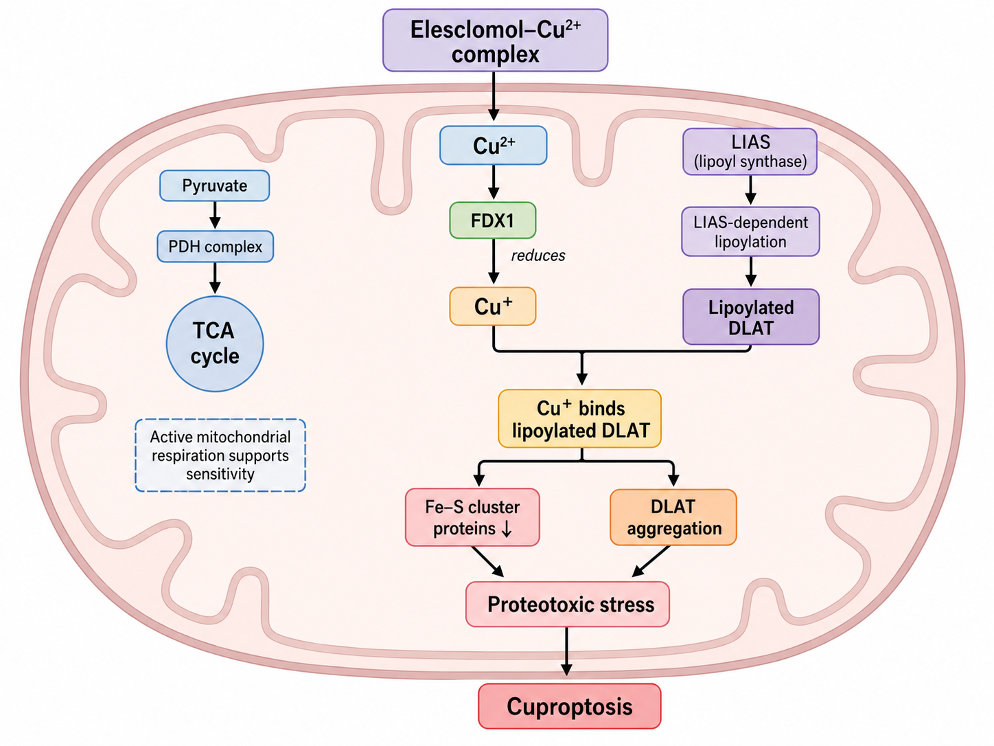
Figure 1 Mitochondrial molecular mechanism of cuproptosis
4 Experimental Evidence for Copper-Ionophore-Induced Cuproptosis
4.1 Copper Dependence and Metal Selectivity
Elesclomol is a lipophilic copper-binding molecule that can form a complex with copper and promote copper entry into cells and mitochondria. Elesclomol alone produces limited effects on the growth of some cells, whereas simultaneous copper supplementation can markedly enhance cytotoxicity, and other metal ions generally cannot fully reproduce this effect, indicating that Elesclomol-associated cell death is strongly copper-dependent. Compounds such as Disulfiram, NSC319726, and 8-Hydroxyquinoline can also alter intracellular copper distribution or produce copper-dependent cytotoxicity under specific conditions, but these compounds also have other pharmacological actions, and reduced cell viability alone cannot establish that they induce cuproptosis. Rescue of copper-ionophore-induced cytotoxicity by copper chelators is an important experiment for demonstrating copper dependence.
4.2 Differences From Apoptosis
In typical Elesclomol-copper models, cell death is not necessarily accompanied by marked Caspase-3 cleavage or Caspase-3/7 activation. Knockout of BAX and BAK1 or treatment with pan-Caspase inhibitors such as Z-VAD-FMK and Boc-D-FMK also cannot completely block copper-dependent cell death, indicating that the core execution process of cuproptosis does not depend on classical mitochondrial apoptosis or the Caspase cascade. However, high-dose or prolonged copper treatment may still secondarily activate apoptosis, so Cleaved Caspase-3, Cleaved PARP, mitochondrial membrane potential, and DNA fragmentation should still be examined in specific experiments.
4.3 Differences From Ferroptosis and Necroptosis
Ferrostatin-1 can inhibit ferroptosis-associated lipid peroxidation but generally cannot effectively eliminate Elesclomol-copper-induced cytotoxicity in typical cuproptosis models. Necrostatin-1 can inhibit RIPK1-dependent necroptosis but also cannot completely block cuproptosis. N-Acetyl-L-Cysteine can supplement cysteine and glutathione and has antioxidant activity, but in some cuproptosis models it does not provide stable rescue comparable to copper chelators, indicating that generalized reactive oxygen species accumulation is not the sole execution mechanism of cuproptosis.
4.4 Dependence on Mitochondrial Respiration
Cells that mainly depend on mitochondrial oxidative metabolism are generally more sensitive to copper ionophores. Transferring cells from high-glucose culture conditions to galactose-containing medium forces them to rely more heavily on mitochondrial oxidative phosphorylation and can enhance the cuproptosis phenotype in some cells. Electron-transport-chain inhibitors and the mitochondrial pyruvate carrier inhibitor UK-5099 can reduce substrate input into the tricarboxylic acid cycle or decrease mitochondrial respiration, thereby reducing the sensitivity of some cells to Elesclomol-copper. FCCP uncouples mitochondrial oxidative phosphorylation, but its action differs from directly blocking pyruvate entry into mitochondria. Therefore, a reduction in mitochondrial membrane potential cannot be simply equated with inhibition of cuproptosis.
4.5 Hypoxia and HIF Signaling
True hypoxia can reduce mitochondrial oxidative metabolism and decrease the sensitivity of some cells to copper ionophores. Roxadustat stabilizes HIF by inhibiting HIF prolyl hydroxylases and can mimic some hypoxia-associated transcriptional responses, but it cannot fully reproduce the effects of true low-oxygen conditions on mitochondrial metabolism and cuproptosis. Therefore, studies of hypoxic regulation of cuproptosis need to distinguish among reduced oxygen concentration, HIF-pathway activation, and mitochondrial-respiration inhibition and should not infer cellular metabolic status solely from HIF stabilization.
5 Cuproptosis-Related Genes and Genetic Evidence
5.1 FDX1 and Lipoic Acid Pathway Genes
CRISPR screening has shown that knockout of genes including FDX1, LIAS, LIPT1, and DLD reduces cellular sensitivity to copper ionophores. These genes jointly participate in mitochondrial lipoic acid synthesis, lipoyl-group transfer, or associated redox processes. Loss of FDX1, LIAS, or LIPT1 reduces lipoylated protein levels, removing some key copper-binding substrates and thereby decreasing DLAT aggregation and cytotoxicity. These findings constitute important genetic evidence that cuproptosis depends on protein lipoylation.
5.2 Pyruvate Dehydrogenase Complex Genes
DLAT, PDHA1, and PDHB are important components of the pyruvate dehydrogenase complex. Knockout of these genes reduces conversion of pyruvate to acetyl-CoA and alters cellular dependence on mitochondrial oxidative metabolism. Loss of these genes can reduce copper-ionophore-induced cytotoxicity, indicating that an intact pyruvate dehydrogenase complex and lipoylated DLAT are important conditions for cuproptosis. This also explains why glycolysis-dependent cells and mitochondrial-respiration-dependent cells exhibit different sensitivities to cuproptosis.
5.3 Metabolic Changes After FDX1 Loss
Loss of FDX1 can alter pyruvate, α-ketoglutarate, succinate, and metabolites associated with lipoic acid synthesis and can reduce cellular respiration. Because FDX1 participates in multiple mitochondrial metabolic processes, the protective effect of FDX1 knockout cannot be attributed solely to reduced copper-reducing capacity and may also involve reduced lipoylated substrates and oxidative metabolic reprogramming. Therefore, FDX1-knockout experiments should be interpreted together with protein lipoylation, cellular oxygen-consumption rate, tricarboxylic acid cycle metabolites, and cellular copper content.
6 Disruption of Copper Homeostasis and Cuproptosis
6.1 SLC31A1 Overexpression Models
Overexpression of SLC31A1 enhances cellular copper uptake and causes more pronounced copper accumulation and cytotoxicity in the presence of exogenous copper, accompanied by changes such as abnormal DLAT aggregation, loss of iron-sulfur cluster proteins, and increased HSP70. Ferroptosis, necroptosis, and apoptosis inhibitors generally cannot stably block copper-dependent death in SLC31A1-overexpressing cells, whereas copper chelators provide marked protection, indicating that chemical copper delivery and genetically increased copper uptake can converge on similar cuproptosis mechanisms.
6.2 ATP7B-Deficient Models
ATP7B is responsible for copper transport and biliary copper excretion in hepatocytes, and ATP7B deficiency causes hepatic copper accumulation and serves as an important model of hereditary disruption of copper homeostasis. ATP7B-deficient tissues may exhibit abnormalities in lipoylated proteins, loss of iron-sulfur cluster proteins, and increased proteotoxic stress, changes that are partly consistent with the molecular phenotype observed in copper-ionophore-treated cells. However, hereditary copper-metabolism disorders also commonly involve oxidative stress, inflammation, lipid-metabolism abnormalities, and damage to multiple organelles, and all tissue injury cannot be attributed to cuproptosis on the basis of a single indicator.
6.3 Shared Features of Chemical and Genetic Models
Copper ionophores, SLC31A1 overexpression, and ATP7B deficiency can all increase cellular or tissue copper load. If these models simultaneously exhibit dependence on FDX1 and the protein lipoylation pathway, DLAT aggregation, loss of iron-sulfur cluster proteins, and rescue by copper chelation, they can be considered to share a common mechanistic basis of cuproptosis.
7 Products for Cuproptosis Induction, Cell-Death Identification, and Mitochondrial Metabolism Research
7.1 Compounds for Copper Delivery and Metal-Ionophore Research
Name | CAS No. | Major Pathway or Target | Characteristics of Action | Main Research Application |
Elesclomol | Copper delivery, FDX1, and mitochondrial copper load | Lipophilic copper-binding molecule that can form an Elesclomol-copper complex and increase mitochondrial copper load | Cuproptosis induction, copper-dependent cytotoxicity, and FDX1-function research | |
Disulfiram | Aldehyde dehydrogenase and copper-dependent proteotoxicity | Can form active complexes with copper and affect protein homeostasis; not a cuproptosis-specific inducer | Copper-dependent cytotoxicity, protein quality control, and ionophore-comparison research | |
NSC319726 | Metal-ion transport and mutant-p53-associated processes | Has metal-ionophore and mutant-p53-reactivation-associated activities, with cytotoxicity influenced by the metal environment | Copper-ionophore comparison, metal-dependent cytotoxicity, and mutant-p53 research | |
8-Hydroxyquinoline | Metal-ion chelation and transmembrane transport | Can bind copper, zinc, and other metal ions and alter their intracellular distribution | Metal ionophore, metal selectivity, and copper-homeostasis research | |
Pyrithione | Transport of zinc and other metal ions | Commonly used as a zinc ionophore and can also alter intracellular distribution of other metal ions | Comparison of copper and zinc selectivity and metal-ionophore control research |
7.2 Compounds for Cell-Death Pathway Identification and Oxidative-Stress Research
Name | CAS No. | Major Pathway or Target | Characteristics of Action | Main Research Application |
ML162 | GPX4 and ferroptosis | Covalently inhibits GPX4 and promotes lipid peroxidation | Ferroptosis positive control and comparison of cuproptosis with ferroptosis | |
Ferrostatin-1 | Lipid peroxidation and ferroptosis | Radical-trapping antioxidant that inhibits multiple ferroptosis models | Exclusion of ferroptosis involvement and lipid-peroxidation rescue experiments | |
DL-Buthionine-(S,R)-Sulfoximine | Glutamate-cysteine ligase and glutathione synthesis | Inhibits glutathione synthesis and reduces cellular antioxidant capacity | Glutathione depletion, oxidative stress, and ferroptosis-sensitivity research | |
Z-VAD-FMK | Caspase family | Cell-permeable irreversible pan-Caspase inhibitor | Exclusion of Caspase-dependent apoptosis and comparison of cell-death pathways | |
Boc-D-FMK | Caspase family | Irreversible pan-Caspase inhibitor | Apoptosis rescue, Caspase dependence, and cuproptosis-mechanism validation | |
Necrostatin-1 | RIPK1 and necroptosis | Inhibits RIPK1 kinase activity and reduces necroptosis | Exclusion of RIPK1-dependent necroptosis | |
N-Acetyl-L-Cysteine | Glutathione synthesis and oxidative stress | Cysteine precursor with antioxidant activity | Oxidative-stress rescue and comparison of whether cuproptosis depends on reactive oxygen species | |
Etoposide | Topoisomerase II and DNA damage | Induces DNA damage, cell-cycle arrest, and apoptosis | Apoptosis positive control and Caspase-activation detection | |
Paclitaxel | Tubulin and mitosis | Stabilizes microtubules and induces mitotic arrest and apoptosis | Mitotic cell-death and apoptosis-control research | |
DPQ | PARP-1 | Inhibits excessive PARP-1 activation | PARP-dependent cell death and NMDA-associated injury-control research | |
Pepstatin A | Aspartic proteases and Cathepsin D | Inhibits multiple aspartic proteases | Lysosomal proteases and lysosome-associated cell-death control research |
7.3 Compounds for Mitochondrial Respiration and Metabolic-Dependence Research
Name | CAS No. | Major Pathway or Target | Characteristics of Action | Main Research Application |
Carbonyl Cyanide-4-(Trifluoromethoxy)phenylhydrazone (FCCP) | Mitochondrial proton gradient and oxidative phosphorylation | Protonophore uncoupler that can reduce mitochondrial membrane potential and increase uncoupled respiration | Mitochondrial membrane potential, maximal oxygen-consumption capacity, and respiratory dependence of cuproptosis | |
UK-5099 | Mitochondrial pyruvate carrier | Inhibits pyruvate entry into mitochondria and reduces pyruvate-driven tricarboxylic acid cycling | Pyruvate metabolism, the PDH complex, and substrate dependence of cuproptosis | |
Roxadustat | HIF prolyl hydroxylases and HIF signaling | Inhibits PHD enzymes and stabilizes HIF transcription factors, mimicking some hypoxic signals | Distinguishing the effects of true hypoxia and HIF stabilization on cuproptosis sensitivity | |
Rotenone | Mitochondrial electron-transport-chain complex I | Inhibits complex I and reduces NADH oxidation and mitochondrial respiration | Electron transport, mitochondrial reactive oxygen species, and respiratory dependence of cuproptosis | |
Oligomycin | Mitochondrial ATP synthase | Inhibits ATP synthase and blocks oxidative-phosphorylation-dependent ATP production | ATP-linked respiration, mitochondrial energy metabolism, and cuproptosis sensitivity |
Reliable identification of cuproptosis requires simultaneous demonstration of copper dependence, protein-lipoylation dependence, and mitochondrial metabolic dependence. Only by integrating cellular copper content, DLAT aggregation, iron-sulfur cluster proteins, HSP70, cellular respiration, and genetic rescue results can cuproptosis be distinguished from generalized copper toxicity and other forms of regulated cell death.
References
[1] Tsvetkov P, Coy S, Petrova B, et al. Copper Induces Cell Death by Targeting Lipoylated TCA Cycle Proteins. Science. 2022;375(6586):1254-1261.
[2] Li SR, Bu LL, Cai L. Cuproptosis: Lipoylated TCA Cycle Proteins-Mediated Novel Cell Death Pathway. Signal Transduct Target Ther. 2022;7(1):158.
[3] Tang D, Chen X, Kroemer G. Cuproptosis: A Copper-Triggered Modality of Mitochondrial Cell Death. Cell Res. 2022;32(5):417-418.
For more related articles, please see below:
[1] Experiments for the morphological evaluation of cell death
[2] Experiments on biochemical analysis of cell death
[3] Staining Principles and Methods for Cell Death, Proliferation, and Metabolic Viability
[4] Cytokines and Inflammation
[5] Microscopic morphologic observation of apoptosis
[6] Cellular Copper Death Assay
[7] Apoptosis Signaling Pathways and Regulated Cell Death Research
[8] Molecular Basis of Ferroptosis-Immune Metabolism Crosstalk


 危险品化学品经营许可证(带存储)