
Structural Organization, Ion Channel Regulation, and Disease Research Applications of Cell Membrane Microdomain Proteins
Structural Organization, Ion Channel Regulation, and Disease Research Applications of Cell Membrane Microdomain Proteins
Cell membrane microdomains are jointly organized by specific lipids, scaffold proteins, and the cytoskeleton. They can restrict membrane protein diffusion and regulate ion channels, transporters, receptor signaling, and vesicular trafficking. Abnormalities in membrane microdomain composition alter membrane mechanics, substance transport, and cellular responses.
Keywords: cell membrane microdomain; lipid raft; Caveolin; Flotillin; Stomatin; STOML3; SPFH domain; CD81; ion channel; membrane transport; cytoskeleton; vesicular trafficking
1 Structural Basis and Functional Characteristics of Cell Membrane Microdomains
1.1 Lipid Composition and Membrane Order
The cell membrane is not a uniform fluid plane. Cholesterol, sphingomyelin, glycosphingolipids, and saturated phospholipids can form relatively ordered local membrane environments and selectively enrich particular membrane proteins. Lipid composition alters membrane thickness, curvature, fluidity, and local mechanical properties, thereby affecting ion channel opening, receptor clustering, and transporter diffusion.
1.2 Dynamic Organization of Membrane Microdomains
Most membrane microdomains are small, short-lived, and dynamic structures. Their formation depends on lipid interactions, scaffold protein oligomerization, and cytoskeletal confinement. Receptor activation, mechanical stimulation, or changes in membrane tension can cause multiple small membrane domains to transiently cluster and form larger signaling or transport platforms.
1.3 Caveolae and Non-Caveolar Membrane Microdomains
Caveolae are flask-shaped membrane invaginations formed jointly by Caveolin and Cavin proteins. They are commonly found in endothelial cells, adipocytes, smooth muscle cells, and muscle cells and participate in mechanical buffering, lipid regulation, and membrane protein trafficking. Non-caveolar membrane microdomains may be formed by Flotillin, Stomatin, tetraspanins, or receptor self-assembly. Although they do not share a uniform morphology, they can also organize signaling complexes.
1.4 Functional Basis of Membrane Microdomains
Membrane microdomains regulate ion channel gating, membrane transport, receptor phosphorylation, endocytic sorting, and cell adhesion by increasing the local concentration of associated proteins, restricting lateral protein diffusion, and altering the local lipid environment. Cholesterol enrichment or detergent insolubility can only indicate membrane-environment characteristics and cannot independently demonstrate that a particular membrane region has a defined physiological function.
2 Classification and Molecular Composition of Membrane Microdomain Scaffold Proteins
2.1 Caveolin and Cavin Proteins
The Caveolin family includes Caveolin-1, Caveolin-2, and Caveolin-3. Caveolin-1 is mainly expressed in endothelial cells, adipocytes, and fibroblasts, whereas Caveolin-3 is mainly expressed in skeletal and cardiac muscle. Cavin proteins are located on the cytoplasmic side of Caveolae and cooperate with Caveolin to stabilize membrane curvature and caveolar structure.
2.2 SPFH Domain Proteins
The SPFH superfamily includes Stomatin, Flotillin, Prohibitin, and related proteins. Their common feature is a conserved SPFH domain that participates in membrane lipid binding, oligomer formation, and organization of membrane protein complexes. Different SPFH proteins localize to the plasma membrane, endosomes, lysosomes, or mitochondria and regulate different functional targets.
2.3 Flotillin Proteins
Flotillin-1 and Flotillin-2 can form homo- or hetero-oligomers and establish membrane-associated platforms within the plasma membrane and endosomal system. Flotillins participate in receptor clustering, clathrin-independent endocytosis, cell adhesion, and signal transduction. Their membrane localization is jointly regulated by lipid modification and oligomerization.
2.4 Tetraspanin-Enriched Microdomains
Tetraspanins such as CD9, CD63, CD81, and CD151 form tetraspanin-enriched microdomains through lateral associations with integrins, receptors, and other tetraspanins. These membrane domains participate in immune synapse formation, cell adhesion, membrane fusion, extracellular vesicle formation, and pathogen entry.
2.5 Membrane-Cytoskeleton Linker Proteins
Annexin proteins bind negatively charged phospholipids in a Ca²⁺-dependent manner and participate in membrane repair, membrane curvature regulation, and vesicle fusion. ERM proteins, Spectrin, and Actin connect membrane microdomains to the cytoskeleton, restrict membrane protein diffusion, and stabilize receptors, ion channels, and adhesion complexes.
Table 1 Structural and Functional Differences Among Major Membrane Microdomain Scaffold Proteins
Protein Type | Representative Members | Major Structural Features | Major Localization | Major Functions |
Caveolin | CAV1, CAV2, CAV3 | Hairpin-like membrane insertion region and oligomerization region | Caveolae and plasma membrane | Membrane curvature, mechanical buffering, and signal regulation |
Cavin | CAVIN1, CAVIN2, CAVIN3 | Cytoplasmic membrane-binding complexes | Cytoplasmic side of Caveolae | Stabilization of caveolar structure and membrane curvature |
Stomatin family | STOM, STOML1, STOML2, STOML3, Podocin | SPFH domain and oligomerization region | Plasma membrane, endosomes, mitochondria, or slit diaphragm | Regulation of channels, transporters, and membrane complexes |
Flotillin | FLOT1, FLOT2 | SPFH domain and lipid-modification regions | Plasma membrane and endosomes | Receptor clustering, endocytosis, and signaling organization |
Tetraspanins | CD9, CD63, CD81, CD151 | Four transmembrane domains | Plasma membrane, endosomes, and extracellular vesicles | Adhesion, membrane fusion, and vesicle formation |
Annexin | ANXA1, ANXA2, ANXA5, ANXA6 | Ca²⁺-dependent phospholipid-binding structure | Inner surface of the plasma membrane and vesicular membranes | Membrane repair, curvature regulation, and vesicular trafficking |
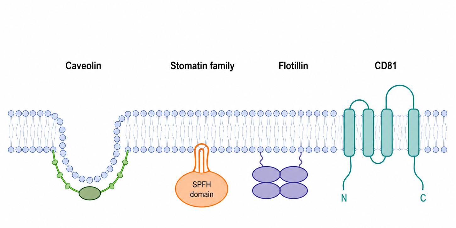
Figure 1. Schematic Membrane Topologies of Major Membrane Microdomain Proteins
3 Membrane Binding and Oligomerization of STOM and SPFH Domain Proteins
3.1 Molecular Composition of the STOM Family
The mammalian Stomatin family mainly includes STOM, STOML1, STOML2, STOML3, and Podocin. STOM is mainly distributed in the plasma membrane and endosomes, STOML1 preferentially localizes to the endosomal-lysosomal system, STOML2 is mainly located in regions associated with the mitochondrial inner membrane, STOML3 participates in the organization of sensory neuron membrane microdomains, and Podocin localizes to the slit diaphragm of glomerular podocytes.
3.2 Hairpin-Like Membrane Insertion Structure
STOM and several related proteins contain a short hydrophobic region that generally inserts into one leaflet of the lipid bilayer in a hairpin-like manner, leaving the main body of the protein on the cytoplasmic side of the membrane. This topology allows the SPFH domain and C-terminal region to interact along the membrane surface with lipids, channels, transporters, and other scaffold proteins.
3.3 Membrane Organization Mediated by the SPFH Domain
The SPFH domain provides an interface for membrane lipid binding and protein interactions but generally does not directly form ion channels. By promoting lateral protein clustering and complex assembly, this domain concentrates ion channels, transporters, or metabolic complexes within specific membrane domains. Different family members vary in their dependence on cholesterol, sphingolipids, or cardiolipin.
3.4 Oligomerization and Formation of Higher-Order Complexes
STOM, STOML3, Flotillin, and other SPFH proteins can form dimers or higher-order oligomers. Oligomerization increases local protein density and stabilizes membrane protein complexes. Mutations in the membrane-binding region, SPFH domain, or C-terminal oligomerization region may cause loss of function because of abnormal localization or complex assembly, even when total protein expression remains unchanged.
3.5 Post-Translational Modifications
Palmitoylation, myristoylation, phosphorylation, and ubiquitination can influence the membrane affinity, stability, and endocytic trafficking of membrane microdomain proteins. Modification patterns differ among proteins, and functional analysis should combine modification-site mutagenesis, mass spectrometry, and subcellular localization experiments.
4 Regulation of Ion Channels and Transporters by Membrane Microdomain Proteins
4.1 Major Mechanisms of Membrane Microdomain-Mediated Channel Regulation
Membrane microdomain proteins can regulate ion channel function through direct channel binding, alteration of channel expression at the plasma membrane, regulation of the local lipid environment, and restriction of lateral diffusion. When changes in total current are detected, alterations in channel number, single-channel open probability, and ion gradients must be distinguished.
4.2 The STOM Family and Acid-Sensing Ion Channels
STOM, STOML1, and STOML3 can regulate certain acid-sensing ion channels, but the direction of regulation differs among family members and ASIC subtypes. Their effects may involve channel membrane localization, gating state, and the local lipid environment and should be jointly validated using electrophysiology, surface-expression assays, and protein-interaction experiments.
4.3 STOML3 and Mechanosensitive Channels
STOML3 can form oligomers on sensory neuron membranes and organize mechanosensitive membrane complexes, thereby enhancing certain responses to mechanical stimulation. STOML3 is not itself a mechanosensitive channel. Instead, it regulates mechanotransduction by influencing the membrane mechanical environment, channel clustering, and complex stability.
4.4 Caveolin and Ion Channels
Caveolin can form complexes with calcium, potassium, sodium, and transient receptor potential channels and regulate their functions through Caveolae localization, the cholesterol environment, and recruitment of signaling enzymes. The direction of regulation may differ among cell types. Channel surface expression, Caveolin-binding status, and electrophysiological parameters should therefore be measured simultaneously.
4.5 Podocin and Podocyte Channels
Podocin can form complexes with slit diaphragm proteins such as Nephrin, CD2AP, and TRPC6. Podocin oligomerization and cholesterol-binding status influence slit diaphragm structure and channel function. NPHS2 abnormalities may simultaneously alter the podocyte cytoskeleton, slit diaphragm integrity, and ion channel regulation.
4.6 Membrane Microdomains and Transporters
STOM can form complexes with glucose transporters in erythrocytes and certain nucleated cells and influence their membrane organization and transport function. Flotillin and Caveolin can also participate in the endocytosis and recycling of various transporters. Transport studies should simultaneously measure substrate uptake, total transporter abundance, and plasma membrane expression.
5 Membrane Microdomains in Cytoskeletal Coupling, Vesicular Trafficking, and Signal Transduction
5.1 Coupling Between Membrane Microdomains and the Cytoskeleton
Actin, Spectrin, and ERM proteins form a cytoskeletal network beneath the plasma membrane that restricts membrane protein diffusion and maintains membrane microdomain stability. Cytoskeletal remodeling alters channel clustering, receptor mobility, and membrane tension. However, cytoskeletal inhibitors also affect cell adhesion, endocytosis, and cell morphology, so genetic methods should be used for confirmation.
5.2 Caveolae and Mechanical Buffering
When cells are stretched or membrane tension increases, some Caveolae flatten and release additional membrane area, thereby reducing the risk of plasma membrane rupture. Loss of Caveolin or Cavin weakens this mechanical buffering capacity and alters mechanosensitive signaling and membrane repair.
5.3 Endocytosis and Membrane Protein Sorting
Caveolin, Flotillin, tetraspanins, and STOM-related proteins can participate in membrane protein endocytosis, endosomal sorting, and membrane recycling. Colocalization of a target protein with endosomal markers cannot distinguish whether the protein is undergoing internalization, recycling, or degradation. Time-course tracking, cell-surface biotinylation, and lysosomal inhibition experiments are required.
5.4 Tetraspanins and Extracellular Vesicles
Tetraspanins such as CD81 are enriched in endosomal and extracellular vesicle membranes and can participate in vesicle formation, cargo sorting, target-cell recognition, and membrane fusion. CD81 is commonly used as an extracellular vesicle-associated marker, but detection of CD81 alone cannot demonstrate that all particles in a sample are exosomes. Particle size, morphology, and additional vesicle markers should also be analyzed.
5.5 Organization of Receptor Signaling Complexes
Membrane microdomains can concentrate receptors, kinases, phosphatases, and small GTPases, thereby altering the initiation rate and duration of signaling. Caveolin, Flotillin, and tetraspanins may promote receptor clustering but may also restrict signaling through sequestration, internalization, or degradation.
5.6 Mitochondrial Membrane Microdomains
STOML2 and Prohibitin can organize respiratory-chain components, lipids, and cristae structure within the mitochondrial inner membrane. STOML2 abnormalities affect respiratory-chain complex stability, oxidative phosphorylation, and mitochondrial stress responses. These structures are organelle membrane microdomains and differ in lipid composition and function from cholesterol-associated plasma membrane domains.
6 Abnormalities of Membrane Microdomain Proteins in Hematological, Neurological, and Tumor-Related Diseases
6.1 Erythrocyte Membranes and Hematological Abnormalities
In erythrocytes, STOM forms complexes with the membrane cytoskeleton, ion channels, and glucose transporters. Marked Stomatin reduction is commonly observed in overhydrated hereditary stomatocytosis, but this reduction is generally a secondary consequence of the primary abnormality in membrane ion permeability. STOM deficiency should therefore not be regarded as the direct cause of all cases.
6.2 Sensory Neurons and Mechanotransduction
STOML3 participates in mechanical-stimulus responses in tactile sensory neurons and nociceptive neurons. Reducing STOML3 expression or inhibiting its oligomerization can weaken certain mechanosensitive responses, making STOML3 suitable for studies of mechanical pain, touch sensation, and membrane organization at sensory nerve endings.
6.3 Membrane Microdomain Signaling in the Nervous System
Caveolin, Flotillin, and tetraspanins can affect the localization of neurotransmitter receptors, ion channels, and synaptic membrane proteins. Changes in membrane cholesterol or scaffold protein abnormalities may alter synaptic transmission and axonal signaling, but membrane microdomain composition differs markedly among neuronal cell types and brain regions.
6.4 Muscle- and Kidney-Related Diseases
CAV3 abnormalities can affect muscle-cell Caveolae and membrane stability and are associated with certain muscular dystrophies and myopathies. NPHS2 abnormalities can impair Podocin membrane localization or oligomerization, disrupt the podocyte slit diaphragm, and cause hereditary proteinuria. NPHS1 abnormalities can directly affect Nephrin-mediated slit diaphragm structure and signaling.
6.5 Tumor Cell Proliferation and Migration
Membrane microdomain proteins such as CAV1, FLOT1, FLOT2, STOML2, and CD81 show abnormal expression in multiple tumor types and influence receptor signaling, cell migration, endocytosis, extracellular vesicles, and energy metabolism. CAV1 may exhibit tumor-promoting or tumor-suppressive effects depending on tumor stage and cellular compartment, and its functional direction cannot be determined solely from increased expression.
6.6 Tumor Metabolism and Therapeutic Responses
STOML2 can support highly metabolic tumor cells by maintaining mitochondrial inner membrane structure and respiratory-chain function. Flotillin and CD81 can influence invasion and therapeutic responses by regulating receptor recycling, vesicle-mediated communication, and cell adhesion. Establishing causality requires gene knockdown, knockout, rescue experiments, and in vivo models.
7 Methods for Studying Membrane Microdomain Protein Interactions and Functions
7.1 Detergent-Resistant Membrane Isolation
Cold Triton X-100 extraction and density-gradient centrifugation are commonly used to isolate detergent-resistant membrane fractions. However, detergents can redistribute membrane lipids and proteins. The presence of a target protein in a low-density fraction only suggests an association with a particular membrane environment and cannot independently demonstrate lipid raft formation in living cells.
7.2 Subcellular Fractionation and Cell-Surface Protein Detection
Membrane protein fractionation, cell-surface biotinylation, and membrane sheet isolation can distinguish total protein expression from plasma membrane expression. Plasma membrane, endosomal, mitochondrial, and cytoplasmic marker proteins should be used to assess fraction purity, and harsh detergents that disrupt native oligomers should be avoided.
7.3 Microscopic Imaging and Membrane Diffusion Analysis
Confocal microscopy is suitable for observing localization at the cellular scale, whereas super-resolution imaging can analyze nanoscale clustering. Single-molecule tracking and fluorescence recovery assays can measure membrane protein diffusion. Colocalization alone does not demonstrate direct protein binding and should be combined with co-immunoprecipitation, proximity assays, or energy-transfer analysis.
7.4 Protein Interactions and Oligomerization
Co-immunoprecipitation is suitable for validating relatively stable protein complexes. Cross-linking and proximity labeling can detect weak interactions and local protein networks, whereas blue native electrophoresis and fluorescence resonance energy transfer can be used to evaluate oligomerization. High-molecular-weight bands may also result from nonspecific aggregation, so deletion mutants and lysis-condition controls should be included.
7.5 Validation of Lipid Dependence
Methyl-β-cyclodextrin is commonly used to reduce membrane cholesterol, whereas Filipin can be used to visualize free cholesterol distribution. Cholesterol depletion broadly alters membrane fluidity, cell viability, and channel surface expression. Cholesterol-repletion groups should therefore be included, and membrane integrity should be assessed simultaneously.
7.6 Ion Channel and Transport Functions
Patch-clamp recording directly measures ion channel currents. Calcium imaging and membrane potential probes are suitable for functional screening, whereas fluorescent or isotopically labeled substrate uptake can evaluate membrane transport. Functional results should be analyzed together with target protein expression at the plasma membrane, membrane microdomain localization, and interactions with scaffold proteins.
7.7 Gene Function and Domain Validation
siRNA and CRISPR/Cas9 can be used to reduce or eliminate membrane microdomain protein expression. Wild-type rescue and mutants defective in membrane binding, oligomerization, or lipid binding can identify critical domains. Knockdown or knockout phenotypes should be confirmed using independent sequences, protein detection, and rescue experiments.
7.8 Extracellular Vesicle and Tetraspanin Research
CD81 fluorescent fusion proteins can be used to trace vesicle formation, release, and intercellular transfer. CD81 knockdown or knockout models can be used to evaluate its role in vesicle biogenesis and target-cell uptake. Overexpression-based tracing may alter protein clustering and vesicle composition and should therefore be compared with endogenous CD81 detection.
8 Products for Studying Cell Membrane Microdomain Structure and Function
Table 2 Products for STOM, Caveolin, Flotillin, and Podocyte Membrane Microdomain Research
Catalog # | Product Name | Grade & Purity | Product Type | Main Application |
OB-1 | ≥98% (HPLC) | Small-molecule inhibitor | Inhibits STOML3 oligomerization for studies of mechanosensitive channels and sensory transduction regulation | |
Recombinant Human STOM Protein | ≥90% (SDS-PAGE) | Recombinant protein | STOM protein-binding studies, antibody evaluation, and in vitro interaction research | |
Recombinant Stomatin Like 2 Antibody | KD Validation | Recombinant antibody | STOML2 expression, mitochondrial localization, and functional studies | |
STOM Human Pre-designed siRNA Set A | — | siRNA | Reduces STOM expression to evaluate ion channel regulation, membrane transport, and membrane microdomain function | |
STOML2 Human Pre-designed siRNA Set A | — | siRNA | Investigates the effects of STOML2 on mitochondrial membrane organization, energy metabolism, and tumor cell function | |
STOML3 Human Pre-designed siRNA Set A | — | siRNA | Investigates STOML3 oligomerization, mechanosensitive channels, and sensory transduction | |
Recombinant Caveolin 1 Antibody | KO Validation | Recombinant antibody | Caveolin-1 expression, localization, and Caveolae structure studies | |
Recombinant Caveolin-2 Antibody | Recombinant, ExactAb™, validated, 0.2 mg/mL | Recombinant antibody | Caveolin-2 expression and Caveolin complex studies | |
Recombinant Caveolin-3 Antibody | Recombinant, ExactAb™, validated, see COA | Recombinant antibody | Muscle-cell Caveolae and Caveolin-3-related disease studies | |
Recombinant Human Caveolin-1 Protein | Carrier-free, ≥95% (SDS-PAGE) | Recombinant protein | Caveolin-1 protein interactions, antibody evaluation, and mechanistic studies | |
Recombinant Flotillin 1 Antibody | Recombinant, ExactAb™, KD Validation, validated, see COA | Recombinant antibody | Flotillin-1 membrane microdomain, endocytosis, and signaling complex studies | |
Recombinant Flotillin 2/ESA Antibody | Recombinant, ExactAb™, validated, 0.6 mg/mL | Recombinant antibody | Flotillin-2 expression, localization, and oligomerization studies | |
NPHS1 Human Pre-designed siRNA Set A | — | siRNA | Reduces Nephrin expression to study podocyte slit diaphragm complexes | |
NPHS2 Human Pre-designed siRNA Set A | — | siRNA | Reduces Podocin expression to study slit diaphragm microdomains and ion channel regulation | |
Nphs1 Mouse Pre-designed siRNA Set A | — | siRNA | Mouse Nephrin gene-function and podocyte disease-model studies |
Table 3 Products for CD81 Tetraspanin-Enriched Microdomain and Extracellular Vesicle Research
Catalog # | Product Name | Grade & Purity | Product Type | Main Application |
CD81 Human Pre-designed siRNA Set A | — | siRNA | Reduces CD81 expression to study tetraspanin-enriched microdomains, adhesion, and vesicle function | |
pLenti-CD81-sgRNA | — | Gene-knockout validation sample | Used to evaluate the specificity of CD81 antibodies in protein detection | |
pLenti-CD81-sgRNA | — | Gene-knockout validation sample | Used to validate CD81 gene expression and nucleic acid detection | |
Lenti-CMV-CD81-EGFP | 10⁹ TU/mL | Lentiviral tracing vector | Green fluorescent tracing of CD81-positive extracellular vesicles | |
Lenti-CMV-CD81-mCherry | 10⁹ TU/mL | Lentiviral tracing vector | Red fluorescent tracing of CD81-positive extracellular vesicles | |
Recombinant CD81/TAPA-1 Antibody | Animal Free, carrier-free, recombinant, ExactAb™, azide-free, validated, high-performance, PBS Only, ≥95% (SDS-PAGE), see COA | Recombinant antibody | CD81 expression, localization, and protein-interaction studies | |
Recombinant CD81/TAPA-1 Antibody (AF488) | ExactAb™, validated, 5 μL/test | Fluorescently labeled antibody | CD81 fluorescence imaging and flow-cytometric phenotyping | |
Recombinant Human CD81 Protein | Animal Free, carrier-free, His-tagged, PBS Only, ≥95% (SDS-PAGE), see COA | Recombinant protein | CD81-binding studies, antibody evaluation, and tetraspanin complex research |
The functions of cell membrane microdomains arise from the coordinated organization of lipids, scaffold proteins, the membrane cytoskeleton, and effector proteins. Combined analysis of protein localization, oligomerization, lipid dependence, membrane trafficking, and functional readouts is required to distinguish changes in membrane protein expression, redistribution among membrane compartments, and alterations in ion channel gating.


 危险品化学品经营许可证(带存储)