
计算溶液所需的质量、体积或浓度。
JC-10线粒体膜电位荧光探针溶液
有货
BioReagent,生物染色剂,适用于荧光分析 级 ,适用于对基线干扰要求严格的色谱和分析工作流程。
-20°C储存。超低温运输 。请查阅批次 COA 获取详细规格。
SDS、COA、产品数据表及规格说明书均可下载。可通过批号查询获取批次 COA。
在色谱分析、有机合成和交叉偶联反应领域已被 0 篇同行评审文献引用。
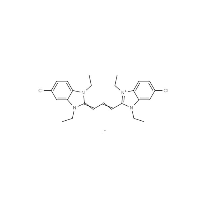
JC‑10 是一种用于检测线粒体膜电位(ΔΨm)的理想荧光探针。当线粒体膜电位较高时,JC‑10 在线粒体基质中聚集,发出红色荧光;当膜电位较低时,JC‑10 以单体形式存在,发出绿色荧光。JC‑10 可选择性地进入线粒体,并随膜电位升高发生可逆的荧光颜色转变(绿色→橙红色)。与 JC‑1 相比,JC‑10 的水溶性更优,而荧光性质相近,因此是 JC‑1 的卓越替代品。
产品用途
仅供科研使用,广泛用于细胞生物学、分子生物学、药理学等研究领域,严禁用于人体。JC‑10 适用于检测细胞、组织或分离线粒体的膜电位变化。
物理性质及指标
外观:液体
分子式: C₂₅H₂₉Cl₂IN₄
分子量:583.34
CAS No.:5563-28-0
浓度:2 mg/mL,相当于3 mM
澄清度:DMSO 中澄清,无杂质
Ex/Em(nm):490/525 nm (单体-绿色);490/590 nm (聚集体-橙红色)
储存条件:-20℃保存,避光、防潮、密封
使用方法:(仅供参考)
一、工作液配制
将 JC-10 溶液回温至室温,用 HBSS(Hanks' Balanced Salt Solution with HEPES)或其他缓冲液,加入 0.02% Pluronic F-127,稀释至终浓度 5~30 μM,涡旋混匀。
二、样品实验方案
• 酶标仪检测
(1)使用待测化合物处理细胞至预定时间诱导凋亡(示例:Jurkat 细胞可采用喜树碱处理 4~6 小时),阴性对照孔(细胞 + 溶剂对照),背景空白孔(无细胞,仅培养基);
(2)对于贴壁细胞,染色前需移除培养基;对于悬浮细胞,染色前离心去除培养基;
(3)96 孔板中每孔加入 100 μL JC-10 工作液;384 孔板中每孔加入 25 μL 工作液;
(4)37℃、5% CO₂培养箱中避光孵育 15~60 min;
注:孵育时间需根据具体细胞类型和浓度进行优化,建议每次实验前确定最佳孵育时长。
【可选】移除 JC-10 工作液后,96 孔板每孔加入 100 μL HHBS 缓冲液(384 孔板 25μL / 孔),再进行检测分析;
(5)在激发 / 发射 = 490/525 nm 和 490/590 nm 处监测荧光变化进行比率分析。
• 荧光显微镜或流式细胞仪检测
(1)使用测试化合物处理细胞至预定时间诱导凋亡(示例:Jurkat 细胞可采用喜树碱处理 4~6 小时);
(2)离心细胞,调整密度至每管 1~5×10⁵个;
(3)使用 500 μL JC-10 工作液重悬细胞;
(4)室温或 37℃避光孵育 10~60min;
注:孵育时间需根据具体细胞类型和浓度进行优化,建议每次实验前确定最佳孵育时长。
【可选】离心移除 JC-10 工作液后,加入适量 HHBS 缓冲液重悬细胞,再进行检测分析;
(5)使用荧光显微镜(FITC/TRITC 滤光片组)或流式细胞仪(FL1/FL2 通道),在激发 / 发射波长 490/525 nm 和 490/590 nm 下监测荧光变化。
注意
1.荧光染料均存在淬灭问题,请尽量注意避光,以减缓荧光淬灭。
2.本产品仅限于专业人员的科学研究用,不得用于临床诊断或治疗,不得用于食品或药品,不得存放于普通住宅内。
3.为了您的安全和健康,请穿实验服并戴一次性手套操作。
4.部分产品我司仅能提供部分信息,我司不保证所提供信息的权威性,以上数据仅供参考交流研究之用。
JC‑10 is an ideal fluorescent probe for detecting mitochondrial membrane potential (ΔΨm). When the mitochondrial membrane potential is high, JC‑10 aggregates in the mitochondrial matrix and emits red fluorescence; when the membrane potential is low, JC‑10 exists as monomers and emits green fluorescence. JC‑10 selectively enters mitochondria and undergoes a reversible fluorescence color shift from green to orange‑red as the membrane potential increases. Compared with JC‑1, JC‑10 exhibits better water solubility while maintaining similar fluorescence properties, making it a superior alternative to JC‑1.
Product Use
For research use only. Widely applied in cell biology, molecular biology, pharmacology, and other research fields. Not for human use. JC‑10 is suitable for detecting membrane potential changes in cells, tissues, or isolated mitochondria.
Physical Properties and Specifications
Appearance: Liquid
Molecular Formula: C₂₅H₂₉Cl₂IN₄
Molecular Weight: 583.34
CAS No. 5563‑28‑0
Concentration: 2 mg/mL (equivalent to 3 mM)
Solubility: Clear in DMSO, free from visible impurities
Excitation/Emission Maxima (nm): 490/525 (monomer, green); 490/590 (aggregate, orange‑red)
Storage Conditions: Store at ‑20°C, protected from light and moisture, in airtight containers
Operating Protocol (For Reference Only)
1. Preparation of Working Solution
Thaw JC-10 solution to room temperature. Dilute it with HBSS (Hanks' Balanced Salt Solution with HEPES) or other buffer supplemented with 0.02% Pluronic F-127 to a final concentration of 5–30 μM, then vortex thoroughly for homogenization.
2. Sample Experimental Protocols
2.1 Microplate Reader Detection
(1) Treat cells with test compounds for scheduled duration to induce apoptosis (Example: Jurkat cells can be treated with camptothecin for 4–6 hours). Set up negative control wells (cells plus solvent control) and background blank wells (medium only without cells).
(2) Remove culture medium from adherent cells before staining; pellet suspension cells by centrifugation to discard medium prior to staining.
(3) Add 100 μL JC-10 working solution per well for 96-well plates, or 25 μL per well for 384-well plates.
(4) Incubate the plates in a 37 ℃ incubator with 5% CO₂ protected from light for 15–60 min.
Note: Incubation time should be optimized according to cell type and concentration; it is recommended to confirm the optimal incubation duration before each experiment.
Note: Incubation time should be optimized according to cell type and concentration; it is recommended to confirm the optimal incubation duration before each experiment.
Optional: After aspirating JC-10 working solution, add 100 μL HHBS buffer per well for 96-well plates (25 μL per well for 384-well plates) before detection and analysis.
(5) Monitor fluorescence signals at excitation/emission wavelengths of 490/525 nm and 490/590 nm for ratiometric analysis.
2.2 Detection via Fluorescence Microscope or Flow Cytometer
(1) Treat cells with test compounds for scheduled duration to induce apoptosis (Example: Jurkat cells can be treated with camptothecin for 4–6 hours).
(2) Centrifuge cells and adjust the cell density to 1–5×10⁵ cells per tube.
(3) Resuspend cell pellets in 500 μL JC-10 working solution.
(4) Incubate the cell suspension protected from light at room temperature or 37 ℃ for 10–60 min.
Note: Incubation time should be optimized according to cell type and concentration; it is recommended to confirm the optimal incubation duration before each experiment.
Optional: Remove JC-10 working solution by centrifugation, resuspend cells with an appropriate volume of HHBS buffer, then proceed to detection and analysis.
(5) Detect fluorescence changes under excitation/emission wavelengths of 490/525 nm and 490/590 nm using a fluorescence microscope equipped with FITC/TRITC filter sets, or a flow cytometer with FL1/FL2 channels.
Precautions
Fluorescent dyes are susceptible to photobleaching. Keep samples away from light to slow down fluorescence quenching.
For research use only by qualified professionals. Not for clinical diagnosis or therapy, food or pharmaceutical applications. Do not store in residential areas.
Wear a lab coat and disposable gloves during operation for your personal safety and health.
We only provide partial technical information for some products and do not guarantee the authenticity and authority of the data. All data above are for reference and academic research only.
通过匹配包装上的批号来查找并下载产品的 COA,每批产品都进行了严格的验证,您可放心使用!
| 批号(Lot Number) | 证书类型 | 货号 |
|---|---|---|
 ZJ26F1040098 |
分析证书 | J1527591 |
我们的级别选择指南涵盖了目录中所有的变体的纯度、稳定剂状态和应用适用性。
查看 BioReagent 级别指南 → 查看 Biological Stain 级别指南 → 查看 for Fluorescence analysis 级别指南 →

 危险品化学品经营许可证(带存储)