线粒体超氧化物是线粒体呼吸链产生的主要活性氧(ROS),其水平直接反映线粒体功能、氧化应激状态及细胞代谢与凋亡进程。MitoSOX Red 是一种新型的,可渗透活细胞,用于线粒体内超氧化物检测的荧光探针,其可快速、特异性地靶向线粒体,从而有选择性地检测线粒体内的超氧化物。MitoSOX Red 进入线粒体后会被超氧化物氧化,而不会被其他活性氧(ROS)或活性氮(RNS)生成系统氧化。被氧化的MitoSOX Red 随后与线粒体/细胞核内的核酸结合,产生强红色荧光。本试剂盒可使用荧光显微镜、激光共聚焦显微镜、荧光酶标仪、流式细胞仪等荧光检测系统进行检测,其激发波长为510nm,发射波长为580nm。
产品组分
M1508403 | 组分 | 100 T | 500 T | 储存条件 |
M1508403A | MitoSOX Red (5mM) | 100 μL | 500μl | -20℃.避光 |
M1508403B | 阳性对照 | 1 mL | 5 mL | -20℃.避光 |
使用方法
1. 检测工作液配制
按照本次用量,用PBS将MitoSOX Red (5mM) 储存液按照 1:1000的比例稀释成5μM的检测工作液,混匀后即为MitoSOX Red染色工作液。备注:具体的稀释比例可以根据实验需求进行调整。
2. 阳性对照的设置(可选)
取状态良好的细胞去除原有培养基,悬浮细胞离心去除培养基,试剂盒中提供的阳性对照推荐按照1:1000的比例加入到细胞培养液中,37℃培养箱处理细胞1h。(若刺激1h,细胞状态变差,可适当降低浓度或者缩短诱导时间;若荧光较弱,则适当延长诱导时间或者增加浓度)。
3. 细胞染色
1)贴壁细胞:处理过后的细胞吸掉培养液,用PBS洗涤2-3次,加入适量 MitoSOX Red 工作液(例如 96 孔板每孔细胞量100 μL,6 孔板每孔细胞量 1 mL),37°C避光孵育 20-30 min。孵育结束后,用PBS清洗2-3次。备注:最佳孵育时间可以根据实验需求进行调整(10-60 min)。
2)悬浮细胞:处理过后的细胞,室温 1000 rpm离心5 min,吸掉培养基,加入适量MitoSOX Red 体积的工作液(推荐细胞密度为1E6-1E7/mL),37°C 避光孵育 20-30 min。孵育结束后,室温 1000 rpm,5 min,吸掉培养基,用PBS清洗一次细胞。备注:最佳MitoSOX Red 孵育时间可以根据实验需求进行调整(10-60 min)。
4. 波长检测
PBS重悬细胞,选择PE通道进行流式检测,如果用荧光显微镜直接观察,MitoSOX Red氧化产物检测条件为 Ex/Em=510nm/580nm。

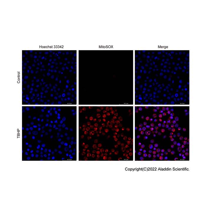
图1. 线粒体超氧化物检测试剂盒(MitoSOX Red)检测Jurkat细胞线粒体内超氧化物的荧光显微镜效果图。Jurkat细胞不处理或用阳性对照处理后,使用本试剂盒和Hoechst 33342(H288601)进行染色。正常的Jurkat细胞中红色荧光几乎观察不到;使用诱导线粒体内超氧化物生成的阳性对照TBHP处理后,线粒体内超氧化物生成增加,MitoSOX Red与超氧化物反应并重新分布,与线粒体及细胞核DNA结合,细胞中线粒体及细胞核的红色荧光显著增强。实际检测效果会因实验条件、检测仪器等的不同而存在差异,图中效果仅供参考。

图2. 线粒体超氧化物检测试剂盒(MitoSOX Red)检测Jurkat细胞线粒体内超氧化物的流式细胞术效果图。Jurkat细胞不处理或用阳性对照处理后,使用本试剂盒进行染色并进行流式细胞术检测。正常的Jurkat细胞中红色荧光很弱(蓝色);使用诱导线粒体内超氧化物生成的阳性对照TBHP处理后,线粒体内超氧化物生成增加,细胞中红色荧光显著增强(红色)。黑色为正常细胞未染色的阴性对照。通过PE通道进行检测。实际检测效果会因实验条件、检测仪器等的不同而存在差异,图中效果仅供参考。
注意事项
1)首次使用可适量分装,避免反复冻融。
2)血清和酚红对本荧光探针的检测有干扰,需避免。
3)MitoSOX Red在与超氧化物反应后可能会重新分布,并与线粒体或细胞核DNA结合,因此观察到红色荧光集中在细胞核属于正常现象。
4)MitoSOX Red的终浓度通常为1-5μM,高难度工作液会产生细胞毒性。
5)荧光染料均存在猝灭问题,请尽量注意避光。
6)配制好的工作液不稳定,建议现配现用。
7)本产品仅限于科学研究用,不得用于临床诊断或治疗,为了您的安全和健康,请穿实验服并戴一次性手套操作
Mitochondrial superoxide is the primary reactive oxygen species (ROS) generated by the mitochondrial respiratory chain, and its abundance serves as a direct indicator of mitochondrial function, oxidative stress status, cellular metabolism, and apoptotic progression. MitoSOX Red is a novel, cell-permeable fluorescent probe specifically developed for the detection of mitochondrial superoxide in live cells. It rapidly and selectively targets mitochondria, allowing for specific quantification of superoxide within the mitochondrial matrix. Upon entry into mitochondria, MitoSOX Red is oxidized exclusively by superoxide, but not by other ROS- or RNS-generating systems. The oxidized probe subsequently binds to nucleic acids in mitochondria and the cell nucleus, emitting intense red fluorescence. This kit is compatible with multiple fluorescence‑based platforms, including fluorescence microscopy, laser scanning confocal microscopy, fluorescence microplate readers, and flow cytometry, with an excitation wavelength of 510 nm and an emission wavelength of 580 nm.
Kit Components
M1508403 | Component | 100 T | 500 T | Storage Condition |
M1508403A | MitoSOX Red (5mM) | 100 μL | 500μl | -20℃. Store in the dark |
M1508403B | Positive Control | 1 mL | 5 mL | -20℃. Store in the dark |
Experimental Protocol
1. Preparation of Staining Working Solution
Prepare a 2.5 μM MitoSOX Red working solution by diluting the 5 mM stock solution with PBS at a ratio of 1:2000. Mix thoroughly before use. Note: The dilution ratio can be optimized according to specific experimental conditions.
2. Positive Control Setup (Optional)
Discard the culture medium from healthy cells (centrifuge suspension cells to remove medium). Add the positive control reagent to the cell culture medium at a ratio of 1:1000, and incubate cells at 37 °C for 1 h. Note: If cell viability declines after 1 h of induction, decrease the concentration or shorten the incubation time; if fluorescence intensity is insufficient, prolong induction or increase the concentration accordingly.
3. Cell Staining
1) Adherent cells
Remove the culture medium, wash cells with PBS 2–3 times, add an appropriate volume of MitoSOX Red working solution (100 μL/well for 96-well plates, 1 mL/well for 6-well plates), and incubate at 37 °C in the dark for 20–30 min. After incubation, wash cells with PBS 2–3 times. Note: Optimal incubation time may be adjusted between 10-60 min based on experimental requirements.
2) Suspension cells
Centrifuge treated cells at 1000 rpm for 5 min at room temperature and discard the medium. Resuspend cells in MitoSOX Red working solution at a density of 1×10⁶–1×10⁷ cells/mL, then incubate at 37 °C in the dark for 20–30 min. After incubation, centrifuge at 1000 rpm for 5 min at room temperature, remove the supernatant, and wash cells once with PBS. Note: Optimal incubation time may be adjusted between 10–60 min based on experimental requirements.
4. Fluorescence Detection
Figure 1. Fluorescence microscopy images of mitochondrial superoxide in Jurkat cells detected with the MitoSOX Red Mitochondrial Superoxide Detection Kit. Jurkat cells were left untreated or treated with positive control, followed by staining with this kit and Hoechst 33342 (H288601). Almost no red fluorescence was observed in normal Jurkat cells. After treatment with TBHP, a positive control that induces mitochondrial superoxide production, the generation of mitochondrial superoxide was increased. MitoSOX Red reacted with superoxide and redistributed, binding to mitochondrial and nuclear DNA, resulting in markedly enhanced red fluorescence in the mitochondria and nucleus of cells. Actual detection results may vary depending on experimental conditions and detection instruments; the results shown in the figure are for reference only.
Figure 2. Flow cytometry results of mitochondrial superoxide in Jurkat cells detected using the MitoSOX Red Mitochondrial Superoxide Detection Kit. Jurkat cells were left untreated or treated with positive control, then stained with the kit and analyzed by flow cytometry. Red fluorescence was very weak in normal Jurkat cells (Blue). After treatment with TBHP, a positive control for inducing mitochondrial superoxide production, mitochondrial superoxide levels increased, accompanied by a significant enhancement of red fluorescence in cells (Red). Black: unstained negative control (normal cells). Detection was performed via the PE channel. Actual detection results may vary with experimental conditions and instruments. The results shown in the figure are for reference only.
Notes
1) Aliquot reagents upon initial use to avoid repeated freeze-thaw cycles.
2) Serum and phenol red interfere with fluorescence detection and must be excluded from the assay system.
3) Following oxidation by superoxide, MitoSOX Red may redistribute and bind to mitochondrial or nuclear DNA; thus, accumulation of red fluorescence in the nucleus is normal.
4) The recommended final concentration of MitoSOX Red is 1-5 μM; excessive concentrations may induce cytotoxicity.
5) Fluorescent probes are susceptible to photobleaching; minimize light exposure throughout all procedures.
6) The working solution is unstable and must be prepared freshly immediately before use.
This product is for scientific research use only and not intended for clinical diagnosis or therapeutic applications. For personal safety, wear a laboratory coat and disposable gloves during handling.

 ZJ26F1039956


 危险品化学品经营许可证(带存储)