
计算溶液所需的质量、体积或浓度。
Masson三色染色试剂盒
有货
用于作为结缔组织染色法之一的Masson三色液,可将细胞质染成红色。Masson三色染色液对结缔组织和非结缔组织进行不同的着色,显示结缔组织各种纤维成分。
Masson三色染色试剂盒主要用于从肌肉组织中区分胶原蛋白。用Bouin’s溶液处理组织切片,可加强最后的染色。用Weigert 铁苏木素对核染色,用比布列希猩红染液对细胞质和肌纤维染色。磷钨酸和磷钼酸处理后,用苯胺蓝对胶原纤维染色。染色后用醋酸漂洗,使色泽更加细腻透明。微波炉方法可加快染色。
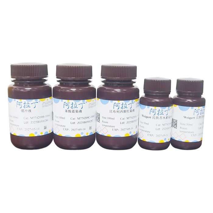
| M774209 | Component | 4×100 mL | Storage |
| M774209A | 比布列西猩红染液 | 100 mL | RT. Store in the dark |
| M774209B | 缓冲液 | 100 mL | RT. Store in the dark |
| M774209C | 苯胺蓝染液 | 100 mL | RT. Store in the dark |
| M774209D | Weigert 氏铁苏木素染液A | 50 mL | RT. Store in the dark |
| M774209E | Weigert 氏铁苏木素染液B | 50 mL | RT. Store in the dark |
使用说明:
1、试剂准备
1.1 Weigert 铁苏木素染液工作液:将Weigert氏铁苏木素染液A与Weigert氏铁苏木素染液B等体积混合,临用前配制。
1.2 Bouin's 溶液:苦味酸1%,甲醛 9%,冰乙酸5%。
2、样品处理
2.1 对于石蜡切片:二甲苯中脱蜡2×5min;无水乙醇5min、90%乙醇2min、70%乙醇2min;PBS 漂洗2min。
2.2 对于冰冻切片:PBS漂洗 2min。
2.3 对于培养细胞:用4%多聚甲醛固定10min以上,PBS洗涤2×2min。
3、Masson 三色染液
3.1 对于福尔马林固定的组织,需使用Bouin's溶液,在56°C下,再固定1h,可改善质量,这一步不是必须的。
3.2 流动自来水冲洗5-10min,以去除多余的黄色。
3.3 用Weigert 铁苏木素染液工作液染色10min。
3.4 流动自来水漂洗10min。
3.5 蒸馏水漂洗。
3.6 用比布列西猩红染液染色5-15min,该溶液可保存至下一次使用。
3.7 蒸馏水快速漂洗3次。
3.8 用缓冲液分化5-15min,直至胶原不显示红色。
3.9 将切片直接转移入苯胺蓝染液中 (不需要漂洗),染色5-10min。蒸馏水直接漂洗,用1%酸酒精溶液分色2-5min。
3.10 蒸馏水漂洗。
3.11 95%乙醇、100%乙醇快速脱水 (可能洗掉染液),二甲苯透明,树脂封片剂封片。
染色结果:
| 细胞质、肌纤维 | 红色 |
| 胶原蛋白 | 蓝色 |
注意事项:
1、需自备4%多聚甲醛、梯度乙醇、二甲苯,中性树胶或其它封片剂。
2、第一次使用本试剂盒时建议先取1-2个样品做预实验。
3、为了您的自身安全,使用试剂前,请做好防护,如穿实验服,带手套等。
Masson’s trichrome stain, one of the connective tissue staining methods, stains cytoplasm red. Masson’s trichrome staining solution differentially stains connective and non‑connective tissues to demonstrate various fibrous components of connective tissue.
The Masson’s trichrome staining kit is mainly used to distinguish collagen from muscle tissue. Pretreatment of tissue sections with Bouin’s solution enhances the final staining result. Nuclei are stained with Weigert’s iron hematoxylin, and cytoplasm and muscle fibers are stained with Biebrich scarlet stain. Following treatment with phosphotungstic acid and phosphomolybdic acid, collagen fibers are stained with aniline blue. Acetic acid rinsing after staining yields finer and more transparent coloration. A microwave protocol can accelerate the staining procedure.
| Component No. | Component Name | Appearance | 4×100ml | Storage |
| M774209A | Biebrich Scarlet Stain | Liquid | 100ml | RT. Store in the dark |
| M774209B | Buffer Solution | Liquid | 100ml | RT. Store in the dark |
| M774209C | Aniline Blue Stain | Liquid | 100ml | RT. Store in the dark |
| M774209D | Weigert’s Iron Hematoxylin Stain A | Liquid | 50ml | RT. Store in the dark |
| M774209E | Weigert’s Iron Hematoxylin Stain B | Liquid | 50ml | RT. Store in the dark |
Instructions for Use:
1. Reagent Preparation
1.1 Working solution of Weigert’s Iron Hematoxylin Stain: Mix Weigert’s Iron Hematoxylin Stain A and Weigert’s Iron Hematoxylin Stain B in equal volumes. Prepare immediately before use.
1.2 Bouin’s Solution: 1% picric acid, 9% formaldehyde, 5% glacial acetic acid.
2. Sample Pretreatment
2.1 Paraffin sections: Deparaffinize in xylene for 2 × 5 min; 5 min in absolute ethanol, 2 min in 90% ethanol, 2 min in 70% ethanol; rinse with PBS for 2 min.
2.2 Frozen sections: Rinse with PBS for 2 min.
2.3 Cultured cells: Fix with 4% paraformaldehyde for no less than 10 min, wash with PBS for 2 × 2 min.
3. Masson’s Trichrome Staining Procedure
3.1 For formalin-fixed tissues, post-fix with Bouin’s solution at 56 °C for 1 h to improve staining quality. This step is optional.
3.2 Rinse under running tap water for 5–10 min to remove excess yellow pigment.
3.3 Stain with working Weigert’s Iron Hematoxylin Stain for 10 min.
3.4 Rinse under running tap water for 10 min.
3.5 Rinse with distilled water.
3.6 Stain with Biebrich Scarlet Stain for 5–15 min. This solution can be stored for subsequent use.
3.7 Quick rinse with distilled water 3 times.
3.8 Differentiate in Buffer Solution for 5–15 min until collagen no longer appears red.
3.9 Transfer sections directly into Aniline Blue Stain (no intermediate rinse) and stain for 5–10 min. Rinse directly with distilled water, then differentiate in 1% acid alcohol for 2–5 min.
3.10 Rinse with distilled water.
3.11 Rapid dehydration in 95% ethanol and absolute ethanol (stain may be eluted), clear with xylene, and mount with resin mounting medium.
Staining Results:
| Cytoplasm and muscle fibers | Red |
| Collagen fibers | Blue |
Precautions:
1. 4% paraformaldehyde, graded ethanol, xylene, neutral balsam or other mounting media are not supplied and need to be prepared by users.
2. It is recommended to perform a preliminary test with 1–2 samples when using this kit for the first time.
3. For personal safety, wear protective equipment such as a lab coat and gloves before handling reagents.
通过匹配包装上的批号来查找并下载产品的 COA,每批产品都进行了严格的验证,您可放心使用!
| 批号(Lot Number) | 证书类型 | 货号 |
|---|---|---|
 ZJ25F0521514 |
分析证书 | M774209 |
我们的级别选择指南涵盖了目录中所有的变体的纯度、稳定剂状态和应用适用性。
查看 BioReagent 级别指南 → 查看 Biological Stain 级别指南 → 查看 for Microscopy 级别指南 →

 危险品化学品经营许可证(带存储)