Platelet-derived growth factor (PDGF) was discovered as a major mitogenic factor present in serum but absent from plasma. It was found to be secreted from the alpha -granules of platelets activated during the coagulation of blood to form serum. Subsequent studies have demonstrated that PDGF is not one molecule but three, each a dimeric combination of two distinct but structurally related peptide chains designated A and B. The dimeric isoforms PDGF-AA, AB and BB are differentially expressed in various cell types and their effects are mediated through two distinct receptors, termed alpha and beta. Differences exist in isoform binding to each receptor. In general, PDGF isoforms are potent mitogens for connective tissue cells, including dermal fibroblasts, glial cells, arterial smooth muscle cells and some epithelial and endothelial cells. In addition to its activity as a mitogen, PDGF is chemotactic for fibroblasts, smooth muscle cells, neutrophils and mononuclear cells. Other reported activities for PDGF include stimulation of granule release by neutrophils and monocytes, facilitation of steroid synthesis by Leydig cells, stimulation of neutrophil phagocytosis, inhibition of natural killer (NK) cell activity, stimulation of collagen synthesis, modulation of thrombospondin expression and secretion, stimulation of collagenase activity and secretion, induction of contraction of rat aorta strips in vitro, and transient induction of T cell IL-2 secretion accompanied by a down-regulation of IL-4 and IFN-gamma production, temporary effects that may allow clonal expansion of antigen-activated B and T helper lymphocytes prior to differentiation. PDGF also appears to be ubiquitous in neurons throughout the CNS, where it is suggested to play an important role in neuron survival and regeneration, and in mediation of glial cell proliferation and differentiation.
生物活性
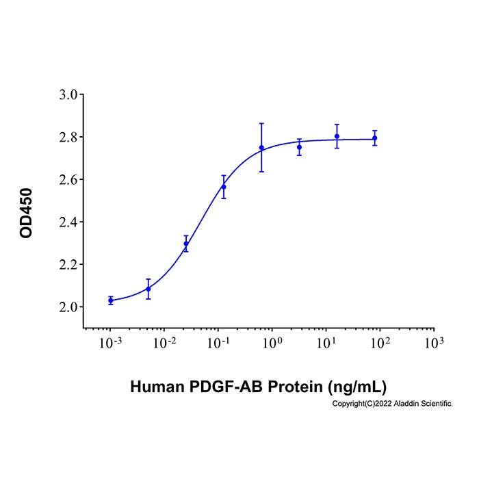
Measured in a cell proliferation assay using NIH‑3T3 mouse embryonic fibroblast cells. The ED50 for this effect is less than 15 ng/mL.
16.0 & 12.1 kDa, under reducing conditions; 28.7 kDa, under non-reducing conditions.
分子类型
蛋白质
产品线
生物产品
储存与运输
物理形态
冻干(Lyophilized)
储存缓冲液
Lyophilized from 4mM HCl.
浓度
见COA
复溶
We recommend that this vial be briefly centrifuged prior to opening to bring the contents to the bottom. Reconstitute at 0.5 mg/mL in sterile distilled water. Stock solutions should be apportioned into working aliquots and stored at ≤ -20 °C. Further dilutions should be made in appropriate buffered solutions.
储存条件
-20°C储存,避免反复冻融
运输条件
超低温运输
稳定性与储存
-20℃保存 12个月;复溶后建议分装,避免反复冻融。
图片
Recombinant Human PDGF-AB Protein (rp181276) - Protein Bioactivity
Measured in a cell proliferation assay using NIH‑3T3 mouse embryonic fibroblast cells. The ED₅₀ for this effect is 0.048 ng/mL.
Recombinant Human PDGF-AB Protein (rp181276) - SDS-PAGE
3 μg/lane of Recombinant Human PDGF-AB Protein was resolved with SDS-PAGE under reducing (R) and non-reducing (N) conditions and visualized by Coomassie® Blue staining, showing the bands at 16.0 &12.1 kDa under reducing conditions and 28.7 kDa under non-reducing conditions.
PDGF-AA and PDGF-BB can form antiparallel heterodimers, which activate PDGFRα (PMID: 20534510).

 ZJ25F1029190


 危险品化学品经营许可证(带存储)