规格或纯度
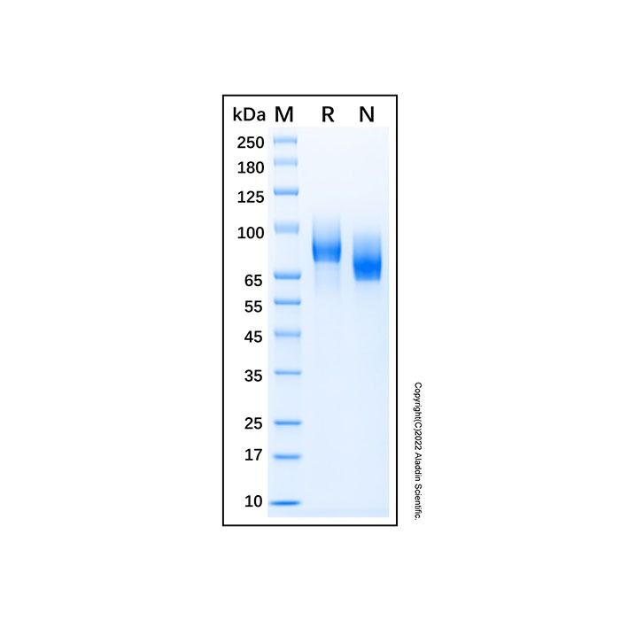
无动物源,无载体,His标签,PBS Only,≥95%(SDS-PAGE)
生化机理
The platelet-derived growth factor (PDGF) family consists of proteins derived from four genes (PDGF-A, -B, -C, and -D) that form disulfide-linked homodimers (PDGF-AA, -BB, -CC, and -DD) and a heterodimer (PDGF-AB). These proteins regulate diverse cellular functions by binding to and inducing the homo- or heterodimerization of two receptors (PDGF R alpha and R beta). Whereas alpha/alpha homo-dimerization is induced by PDGF-AA, -BB, -CC, and -AB, alpha/beta hetero-dimerization is induced by PDGF-AB, -BB, -CC, and -DD, and beta/beta homo-dimerization is induced only by PDGF-BB and -DD. Both PDGF R alpha and R beta are members of the class III subfamily of receptor tyrosine kinases (RTK) that also includes the receptors for M-CSF, SCF, and Flt3-ligand. All class III RTKs are characterized by the presence of five immunoglobulin-like domains in their extracellular region and a split kinase domain in their intracellular region. The extracellular domain of human PDGF R beta contains four disulfide bonds and shares a 79% sequence identity with mouse and rat PDGF R beta. Ligand-induced receptor dimerization results in autophosphorylation in trans, leading to the activation of several intracellular signaling pathways that can result in cell proliferation, cell survival, cytoskeletal rearrangement, and cell migration. Many cell types, including fibroblasts and smooth muscle cells, express both the alpha and beta receptors. Others have only the alpha receptors (oligodendrocyte progenitor cells, mesothelial cells, liver sinusoidal endothelial cells, astrocytes, platelets, and megakaryocytes) or only the beta receptors (myoblasts, capillary endothelial cells, pericytes, T cells, myeloid hematopoietic cells, and macrophages). A soluble PDGF R alpha has been detected in normal human plasma and serum as well as in the conditioned medium of the human osteosarcoma cell line MG-63. Both the recombinant mouse and human soluble PDGF R alpha bind PDGF with high affinity and are potent PDGF antagonists.
Post-translational: Autophosphorylated on tyrosine residues upon ligand binding. Autophosphorylation occurs in trans, i.e. one subunit of the dimeric receptor phosphorylates tyrosine residues on the other subunit. Phosphorylation at Tyr-579, and to a lesser degree, at Tyr-581, is important for interaction with SRC family kinases. Phosphorylation at Tyr-740 and Tyr-751 is important for interaction with PIK3R1. Phosphorylation at Tyr-751 is important for interaction with NCK1. Phosphorylation at Tyr-771 and Tyr-857 is important for interaction with RASA1/GAP. Phosphorylation at Tyr-857 is important for efficient phosphorylation of PLCG1 and PTPN11, resulting in increased phosphorylation of AKT1, MAPK1/ERK2 and/or MAPK3/ERK1, PDCD6IP/ALIX and STAM, and in increased cell proliferation. Phosphorylation at Tyr-1009 is important for interaction with PTPN11. Phosphorylation at Tyr-1009 and Tyr-1021 is important for interaction with PLCG1. Phosphorylation at Tyr-1021 is important for interaction with CBL; PLCG1 and CBL compete for the same binding site. Dephosphorylated by PTPRJ at Tyr-751, Tyr-857, Tyr-1009 and Tyr-1021. Dephosphorylated by PTPN2 at Tyr-579 and Tyr-1021. N-glycosylated. Ubiquitinated. After autophosphorylation, the receptor is polyubiquitinated, leading to its degradation.

 ZJ24F1215824


 危险品化学品经营许可证(带存储)