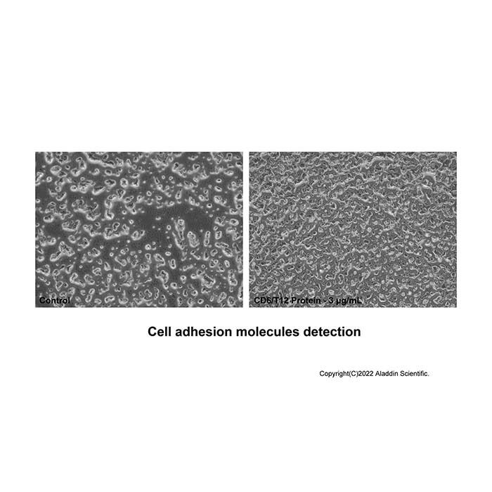
Measured by the ability of the immobilized protein to support the adhesion of Jurkat human acute T cell leukemia cells. When 8×10E4 cells/well are added to CD6-Fc coated plates (3 μg/mL and 100 μL/well) in the presence of 10μg/ml PHA, approximately > 50% cells will adhere specifically after 60 minutes at 37℃.
CD6 is a member of the group B scavenger receptor cysteine-rich (SRCR) superfamily. CD6 is a type I membrane glycoprotein and contains three extracellular SRCR domains. CD6 is expressed at low levels on immature thymocytes and at high levels on mature thymocytes. The majority of peripheral blood T cells, a subset of B cells, and a subset of neuronal cells express CD6. CD6 is a 668 amino acid protein with a 24 amino acid predicted signal sequence, a 374 amino acid extracellular domain, and a 244 amino acid transmembrane region. The 626 amino acid murine homolog has also been identified. The human and murine proteins share 70% amino acid identity over their full-lengths. The role of CD6 has not been fully elucidated. However, it appears to play a role as both a co-stimulatory molecule in T cell activation and as an adhesion receptor. Studies demonstrating a mitogenic effect for T cells with some CD6 specific monoclonal antibodies, in conjunction with either accessory cells or PMA and anti-CD2 mAb, support the concept of CD6 as a co-stimulatory molecule. Anti-CD6 monoclonal antibody has been used as an immunosuppressive agent for patients undergoing kidney or bone marrow allograft rejection. It has also been used to remove CD6+ T cells from donor bone marrow prior to allogeneic bone marrow transplantation. Other studies have demonstrated an adhesive role for CD6, it has been demonstrated to bind the activated leukocyte cell adhesion molecule (ALCAM, CD166). CD6/ALCAM interactions have been postulated to play a role in thymocyte development. The presence of ALCAM on neuronal cells may provide a mechanism of interaction between CD6+ T cells and ALCAM+ neuronal cells. Phosphorylation of the CD6 molecule appears to play a role in CD6-mediated signal transduction. Serine and threonine residues become hyperphosphorylated and tyrosine residues become phosphorylated when T cells are activated with anti-CD6 mAb in conjunction with PMA, anti-CD2, or anti-CD3 mAb. The CD6 intracellular domain contains regions that can interact with SH2 or SH3 containing proteins. However, the signaling pathways have not been elucidated.
Post-translational: After T-cell activation, becomes hyperphosphorylated on Ser and Thr residues and phosphorylated on Tyr residues. Glycosylated.
生物活性
Measured by the ability of the immobilized protein to support the adhesion of Jurkat human acute T cell leukemia cells. When 8×10E4 cells/well are added to CD6-Fc coated plates (3 μg/mL and 100 μL/well) in the presence of 10μg/ml PHA, approximately > 50% cells will adhere specifically after 60 minutes at 37℃.
56.6-93.7 kDa, under reducing conditions; 54.1 & 57.0-98.1 kDa, under non-reducing conditions.
分子类型
蛋白质
产品线
生物产品
储存与运输
物理形态
冻干(Lyophilized)
储存缓冲液
Lyophilized from PBS, pH 7.4
浓度
见COA
复溶
We recommend that this vial be briefly centrifuged prior to opening to bring the contents to the bottom. Reconstitute at 1.0 mg/mL in sterile distilled water. Stock solutions should be apportioned into working aliquots and stored at ≤ -20 °C. Further dilutions should be made in appropriate buffered solutions.
储存条件
-20°C储存,避免反复冻融
运输条件
超低温运输
稳定性与储存
Store at -20°C for 1 year. Avoid freeze/thaw cycle.
图片
Recombinant Human CD6 Protein (rp187152) - Bioactivity Measured by the ability of the immobilized protein to support the adhesion of Jurkat human acute T cell leukemia cells. When 8×10E4 cells/well are added to CD6-Fc coated plates (3 μg/mL and 100 μL/well) in the presence of 10μg/ml PHA, approximately > 50% cells will adhere specifically after 60 minutes at 37℃.
Recombinant Human CD6 Protein (rp187152) - SDS-PAGE 3 μg/lane of Recombinant Human CD6 Protein was resolved with SDS-PAGE under reducing (R) and non-reducing (N) conditions and visualized by Coomassie® Blue staining, showing a band at 56.6-93.7 kDa under reducing conditions and 54.1 & 57.0-98.1 kDa under non-reducing conditions. PTM shows phosphoserine modification (Uniprot ID: P30203).

 ZJ25F0319294


 危险品化学品经营许可证(带存储)