
计算溶液所需的质量、体积或浓度。
为什么选择此级别
提供 ≥99% 纯度,适用于对基线干扰要求严格的色谱和分析工作流程。
储存与运输
2-8°C储存,干燥。低温运输 。请查阅批次 COA 获取详细规格。
质量文档
SDS、COA、产品数据表及规格说明书均可下载。可通过批号查询获取批次 COA。
文献证明
在色谱分析、有机合成和交叉偶联反应领域已被 0 篇同行评审文献引用。
概述
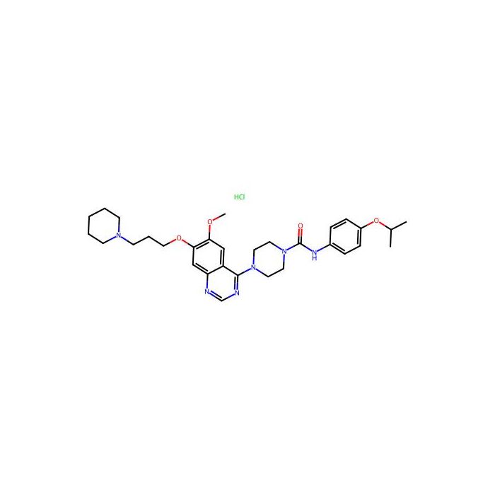
Tandutinib hydrochloride (MLN518 hydrochloride) is a potent and selective inhibitor of the FLT3 with an IC 50 of 0.22 μM, and also inhibits c-Kit and PDGFR with IC 50 s of 0.17 μM and 0.20 μM, respectively. Tandutinib hydrochloride can be used for acute myelogenous leukemia (AML) Tandutinib hydrochloride has the ability to cross the blood-brain barrier
In Vitro
Tandutinib (0-3 μM; 30 minutes; Ba/F3 cells) treatment inhibits IL-3-independent cell growth and FLT3-ITD autophosphorylation with an IC 50 of 10-100 nM in Ba/F3 cells expressing different FLT3-ITD mutants. Tandutinib (1 μM; 24-96 hours; Molm-14 and THP-1 AML cells) treatment induces apoptosis in FLT3-ITD-positive AML cells. In human FLT3-ITD-positive AML cell lines, Tandutinib inhibits FLT3-ITD phosphorylation ( IC 50 of ~30 nM). As with Erk2, a constitutively high level of Akt phosphorylation is readily detected and is efficiently blocked by pretreatment of the Molm-14 cells with 100-300 nM Tandutinib. Tandutinib inhibits cell proliferation of the FLT3-ITD-positive Molm-13 and Molm-14 with an IC 50 of 10 nM. And signaling through the MAP kinase and PI3 kinase pathways. MCE has not independently confirmed the accuracy of these methods. They are for reference only. Apoptosis AnalysisCell Line: Molm-14 and THP-1 AML cells Concentration: 1 μM Incubation Time: 24 hours, 48 hours, 72 hours, 96 hours Result: Induced apoptosis in FLT3-ITD-positive AML cells. Western Blot AnalysisCell Line: Ba/F3 cells Concentration: 0 μM, 0.003 μM, 0.01 μM, 0.03 μM, 0.1 μM, 1 μM and 3 μM Incubation Time: 30 minutes Result: In Ba/F3 cells expressing different FLT3-ITD mutants, inhibited IL-3-independent cell growth and FLT3-ITD autophosphorylation.
In Vivo
Tandutinib (60 mg/kg; oral gavage; daily; for 35 days; athymic nude mice) treatment causes a statistically significant increase in survival that was extended on average by 20 days . MCE has not independently confirmed the accuracy of these methods. They are for reference only. Animal Model: Athymic nude mice injected with Ba/F3 cells Dosage: 60 mg/kg Administration: Oral gavage; daily; for 35 days Result: Caused a statistically significant increase in survival that was extended on average by 20 days.
Form:Solid
IC50& Target:FLT3 0.22 μM (IC 50 ) c-Kit 0.17 μM (IC 50 ) PDGFR 0.2 μM (IC 50 )
规格
别名
盐酸坦度替尼
规格或纯度
≥99%
英文名称
Tandutinib hydrochloride
生化机理
盐酸坦度替尼(MLN518 盐酸盐)是一种强效的 FLT3 选择性抑制剂,IC 50 为 0.22 μM,还能抑制 c-Kit 和 PDGFR,IC 50 分别为 0.17 μM 和 0.20 μM。盐酸坦度替尼可用于治疗急性髓系白血病。
储存条件
2-8°C储存,干燥
运输条件
低温运输
纯度
≥99%
名称和识别符
分子类型
小分子
Smiles
O=C(N1CCN(C2=C(C(C=C3OCCCN4CCCCC4)=NC=N2)C=C3OC)CC1)NC5=CC=C(OC(C)C)C=C5.Cl
分子量
599.16
技术文档
高级数据
化学和物理性质
溶解性
H2O : 100 mg/mL (166.90 mM; Need ultrasonic) DMSO : ≥ 100 mg/mL (166.90 mM)
质检证书(CoA,COO,BSE/TSE 和分析图谱)
C of A & Other Certificates(BSE/TSE, COO):
输入批号以搜索分析图谱:
溶液计算器
产品仅提供给经过验证的企业、机构和合格的专业人员用于研发。不适用于人类、动物、诊断或治疗。
Copyright © 2023至今 上海阿拉丁生化科技股份有限公司 保留所有权利。


 危险品化学品经营许可证(带存储)