人的c-Myc基因是Myc基因家族的重要成员,与多种肿瘤的发生发展密切相关。其编码的Myc蛋白分子量为50–70 kDa,而Myc标签是源自该蛋白E424–L433区域的十肽序列(氨基酸序列:EQKLISEEDL),分子量约1.2 kDa。通过重组DNA技术,Myc标签可融合于目的蛋白的N端或C端,能有效辅助目的蛋白的亲和纯化与表达检测,且对目的蛋白天然功能的干扰较小。
Anti-Myc抗体可特异性识别与Myc标签融合表达的目的蛋白,广泛应用于该类融合蛋白的表达验证、细胞内定位分析,同时可用于融合蛋白的亲和纯化、定性及定量检测。本公司研发的Anti-Myc抗体亲和力高、特异性强,适配Western blot(WB)、免疫沉淀(IP)等蛋白表达检测及蛋白互作相关实验。
本产品所用高聚物磁珠,采用先进高分子聚合技术将超顺磁性材料与高分子基质完美结合,兼具快速磁响应性与优异的微球分散性,同时具备极低的非特异性吸附特性及丰富的配体结合位点,可高效适配Anti-Myc抗体偶联及后续Myc标签融合蛋白的捕获实验。磁珠具体性能参数详见下表。
技术参数
| 项目 | 技术参数
|
微球基质
| 高聚物磁性微球
|
配体
| Anti-Myc重组标签抗体(人源化)
|
粒径
| 1 μm
|
磁珠浓度
| 10 mg/mL
|
储存缓冲液
| Tris (pH 8.0), 0.01% Tween 20, 0.1% ProClin 300. |
使用方法
本操作流程主要为蛋白互作实验,每次反应使用20 μL UltraBio™ Anti-Myc Magnetic Beads,使用流程可参考图1。

图1:UltraBio™ Anti-Myc Magnetic Beads使用流程图
1. 推荐缓冲液(自备)
缓冲液名称
| 试剂配制
|
洗杂缓冲液
| 20 mM Na₂HPO₄, 0.15 M NaCl, 0.05% (v/v) Tween-20, pH 7.4
|
洗脱液
| 0.1 M Glycine, 0.15 M NaCl, pH 3.0
|
中和缓冲液
| 1 M Tris-HCl, pH 8.0
|
2. 磁珠漂洗
2.1 取20 μL混匀后的磁珠置于1.5 mL离心管中,加入500 μL洗杂缓冲液,轻柔上下颠倒混匀5–10次(勿涡旋)。
2.2 将离心管置于磁力架上,待磁珠完全吸附(约30 s–1 min)后,吸弃全部上清(避免吸到磁珠)。
2.3 重复上述步骤4次,总计完成5次漂洗,最后一次漂洗后尽量吸尽上清,保留磁珠备用。
3. 样品孵育
3.1 向上述漂洗好的磁珠中加入500-1000 μL 的细胞裂解样品,常温或4℃下孵育60 min(建议在旋转混匀仪上孵育,使样品和磁珠充分接触)。
3.2 孵育结束后,将离心管置于磁力架上,待磁珠全部吸附后,将上清转移至新的离心管中,留样备检。
3.3 向离心管中加入500 μL 洗杂缓冲液,轻轻颠倒混匀5-10次后将离心管置于磁力架上,待磁珠完全吸附后弃去上清。
3.4 重复操作5次。
注:孵育温度和时间范围推荐为:室温、0.5 h-2 h或者4℃、1 h-4 h,根据实际的结合效果适当调整孵育时间,一般情况下不建议过夜孵育,过夜孵育可能会引起过多的非特异性吸附。磁珠每次洗涤,加入洗杂缓冲液后可以冰上静置3-5 min。
4. 蛋白洗脱
4.1 洗脱液洗脱
每20 μL 原始磁珠体积加入40 μL 洗脱液,常温或4℃洗脱5-10 min,期间轻轻混匀3-5次。孵育结束后将离心管置于磁力架上,待磁珠完全吸附后,吸取上清至新的离心管中,加入少量的中和缓冲液将pH调至中性,最后进行Western检测。
4.2 SDS-PAGE上样缓冲液洗脱
每20 μL 原始磁珠体积加入60 μL的1×SDS Loading Buffer,轻摇摇晃混匀后95-100℃高温处理5-10 min。将离心管置于磁力架上分离10 s,取上清进行SDS-PAGE电泳或进行Western检测。
注意事项
1. 本产品保存在2-8℃,不可冻结保存;
2. 本产品出现轻微团聚属正常现象,可正常使用;
3. 本产品不要使用含有DTT等还原剂的细胞裂解液样品,DTT可能会导致抗体配体的脱落;
4. 本产品能耐受≤2 M尿素,高浓度的尿素会导致抗体配体脱落;
5. 在清洗和洗脱时避免吸取到磁珠,清洗时吸取到磁珠会影响最终的载量,洗脱时吸取到磁珠会影响目的蛋白的质量和电泳效果,第一次洗脱后可以将洗脱液置于新的离心管中,再次置于磁力架上进行吸附。
常见问题及解答
Q:聚合物磁珠和琼脂糖磁珠有何区别?
A:
- 琼脂糖磁珠是带孔微球,粒径在30-100 μm,交联时会有一部分配体会进入球孔内部,所以偶联载量相对较高,适用于蛋白的快速纯化;而聚合物磁珠通常粒径为1-3 μm以下,配体主要分布在微球表面,这种类型的磁珠能够快速结合蛋白,空间位阻影响小,适用于蛋白互作实验,但其载量不高;
- 推荐使用琼脂糖磁珠进行蛋白纯化实验,使用聚合物磁珠进行蛋白互作实验如IP、Co-IP和Pull-down实验等。
Q:为何结合不到目的蛋白?
A:
- 目的蛋白无表达或表达量过低:建议纯化前用 Western blot (WB) 检测原始表达量,WB 无信号则难以捕获;
- 样本含高浓度还原剂(如 DTT、β-ME):破坏抗体二硫键导致配体脱落,需选用无还原剂的温和裂解液;
- Flag 标签被空间位阻遮挡:融合蛋白折叠导致标签隐藏,可尝试将标签移至蛋白 N 端 / C 端或增加柔性连接肽(如 (GGS)ₙ);
- 孵育条件不足:温度 / 时间未优化(如室温孵育 < 30 min、4℃孵育 < 1 h),或未旋转混匀导致结合不充分;
- 磁珠用量不足:按样本中目的蛋白量调整磁珠体积(通常 20 μL 磁珠对应 1–5 mg 总蛋白);
- 洗涤过于剧烈:剧烈涡旋导致结合蛋白脱落,建议轻柔颠倒混匀而非涡旋。
实例
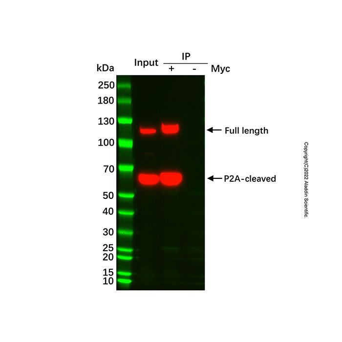
1. Myc tag IP实验:样品为胞内表达蛋白,含有His和Myc标签;
2. 取0.5 mL 悬浮细胞(≈1.5×10⁶个)常温下1×PBS(pH 7.4)清洗三次后快速进行裂解、离心;
3. 加入20 μL 已经清洗过的磁珠于离心后的上清中摇晃孵育30 min;
4. 将含有磁珠的样品管转移到磁力架上,分离磁珠和液体,用移液器吸走上清,留下磁珠;
5. 用洗杂缓冲液清洗5次后加入2× SDS Loading Buffer 99℃高温处理10 min后进行WB;
6. 检测抗体为Anti-His-HRP mAb,阴性对照(-)为未偶联 Anti-Myc 抗体的空白高聚物磁性微球与样本孵育,结果请见图2。

图2:通过UltraBio™ Anti-Myc Magnetic Beads进行IP后的WB图
图2表明UltraBio™ Anti-Myc Magnetic Beads能够很好的捕获样品中的Myc标签融合蛋白,同时没有非特异性的蛋白结合。
The human c-Myc gene is an important member of the Myc gene family and is closely related to the occurrence and development of various tumors. The Myc protein encoded by it has a molecular weight of 50–70 kDa, while the Myc tag is a decapeptide sequence derived from the E424–L433 region of this protein (amino acid sequence: EQKLISEEDL) with a molecular weight of approximately 1.2 kDa. Through recombinant DNA technology, the Myc tag can be fused to the N-terminus or C-terminus of the target protein, which can effectively assist in the affinity purification and expression detection of the target protein, and has minimal interference with the natural function of the target protein.
Anti-Myc antibody can specifically recognize target proteins fused with the Myc tag, and is widely used for the expression verification, intracellular localization analysis of such fusion proteins, as well as the affinity purification, qualitative and quantitative detection of fusion proteins. The Anti-Myc antibody developed by our company has high affinity and strong specificity, and is compatible with protein expression detection and protein-protein interaction related experiments such as Western blot (WB) and Immunoprecipitation (IP).
The polymer magnetic beads used in this product perfectly combine superparamagnetic materials and polymer matrices through advanced polymer polymerization technology. They have faster magnetic responsiveness while maintaining excellent microsphere dispersibility, extremely low non-specific adsorption, and more abundant ligand binding sites. These beads can be efficiently adapted for the conjugation of Anti-Myc antibody and subsequent capture experiments of Myc-tagged fusion proteins. For detailed performance parameters of the magnetic beads, see Table below.
Technical Parameters
Parameter
| Specification
|
Bead Matrix
| Polymer magnetic beads
|
Ligand
| Anti-Myc recombinant tag antibody (humanized)
|
Particle Size
| 1 μm
|
Bead Concentration
| 10 mg/mL
|
Storage Buffer
| Tris (pH 8.0), 0.01% Tween 20, 0.1% ProClin 300. |
Usage Instructions
This protocol is primarily designed for protein interaction experiments. Each reaction uses 20 µL of UltraBio™ Anti-Myc Magnetic Beads. The workflow can be referenced in Figure 1.

Figure 1: Workflow for using UltraBio™ Anti-Myc Magnetic Beads
1. Recommended Buffers (Self-prepared)
Buffer Name
| Preparation
|
Wash Buffer
| 20 mM Na₂HPO₄, 0.15 M NaCl, 0.05% (v/v) Tween-20, pH 7.4
|
Elution Buffer
| 0.1 M Glycine, 0.15 M NaCl, pH 3.0
|
Neutralization Buffer
| 1 M Tris-HCl, pH 8.0
|
2. Bead Washing
2.1 Transfer 20 μL of thoroughly resuspended magnetic beads to a 1.5 mL microcentrifuge tube. Add 500 μL of Wash Buffer and gently invert the tube 5–10 times to mix (avoid vortexing).
2.2 Place the tube on a magnetic stand. Once the beads are fully captured (approximately 30 seconds to 1 minute), carefully aspirate and discard all supernatant (avoid disturbing the bead pellet).
2.3 Repeat steps 2.1 and 2.2 four more times, for a total of five washes. After the final wash, remove as much supernatant as possible, leaving the beads ready for use.
3. Sample Incubation
3.1 Add 500–1000 μL of your cell lysate sample to the washed beads. Incubate at room temperature or 4°C for 60 minutes (incubation on a rotary mixer is recommended for thorough mixing).
3.2 After incubation, place the tube on the magnetic stand. Once all beads are captured, transfer the supernatant to a new tube (keep for later analysis if needed).
3.3 Add 500 μL of Wash Buffer to the tube with the beads, gently invert 5–10 times, place on the magnetic stand, and discard the supernatant once beads are captured.
3.4 Repeat step 3.3 five times.
Note: Recommended incubation temperature and duration: Room temperature for 0.5–2 hours or 4°C for 1–4 hours. Optimize incubation time based on actual binding efficiency. Overnight incubation is generally not recommended as it may increase non-specific binding. During each wash step, after adding Wash Buffer, the beads can be left on ice for 3–5 minutes.
4. Protein Elution
4.1 Elution Buffer Method
Add 40 μL of Elution Buffer per 20 μL of original bead volume. Incubate at room temperature or 4°C for 5–10 minutes, gently mixing 3–5 times during incubation. Place the tube on the magnetic stand. Once beads are captured, transfer the supernatant to a new tube. Add a small amount of Neutralization Buffer to adjust the pH to neutral, then proceed to Western blot analysis.
4.2 SDS-PAGE Loading Buffer Method
Add 60 μL of 1× SDS Loading Buffer per 20 μL of original bead volume. Mix gently and heat at 95–100°C for 5–10 minutes. Place the tube on the magnetic stand for 10 seconds, then collect the supernatant for SDS-PAGE electrophoresis or Western blot analysis.
Precautions:
1. Storage: Store this product at 2–8°C. Do not freeze.
2. Appearance: Minor aggregation is normal and does not affect performance.
3. Reducing Agents: Do not use cell lysis samples containing high concentrations of reducing agents (e.g., DTT), as these may cause antibody ligand detachment.
4. Urea Tolerance: This product can tolerate urea concentrations ≤ 2 M. Higher concentrations may cause antibody ligand detachment.
5. Handling: Avoid aspirating beads during washing and elution steps. Aspirating beads during washing reduces final capacity; aspirating beads during elution affects protein quality and electrophoresis results. After the first elution, you can transfer the eluate to a new tube and place it on the magnetic stand again to capture any beads accidentally transferred.
Frequently Asked Questions (FAQ)
Q: What is the difference between polymer magnetic beads and agarose magnetic beads?
A:
Agarose beads are porous microspheres with a particle size of 30–100 μm. During coupling, some ligands enter the pores, resulting in relatively high binding capacity, making them suitable for rapid protein purification.
Polymer beads typically have a particle size below 1–3 μm, with ligands mainly distributed on the surface. This type of bead allows rapid protein binding with minimal steric hindrance, making them suitable for protein interaction experiments like IP, Co-IP, and Pull-down, although their binding capacity is not as high.
We recommend using agarose beads for protein purification experiments and polymer beads for protein interaction experiments such as IP, Co-IP, and Pull-down.
Q: Why is the target protein not binding?
A:
No or low expression of the target protein: It is recommended to check the original expression level by Western blot (WB) before purification. If there is no WB signal, capturing the protein will be difficult.
Sample contains high concentrations of reducing agents (e.g., DTT, β-ME): These can break antibody disulfide bonds, causing ligand detachment. Use mild lysis buffers without reducing agents.
Flag tag is sterically hindered: Protein folding may hide the tag. Try moving the tag to the N-terminus/C-terminus or adding a flexible linker peptide (e.g., (GGS)ₙ).
Insufficient incubation conditions: Temperature/time not optimized (e.g., room temperature incubation < 30 min, 4°C incubation < 1 h), or lack of rotation/mixing leading to incomplete binding.
Insufficient bead amount: Adjust the bead volume according to the amount of target protein in the sample (typically 20 μL beads for 1–5 mg total protein).
Excessively vigorous washing: Vortexing can cause bound protein to detach. Gently invert the tube instead.
Application Example
1. Myc tag IP Experiment: The sample is an intracellularly expressed protein containing both His and Myc tags.
2. Take 0.5 mL of suspension cells (≈1.5×10⁶ cells), wash three times with 1×PBS (pH 7.4) at room temperature, then quickly perform lysis and centrifugation.
3. Add 20 µL of pre-washed magnetic beads to the post-centrifugation supernatant and incubate with shaking for 30 minutes.
4. Transfer the tube containing the beads to a magnetic stand to separate beads from liquid. Aspirate the supernatant, leaving the beads.
5. Wash with Wash Buffer five times, then add 2× SDS Loading Buffer and heat at 99°C for 10 minutes before proceeding to WB.
6. The detection antibody used was Anti-His-HRP mAb. The negative control (-) used blank polymer magnetic beads (not conjugated with Anti-Myc antibody) incubated with the sample. Results are shown in Figure 2.
Figure 2: Western blot after IP using UltraBio™ Anti-Myc Magnetic Beads.
Figure 2 demonstrates that UltraBio™ Anti-Myc Magnetic Beads effectively capture the Myc-tagged fusion protein from the sample, with no non-specific protein binding.

 ZJ26F0434492


 危险品化学品经营许可证(带存储)