Mouse anti Human ASRGL1 Antibody, Monoclonal (K52014_9F2), could be used for WB, IHC and so on.
规格
产品名称
ASRGL1 Mouse mAb
别名
ASRGL1抗体 | ASRGL1小鼠单克隆抗体 | ASRGL1小鼠单抗
英文别名
ALP antibody | ALP1 antibody | ASGL1antibody | ASGL1_HUMAN antibody | Asparaginase like 1 antibody | Asparaginase like 1 protein antibody | Asparaginase like protein 1antibody | Asparaginase-like protein 1 antibody | ASRGL 1 antibody | Asrgl1 antibody | B
规格或纯度
无载体, ExactAb™, 已验证, 1.0 mg/mL
生化机理
Has both L-asparaginase and beta-aspartyl peptidase activity. May be involved in the production of L-aspartate, which can act as an excitatory neurotransmitter in some brain regions. Is highly active with L-Asp beta-methyl ester. Besides, has catalytic activity toward beta-aspartyl dipeptides and their methyl esters, including beta-L-Asp-L-Phe, beta-L-Asp-L-Phe methyl ester (aspartame), beta-L-Asp-L-Ala, beta-L-Asp-L-Leu and beta-L-Asp-L-Lys. Does not have aspartylglucosaminidase activity and is inactive toward GlcNAc-L-Asn. Likewise, has no activity toward glutamine.
宿主种属
小鼠(Mouse)
特异性
ASRGL1
种属反应性
人(Human)
免疫原
Recombinant protein of human ASRGL1 (AA 1-308).
阳性对照
WB: Hep G2 and Jurkat cell lysates. IHC: Human endometrium tissue.
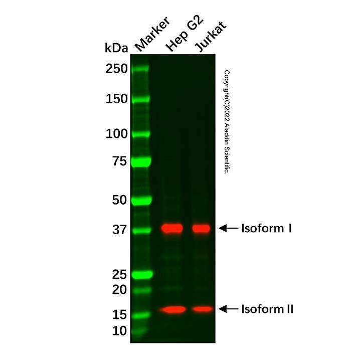
ASRGL1 Mouse mAb (Ab089867) - Western Blot All lanes: ASRGL1 Mouse mAb (Ab089867) at 1/1000 dilution Samples: Lysates at 20 µg per lane Secondary: Goat Anti-Mouse IgG H&L (HRP) (Ab179001) at 1/20000 dilution
Predicted band size: 32 kDa Observed band size: 16, 38 kDa ASRGL1 Mouse mAb (Ab089867) recognizes two isoforms of ASRGL1. Exposure time: 60.0 s
ASRGL1 Mouse mAb (Ab089867) - IHC IHC analysis of endometrium tissue by ASRGL1 Mouse mAb (Ab089867). IHC was performed using sections of the formalin-fixed paraffin-embedded endometrium tissue. Antigen was retrieved through addition of boiling Tris/EDTA buffer pH 9.0 in a pressure cooker for 3 min. Endogenous peroxidase activity was quenched by incubating the sections with 3% H2O2 for 30 min at room temperature. The sections were then incubated with ASRGL1 Mouse mAb (Ab089867) at 1 µg/mL at room temperature for 1 h. Poly-peroxidase conjugated goat anti-mouse IgG was used as the secondary antibody. Diaminobenzidine was used as the chromogen. The section was counterstained with hematoxylin. A tissue section incubated with phosphate-buffered saline followed by incubation with the secondary antibody was used as the background control. Result: Glandular cells are positively stained at the cytoplasm.

 ZJ24F1012501


 危险品化学品经营许可证(带存储)