p10 protein|p11|S100 calcium-binding protein A10|42C antibody|AA409961 antibody|AL024248 antibody|Annexin II ligand antibody|Annexin II tetramer (AIIt) p11 subunit antibody|Annexin II, light chain antibody|CAL12 antibody|Calpactin I light chain antibody|Calpactin I, p11 subunit antibody|Calpactin-1 light chain antibody|Cellular ligand of annexin II antibody|Nerve growth factor-induced protein 42C antibody|p10 protein antibody|p11 antibody|Protein S100 A10 antibody|Protein S100-A10 antibody|S100 calcium binding protein A10 (calpactin) antibody|S100 calcium binding protein A10 antibody|S100 calcium-binding protein A10 antibody|S100a10 antibody|annexin II ligand, calpactin I, light polypeptide|ANX2L|CAL1L|GP11|CLP11|p10|P11|protein S100-A10|S100 calcium binding protein A10 (annexin II ligand, calpactin I, lightpolypeptide (p11))|S100 calcium binding protein A10|S100 calcium-binding protein A10 (annexin II ligand, calpactin I, lightpolypeptide (p11))|S100A10
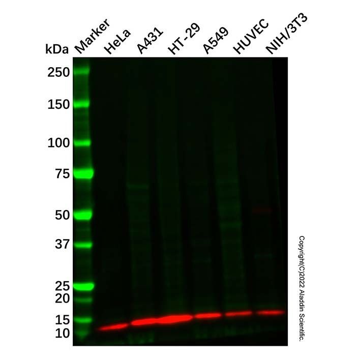
S100A10 Mouse mAb (Ab126219) - Western Blot All lanes: S100A10 Mouse mAb (Ab126219) at 1/1000 dilution Samples: Lysates at 20 µg per lane Secondary: Goat Anti-Mouse IgG H&L (HRP) (Ab179001) at 1/20000 dilution
Predicted band size: 11 kDa Observed band size: 13 kDa Exposure time: 58.4 s
S100A10 Mouse mAb (Ab126219) - IF/ICC Immunofluorescent analysis of S100-A10 staining in HeLa cells. Formalin-fixed cells were permeabilized with 0.1% Triton X-100 in TBS for 5-10 minutes and blocked with 3% BSA-PBS for 30 minutes at room temperature. Cells were probed with S100A10 Mouse mAb (Ab126219) in 3% BSA-PBS and incubated overnight at 4 °C in a hidified chamber. Cells were washed with PBST and incubated with a AF488-conjugated secondary antibody (green) in PBS at room temperature in the dark. DAPI was used to stain the cell nuclei (blue).

 ZJ24F1216036


 危险品化学品经营许可证(带存储)