Mouse anti Human UBR7 Antibody, Monoclonal (K49039_15G1), could be used for WB, IHC, IP and so on. Application: WB: 1/1000 IHC: 1/1000 IP: 1/100 Protein Relevance: UBR7 is a putative E3 ubiquitin-protein ligase which is a component of the N-end rule pathway. Recognizes and binds to proteins bearing specific amino-terminal residues that are destabilizing according to the N-end rule, leading to their ubiquitination and subsequent degradation.
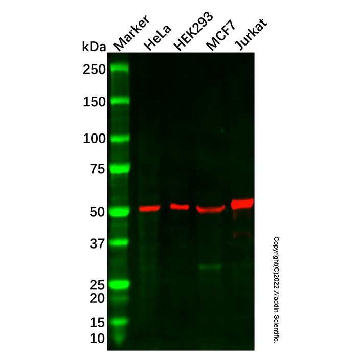
UBR7 Mouse mAb (Ab133358) - Western Blot All lanes: UBR7 Mouse mAb (Ab133358) at 1/1000 dilution Samples: Lysates at 20 µg per lane Secondary: Goat Anti-Mouse IgG H&L (HRP) (Ab179001) at 1/20000 dilution
Predicted band size: 48 kDa Observed band size: 52 kDa Exposure time: 47.8 s
UBR7 Mouse mAb (Ab133358) - IP Immunoprecipitation was performed by incubation of 2.5 µg of UBR7 Mouse mAb (Ab133358) with T-47D lysate containing 200 µg of total protein. After absorption with Protein G beads, the mixture was run on 6-18% SDS-PAGE and blotted onto nitrocellulose membrane. UBR7 Mouse mAb (Ab133358) at 1 µg/mL was used as the primary antibody and peroxidase conjugated rabbit anti-mouse IgG (Light chain specific) was used as the secondary antibody. Lane 1: T-47D lysateLane 2: UBR7 immunoprecipitated from T-47D lysate by UBR7 Mouse mAb (Ab133358) Lane 3: The isotype control antibody instead of UBR7 Mouse mAb (Ab133358) in T-47D lysate.Result: UBR7 Mouse mAb (Ab133358) can immunoprecipitate UBR7.
UBR7 Mouse mAb (Ab133358) - IHC IHC analysis of testis tissue by UBR7 Mouse mAb (Ab133358).IHC was performed using sections of the formalin-fixed paraffin-embedded testis tissue. Antigen was retrieved through addition of boiling Tris/EDTA buffer pH 9.0 in a pressure cooker for 3 min. Endogenous peroxidase activity was quenched by incubating the sections with 3% H2O2 for 30 min at room temperature. The sections were then incubated with UBR7 Mouse mAb (Ab133358) at 1 µg/mL at room temperature for 1 h. Poly-peroxidase conjugated goat anti-mouse IgG was used as the secondary antibody. Diaminobenzidine was used as the chromogen. The section was counterstained with hematoxylin. A tissue section incubated with phosphate-buffered saline followed by incubation with the secondary antibody was used as the background control.Result: Cells in seminiferous ducts are positively stained at the nuclei.

 ZJ24F0708511


 危险品化学品经营许可证(带存储)