Application Flow: 0.01 μg/mL- 2.0 μg/mL ICC/IF: 0.5 μg/mL Mouse anti BrdU antibody, clone 4C1, is suitable for detecting incorporated BrdU in a wide variety of cell types.
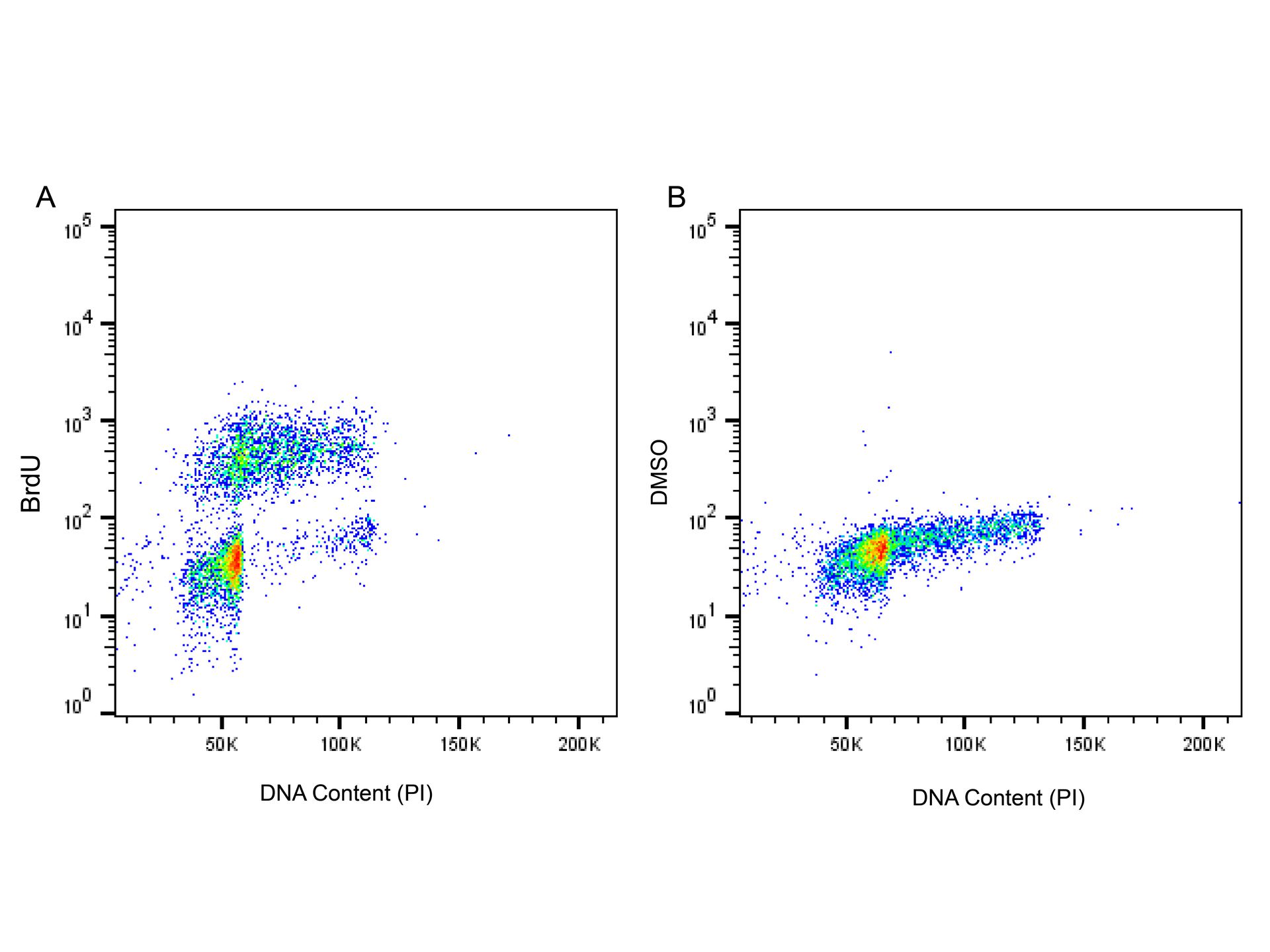
BrdU Mouse mAb (Ab176205) - Flow Cytometry Dot plot showing BrdU-treated (A) and DMSO-treated (B) Jurkat cells stained with Ab176205. Cells were incubated with 10 µM BrdU for 1 hour prior to being harvested. And then fixed in 100% methanol for 20 minutes at -20℃. Once fixed, pellets were acid denatured with 2M HCl for 30 minutes at room temperature.
Cells were stained with 0.1 μg/mL BrdU Mouse mAb (Ab176205) for 1 hour at room temperature. Goat Anti-Mouse IgG H&L (FITC) (Ab156254) was used as a secondary antibody at a 1/1000 dilution. Propidium Iodide was used to stain total DNA.
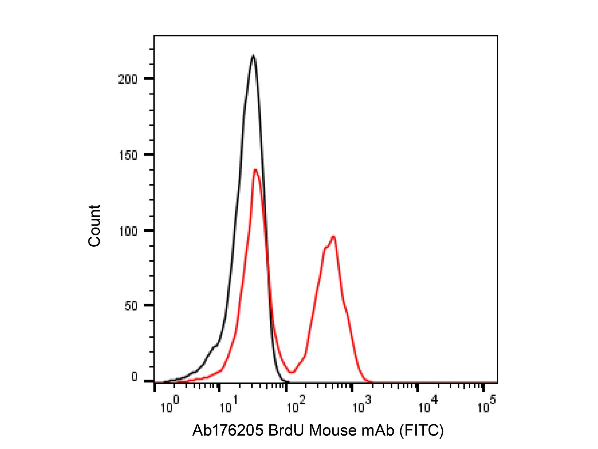
BrdU Mouse mAb (Ab176205) - Flow Cytometry Flow cytometry analysis of BrdU (red line) in Jurkat cells. The cells were treated with 10μM BrdU for 1 hour. And then fixed in 100% methanol for 20 minutes at -20℃. An acid denaturation method was used to prepare the cells before labeling with BrdU Mouse mAb (Ab176205) to detect the incorporated BrdU. Cells were then incubated with Goat Anti-Mouse IgG H&L (FITC) (Ab156254) at a dilution of 1/1000 for 30 minutes at room temperature in the dark. Unlabeled sample (black line) was used as a negative control.
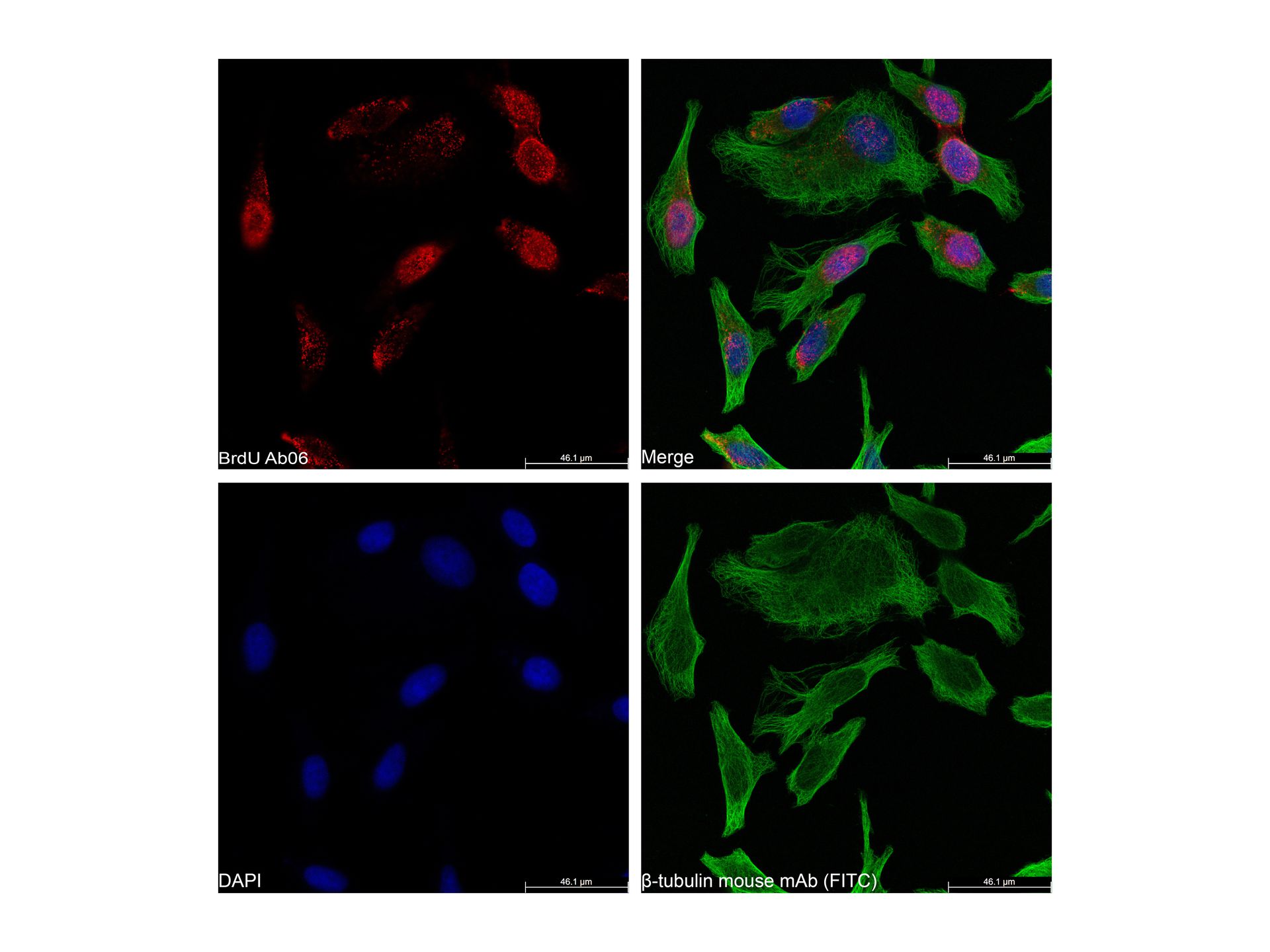
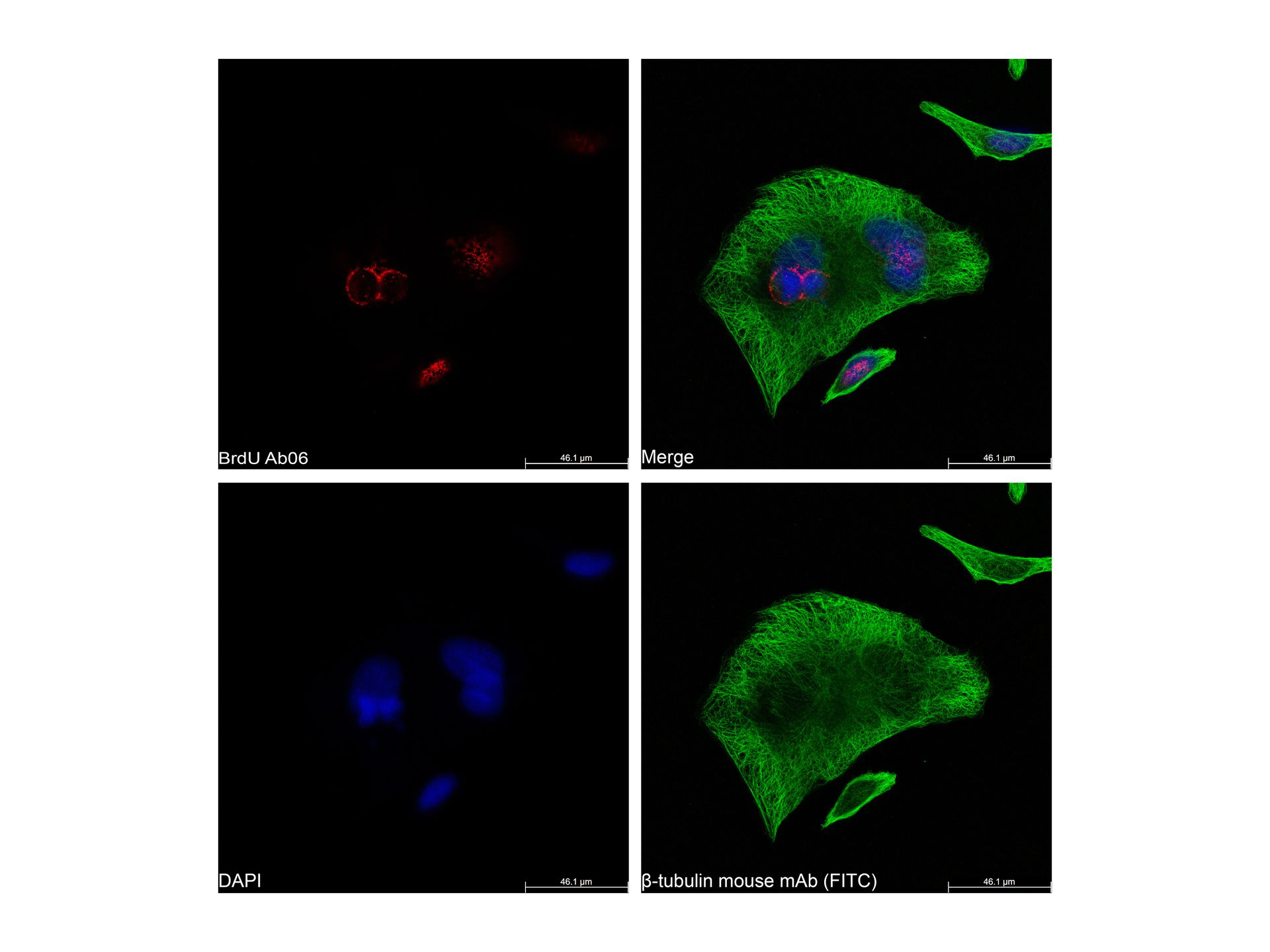
BrdU Mouse mAb (Ab176205) - ICC/IF IF analysis of HeLa cells labeling BrdU with BrdU Mouse mAb (Ab176205) at 0.5 μg/mL. The cells were fixed with 100% methanol for 5 minutes and then subjected to acid hydrolysis using 2M HCl for 30 minutes at room temperature to denature the DNA. They were then blocked with 2% BSA in PBS for 1 hour and incubated overnight at 4℃ with BrdU Mouse mAb (Ab176205) at 0.5 μg/mL.
beta Tubulin Mouse mAb (Ab155823) (FITC) (shown in green) counterstained with the cells after Goat Anti-Mouse Cy5 (Ab156257) (shown in red) incubation. Nuclear DNA was labelled with DAPI (shown in blue).
Images were taken on the confocal laser scanning microscope.


 ZJ24F0809940


 危险品化学品经营许可证(带存储)