Binds peptides derived from antigens that access the endocytic route of antigen presenting cells (APC) and presents them on the cell surface for recognition by the CD4 T-cells. The peptide binding cleft accomodates peptides of 10-30 residues. The peptides presented by MHC class II molecules are generated mostly by degradation of proteins that access the endocytic route, where they are processed by lysosomal proteases and other hydrolases. Exogenous antigens that have been endocytosed by the APC are thus readily available for presentation via MHC II molecules, and for this reason this antigen presentation pathway is usually referred to as exogenous. As membrane proteins on their way to degradation in lysosomes as part of their normal turn-over are also contained in the endosomal/lysosomal compartments, exogenous antigens must compete with those derived from endogenous components. Autophagy is also a source of endogenous peptides, autophagosomes constitutively fuse with MHC class II loading compartments. In addition to APCs, other cells of the gastrointestinal tract, such as epithelial cells, express MHC class II molecules and CD74 and act as APCs, which is an unusual trait of the GI tract. To produce a MHC class II molecule that presents an antigen, three MHC class II molecules (heterodimers of an alpha and a beta chain) associate with a CD74 trimer in the ER to form an heterononamer. Soon after the entry of this complex into the endosomal/lysosomal system where antigen processing occurs, CD74 undergoes a sequential degradation by various proteases, including CTSS and CTSL, leaving a small fragment termed CLIP (class-II-associated invariant chain peptide). The removal of CLIP is facilitated by HLA-DM via direct binding to the alpha-beta-CLIP complex so that CLIP is released. HLA-DM stabilizes MHC class II molecules until primary high affinity antigenic peptides are bound. The MHC II molecule bound to a peptide is then transported to the cell membrane surface. In B cells, the interaction between HLA-DM and MHC class II molecules is regulated by HLA-DO. Primary dendritic cells (DCs) also to express HLA-DO. Lysosomal miroenvironment has been implicated in the regulation of antigen loading into MHC II molecules, increased acidification produces increased proteolysis and efficient peptide loading.
宿主种属
大鼠(Rat)
特异性
MHC Class II
种属反应性
小鼠(Mouse)
免疫原
Activated C57BL/6 mouse spleen cells
阳性对照
Flow: C57BL/6 mouse splenocytes
偶联
AF405
Excitation(nm)
Violet Laser(405nm)
Emission(nm)
422nm
Ex/Em(nm)
Ex:401nm;Em:422nm
克隆类型
重组抗体
Format
Whole IgG
轻链亚型
kappa
纯化方法
Protein A purified
浓度
0.5 mg/mL
分子类型
抗体
产品线
生物产品
储存与运输
储存缓冲液
Liquid in PBS pH 7.4, containing 1% BSA and 0.05% Proclin300.
防腐剂
0.05% Proclin300
储存条件
2-8°C储存,避光,禁止冷冻
运输条件
低温运输,禁止冷冻
稳定性与储存
The antibody solution should be stored undiluted between 2°C and 8°C, and protected from prolonged exposure to light. Do not freeze.
图片
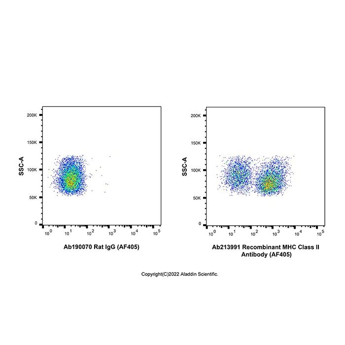
Recombinant MHC Class II Antibody (AF405) (Ab213991) - Flow Cytometry Flow cytometry analysis of MHC Class II expression by C57BL/6 mouse splenocytes (right panel) compared to an isotype control (left panel). Cells were stained with Recombinant MHC Class II Antibody (AF405) (Ab213991) at 0.2μg/test for 1 hour at 4°C. Left panel - Rat IgG (AF405) (Ab190070).
Recombinant MHC Class II Antibody (AF405) (Ab213991) - Flow Cytometry Flow cytometry analysis of MHC Class II expression by C57BL/6 mouse splenocytes (red) compared to an isotype control (blue). Cells were stained with Recombinant MHC Class II Antibody (AF405) (Ab213991) at 0.2μg/test for 1 hour at 4°C. Blue - Rat IgG (AF405) (Ab190070). Black - Unlabelled control, cells without incubation with primary antibody.
应用
应用名称
稀释比例
Flow Cytometry
≤0.3μg/test
For flow cytometric staining, the suggested use of this reagent is ≤0.3 μg per million cells in 100 µL staining volume.

 ZJ25F0318895


 危险品化学品经营许可证(带存储)