
计算溶液所需的质量、体积或浓度。
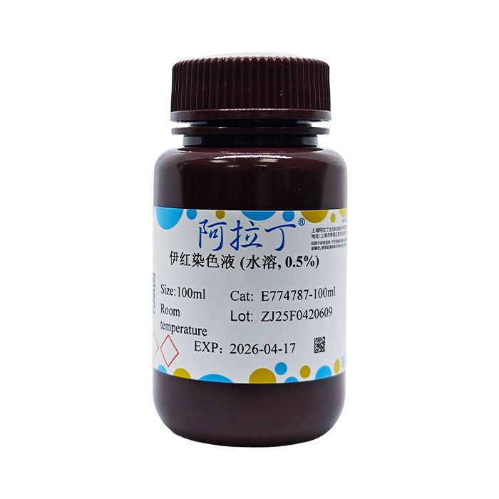
伊红染色液 (水溶, 0.5%)
有货
伊红 (Eosin) 又称曙红,属人工合成染料中的呫吨类染料,为桃红色或粉红色的粉末,最适宜与苏木精配合染色以显示正常或病理组织的形态结构。伊红染色液 (水溶,0.5%) 采用进口伊红染料,操作简单,不使用汞、甲醇等有毒试剂,可以用于组织切片或培养细胞的染色。可以和免疫荧光染色或免疫组化染色配合使用。一方面可以在伊红染色后进行免疫荧光染色或其它染料的染色,另一方面也可以在免疫组化染色后再进行伊红复染。本染色液可以重复使用,直至认为效果不佳时再换用新的染色液,染色后细胞浆呈粉红色或红色。
使用说明:
一、样本处理
(一)石蜡切片处理
1、切片烤片30-60min,二甲苯 (I) 脱蜡5-10min。
2、二甲苯 (II) 脱蜡5-10min。
3、无水乙醇洗二甲苯1-5min。
4、95%的乙醇1-5min。
5、75%的乙醇1-5min。
6、自来水或蒸馏水冲洗。
(二)冰冻切片处理
1、无需脱蜡,固定液固定后直接用蒸馏水冲洗2-3min.
(三)细胞样本处理
1、4%多聚甲醛固定10-20min。
2、自来水冲洗2次,每次2min。
3、蒸馏水冲洗2次,每次2min。
二、染色
(一)伊红染色液染色2-5min (根据染色结果和要求调整时间)。
如果需要直接观察,可以用70%乙醇洗涤2次。如需脱水、透明后封片按后续步骤进行,70%乙醇洗涤后仍可按照后续步骤进行脱水、透明和封片处理。
注:如果用于免疫组化等染色后的复染,参考上述步骤在其它染色完成后进行伊红染色。
三、脱水、透明、封固 (可选)
(一)95%乙醇 (I) 洗涤0.5-2min。
(二)95%乙醇 (II) 脱水2-5min。
(三)无水乙醇 (I) 脱水2-5min。
(四)无水乙醇 (II) 脱水2-5min。
(五)二甲苯 (I) 透明1min。
(六)二甲苯 (II) 透明1min,中性树脂货其他封片剂封片。
(七)显微镜下观察,细胞浆呈粉红色或红色。
四、进行其它染色
(一)如果进行免疫荧光染色或进行Hoechst等荧光染料的染色,在伊红染色液染色后70%乙醇洗涤2次。
(二)PBS或生理盐水或TBS或TBST等用于免疫染色或荧光染料染色的溶液浸泡5min。
(三)进行免疫荧光染色或其它荧光染料的染色。
注意事项:
1、如果需要脱水、透明和封片处理,需自备二甲苯,中性树胶或其它封片剂。如果样品是石蜡切片,需自备90%乙醇,无水乙醇以及二甲苯。
2、为了您的安全和健康,请穿实验服并戴一次性手套操作。
染色结果:
| 细胞质、纤维 | 红色 |
Eosin, also called erythrosin, is a xanthene dye among synthetic dyes. It is a peach-red or pink powder, and is most suitable for use in combination with hematoxylin to display the morphological structure of normal or pathological tissues.The Eosin Staining Solution (water-soluble, 0.5%) is prepared with imported eosin dye. It is simple to operate and does not use toxic reagents such as mercury or methanol. It can be used for staining tissue sections or cultured cells, and can be used in combination with immunofluorescence staining or immunohistochemical staining. On the one hand, immunofluorescence staining or staining with other dyes can be performed after eosin staining; on the other hand, eosin counterstaining can be performed after immunohistochemical staining. This staining solution can be reused until the effect is considered unsatisfactory, at which point a fresh staining solution should be used instead. After staining, the cytoplasm appears pink or red.
Instructions:
I. Sample Preparation
(I) Paraffin Section Processing
1. Bake the sections for 30-60 min, then deparaffinize with xylene (I) for 5-10 min.
2. Deparaffinize with xylene (II) for 5-10 min.
3. Wash off the xylene with anhydrous ethanol for 1-5 min.
4. Treat with 95% ethanol for 1-5 min.
5. Treat with 75% ethanol for 1-5 min.
6. Rinse with tap water or distilled water.
(II) Frozen Section Processing
1. Deparaffinization is not required. After fixation with the fixative, rinse directly with distilled water for 2-3 min.
(III) Cell Sample Processing
1. Fix with 4% paraformaldehyde for 10-20 min.
2. Rinse with tap water twice, 2 min each time.
3. Rinse with distilled water twice, 2 min each time.
II. Staining
(I) Stain with the Eosin Staining Solution for 2-5 min (adjust the time according to the staining results and requirements).
If direct observation is required, wash twice with 70% ethanol. If dehydration, clearing and mounting are required, proceed with the subsequent steps; after washing with 70% ethanol, the subsequent steps of dehydration, clearing and mounting can still be followed.
Note: If it is used for counterstaining after staining such as immunohistochemistry, refer to the above steps and perform eosin staining after the other staining has been completed.
III. Dehydration, Clearing and Mounting (Optional)
(I) Wash with 95% ethanol (I) for 0.5-2 min.
(II) Dehydrate with 95% ethanol (II) for 2-5 min.
(III) Dehydrate with anhydrous ethanol (I) for 2-5 min.
(IV) Dehydrate with anhydrous ethanol (II) for 2-5 min.
(V) Clear with xylene (I) for 1 min.
(VI) Clear with xylene (II) for 1 min, then mount with neutral resin or other mounting media.
(VII) Observe under a microscope; the cytoplasm appears pink or red.
IV. Performing Other Staining
(I) If immunofluorescence staining or staining with fluorescent dyes such as Hoechst is to be performed, wash twice with 70% ethanol after staining with the Eosin Staining Solution.
(II) Soak in a solution used for immunostaining or fluorescent dye staining, such as PBS, normal saline, TBS or TBST, for 5 min.
(III) Perform immunofluorescence staining or staining with other fluorescent dyes.
Precautions:
1. If dehydration, clearing and mounting are required, prepare xylene, neutral gum or other mounting media yourself. If the sample is a paraffin section, prepare 90% ethanol, anhydrous ethanol and xylene yourself.
2. For your safety and health, please wear a laboratory coat and disposable gloves when operating.
Staining results
| Cytoplasm, fibers | Red |
通过匹配包装上的批号来查找并下载产品的 COA,每批产品都进行了严格的验证,您可放心使用!
| 批号(Lot Number) | 证书类型 | 货号 |
|---|---|---|
 ZJ25F0420608 |
分析证书 | E774787 |
| ZJ25F0420609 |
分析证书 | E774787 |
我们的级别选择指南涵盖了目录中所有的变体的纯度、稳定剂状态和应用适用性。
查看 BioReagent 级别指南 → 查看 Biological Stain 级别指南 → 查看 for Microscopy 级别指南 →

 危险品化学品经营许可证(带存储)