
HMGB1-Mediated Damage-Associated Inflammatory Signaling and Tumor Immune Regulation
HMGB1-Mediated Damage-Associated Inflammatory Signaling and Tumor Immune Regulation
HMGB1 converts cellular damage into inflammatory and chemotactic signals and regulates tumor immunity, tumor stemness, and therapy tolerance through TLRs, RAGE, and CXCR4.
Keywords: HMGB1; damage-associated molecular pattern; DAMP; TLR4; RAGE; CXCL12; CXCR4; NF-κB; MAPK; tumor immunity; tumor stemness; chemoresistance
1 Molecular States of HMGB1 and Its Damage-Associated Molecular Pattern Function
1.1 Structure and Intracellular Function of HMGB1
High mobility group box 1 (HMGB1) is a non-histone chromatin-binding protein composed of an A box, a B box, and an acidic C-terminal tail enriched in acidic amino acids.
(1) A box
The A box participates in DNA structure recognition and protein interactions. Free A box can competitively inhibit the extracellular pro-inflammatory activity of full-length HMGB1 in some experimental systems.
(2) B box
The B box contains an important pro-inflammatory functional region of HMGB1 and can participate in activation of receptor complexes such as TLR4/MD-2.
(3) Acidic C-terminal tail
The acidic C-terminal tail regulates HMGB1 conformation and its binding ability to DNA and other proteins.
In the nucleus, HMGB1 can bind bent or damaged DNA and participate in chromatin conformation regulation, transcription, and DNA damage response. After HMGB1 translocates to the cytoplasm or is released into the extracellular space, its functional focus shifts from chromatin regulation to inflammatory, chemotactic, and tissue injury signal transmission.
1.2 Redox State Determines the Extracellular Function of HMGB1
HMGB1 contains three cysteine residues, Cys23, Cys45, and Cys106. Different oxidation states correspond to different receptor preferences and biological functions.
Table 1. HMGB1 Redox States and Main Functions
HMGB1 State | Cysteine Features | Main Functional Axis | Main Effects |
Fully reduced HMGB1 | Cys23, Cys45, and Cys106 remain reduced | HMGB1-CXCL12-CXCR4 | Enhances migration of immune cells, progenitor cells, and some tumor cells |
Disulfide HMGB1 | Cys23 and Cys45 form a disulfide bond, while Cys106 remains reduced | HMGB1-TLR4/MD-2 | Induces expression of inflammatory cytokines and chemokines |
Terminally oxidized HMGB1 | Cysteine residues are further oxidized | Reduced receptor activation capacity | Chemotactic and pro-inflammatory activities are markedly weakened |
1.3 DAMP Function of HMGB1
After cellular injury, extracellular HMGB1 can act as a damage-associated molecular pattern, converting cellular and tissue damage into innate immune signals.
(1) Activates macrophages, dendritic cells, and neutrophils.
(2) Promotes the expression of inflammatory cytokines, chemokines, and adhesion molecules.
(3) Regulates immune cell migration toward injured regions or the tumor microenvironment.
(4) Participates in chronic inflammation, vascular changes, and tissue remodeling when persistently released.
2 HMGB1 Release Mechanisms and Extracellular Inflammatory Effects
2.1 Passive Release Caused by Injury and Lytic Cell Death
Necrosis, pyroptosis, and other forms of cell death accompanied by plasma membrane rupture can release nuclear HMGB1 into the extracellular space. Tumor hypoxia, mechanical injury, chemotherapy, and radiotherapy may all increase passive HMGB1 release.
Early apoptotic cells usually still maintain plasma membrane integrity, and HMGB1 does not immediately leak out in large amounts. If apoptotic cells are not cleared in time and undergo secondary membrane rupture, HMGB1 can also be released extracellularly. Different modes of cell death and oxidative environments can alter the molecular state and receptor activity of HMGB1.
2.2 Active Release from Immune Cells
After stimulation by pathogen components, inflammatory cytokines, or tissue injury signals, monocytes, macrophages, and dendritic cells can promote HMGB1 translocation from the nucleus to the cytoplasm and release it through non-classical secretory pathways.
(1) Post-translational modifications
Modifications such as lysine acetylation can reduce the nuclear localization capacity of HMGB1 and promote its accumulation in the cytoplasm.
(2) Cellular stress
Oxidative stress, DNA damage, and inflammatory signaling can regulate HMGB1 nuclear-cytoplasmic translocation and extracellular release.
(3) Non-classical secretory pathways
Secretory lysosomes, extracellular vesicles, and changes in membrane permeability can all participate in HMGB1 release.
2.3 Main Effector Cells of Extracellular HMGB1
(1) Macrophages
HMGB1 can induce the expression of inflammatory cytokines and chemokines and alter macrophage functional states in the tumor microenvironment.
(2) Dendritic cells
Therapy-induced HMGB1 release can influence dendritic cell uptake, processing, and presentation of tumor antigens and participate in adaptive immune initiation after immunogenic cell death.
(3) Vascular endothelial cells
HMGB1 can promote endothelial cell activation, adhesion molecule expression, and changes in vascular permeability, providing conditions for immune cell recruitment and tumor vascular remodeling.
(4) Tumor cells and stromal cells
Tumor cells, fibroblasts, and other stromal cells can respond to HMGB1 through RAGE-, TLR-, or CXCR4-related signaling, altering proliferation, migration, and therapy sensitivity.
3 HMGB1-TLRs/RAGE Signaling and Activation of NF-κB and MAPK Pathways
3.1 TLR4/MD-2-Mediated Inflammatory Response
Disulfide HMGB1 can interact with MD-2 and activate the TLR4 receptor complex. TLR4 transmits signals downstream through molecules such as MyD88, IRAK, and TRAF6, further activating IKK/NF-κB and MAPK pathways.
(1) Promotes the expression of inflammatory cytokines such as TNF, IL-1β, and IL-6.
(2) Enhances dendritic cell maturation and antigen presentation.
(3) Increases chemokine and adhesion molecule expression.
(4) Promotes inflammatory cell recruitment into injured tissues or tumor tissues.
TLR2 may also participate in cellular responses induced by some HMGB1 complexes. Endotoxin, nucleic acids, or other bound molecules in recombinant HMGB1 can interfere with TLR experiments, so endotoxin controls and receptor blockade groups should be included.
3.2 RAGE-Mediated Sustained Signaling
The receptor for advanced glycation end products, RAGE, is encoded by the AGER gene and can be expressed in myeloid cells, vascular endothelial cells, and many tumor cells. After HMGB1 binds RAGE, it can regulate NF-κB, ERK, p38, PI3K/AKT, and other signaling pathways.
(1) Promotes survival, proliferation, migration, and invasion of some tumor cells.
(2) Regulates the function of tumor-associated macrophages and stromal cells.
(3) Participates in angiogenesis, lymphangiogenesis, and tumor tissue remodeling.
(4) Affects autophagy, mitochondrial dynamics, and therapy tolerance.
3.3 NF-κB and MAPK Signal Output
(1) NF-κB pathway
IKK activation promotes IκBα phosphorylation and degradation, allowing p65/p50 to enter the nucleus and regulate the expression of inflammatory cytokines, chemokines, anti-apoptotic proteins, and immunoregulatory molecules.
(2) p38 MAPK pathway
The p38 MAPK pathway participates in inflammatory cytokine transcription, mRNA stability, and cellular stress responses and is closely related to inflammatory activation of myeloid cells.
(3) ERK pathway
The ERK pathway participates in tumor cell proliferation, migration, mitochondrial fission, and some therapy tolerance processes.
(4) JNK/AP-1 pathway
The JNK/AP-1 pathway participates in stress responses and inflammatory gene transcription and can cooperate with NF-κB to regulate HMGB1-mediated inflammatory output.
4 HMGB1-CXCL12-CXCR4 Axis Regulates Cell Migration
4.1 HMGB1 Forms a Heterocomplex with CXCL12
Fully reduced HMGB1 can form a heterocomplex with CXCL12 and enhance the interaction between CXCL12 and CXCR4, allowing cells to generate stronger chemotactic responses at lower CXCL12 concentrations.
The HMGB1-CXCL12 complex mainly enhances CXCR4-dependent cell migration, which differs from the induction of inflammatory cytokines by disulfide HMGB1 through TLR4. In experiments, cell migration and inflammatory cytokine release should be evaluated separately.
4.2 Migration of Immune Cells and Repair Cells
(1) Recruits monocytes and other immune cells into injured regions.
(2) Regulates lymphocyte localization in inflammatory and tumor tissues.
(3) Recruits mesenchymal stem cells and progenitor cells related to tissue repair.
(4) Enhances chemotactic migration of CXCR4-positive tumor cells.
4.3 Tumor Migration and Spatial Distribution of Immune Cells
CXCR4-positive tumor cells can respond to CXCL12 concentration gradients and migrate toward tissues rich in CXCL12, such as lymph nodes, bone marrow, lung, or liver. After HMGB1 enhances CXCL12/CXCR4 signaling, migration and invasion capacity may increase in some tumor cells.
The CXCL12/CXCR4 axis can also affect the distribution of regulatory T cells, myeloid-derived suppressor cells, macrophages, and effector T cells in tumor tissues. Related research can combine Transwell migration assays, chemotaxis assays, and analysis of immune cell infiltration in tumor tissues.
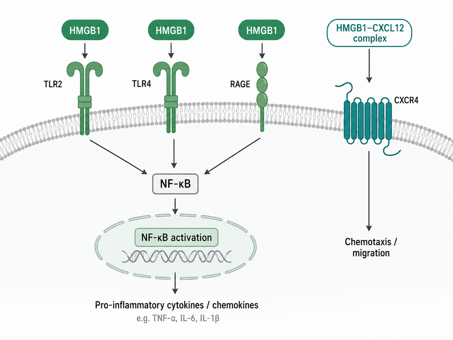
Figure 1. HMGB1 receptor signaling and its inflammatory and chemotactic effects
5 HMGB1 Regulates Tumor Inflammation, Tumor Stemness, and Therapy Tolerance
5.1 Dual Roles of HMGB1 in Tumor Immunity
(1) Promotion of antitumor immunity
Some chemotherapy, radiotherapy, and other forms of immunogenic cell death can induce HMGB1 release, promote dendritic cell processing of tumor antigens, and support tumor-specific T cell responses.
(2) Maintenance of pro-tumor inflammation
When tumor tissues persistently release HMGB1, RAGE and TLRs can maintain inflammatory cytokine expression, vascular changes, and stromal remodeling.
(3) Promotion of immunosuppression
Chronic HMGB1 signaling can alter the state of tumor-associated macrophages and other myeloid cells, limiting effector T cell infiltration and function.
5.2 Tumor Stemness and Self-Renewal
HMGB1 can affect survival, self-renewal, and tumorigenic capacity in some tumor cells through RAGE, TLR4, ERK, PI3K/AKT, and other signaling pathways. Its effects are tumor type- and cell context-dependent.
HMGB1-related tumor stemness research can detect:
(1) Stemness markers such as SOX2, NANOG, and OCT4.
(2) Tumor sphere formation and serial passaging capacity.
(3) Limiting dilution tumorigenic capacity.
(4) Changes in stemness phenotypes after HMGB1, TLR4, or RAGE intervention.
5.3 HMGB1-Mediated Chemoresistance
(1) Protective autophagy
Cytoplasmic or extracellular HMGB1 can promote autophagy-related responses, providing metabolic adaptation and organelle quality control for damaged tumor cells and reducing the sensitivity of some tumors to therapy.
(2) Nucleophagy
In some tumor models, HMGB1 can participate in nucleophagy and DNA damage handling, helping tumor cells tolerate chemotherapy-induced nuclear damage.
(3) Mitochondrial fission
HMGB1 released from dying tumor cells can regulate DRP1 activity and mitochondrial fission through RAGE-ERK signaling, supporting the survival and regrowth of residual tumor cells.
(4) Microenvironmental protection
HMGB1 can regulate macrophages and tumor-associated fibroblasts, providing support for tumor cell survival, repair, and therapy escape.
6 HMGB1-Targeted Intervention and Immune Checkpoint Therapy
6.1 Direct HMGB1 Intervention
(1) HMGB1 neutralization or pharmacological inhibition
Blocking extracellular HMGB1 activity can be used to verify whether inflammatory factor release, cell migration, and therapy tolerance depend on HMGB1.
(2) HMGB1 gene intervention
siRNA or gene knockout can be used to reduce overall HMGB1 expression and determine whether a phenotype depends on HMGB1.
(3) Inhibition of nuclear-cytoplasmic translocation and release
Regulating HMGB1 acetylation, oxidative stress, or non-classical secretion can reduce extracellular HMGB1 accumulation.
(4) Interference with the HMGB1-CXCL12 complex
Selective reduction of CXCR4-dependent chemotaxis can be used to evaluate the contribution of this complex to immune cell recruitment and tumor migration.
6.2 Receptor and Downstream Signal Intervention
(1) Blocking TLR4/MD-2 to inhibit inflammatory cytokine expression mediated by disulfide HMGB1.
(2) Blocking RAGE to reduce sustained NF-κB, MAPK, tumor cell survival, and therapy tolerance signaling.
(3) Inhibiting CXCR4 to restrict directional migration of tumor cells or immunosuppressive cells.
(4) Inhibiting IKK/NF-κB, ERK, or p38 to verify the role of specific downstream branches in inflammatory and tumor phenotypes.
6.3 Combined Mechanisms of HMGB1 and Immune Checkpoint Therapy
(1) Immune activation during early treatment
After chemotherapy or radiotherapy induces tumor cell death, HMGB1 release can promote dendritic cell antigen processing and provide a basis for antigen release and innate immune activation for immune checkpoint blockade such as PD-1/PD-L1 targeting.
(2) Immunosuppression mediated by chronic signaling
Persistent HMGB1/RAGE or HMGB1/TLR signaling can maintain myeloid cell accumulation, chronic inflammation, and tumor cell survival, limiting the efficacy of immune checkpoint therapy.
(3) Intervention window
The timing and intensity of HMGB1 blockade need to be determined according to treatment modality, HMGB1 redox state, dominant receptor, and major effector cells in the tumor microenvironment.
7 Products Related to HMGB1 Damage-Associated Inflammation and Tumor Immunity Research
Research Module | Cat. No. | Product Name | Grade & Purity | Main Application |
HMGB1 gene intervention | HMGB1 Human Pre-designed siRNA Set A |
| Knocks down endogenous HMGB1 to validate HMGB1 dependence of the phenotype | |
HMGB1 expression detection | HMGB1 Mouse mAb | Carrier-free, ExactAb™, azide-free, validated, high performance, PBS Only, ≥95%(SDS-PAGE), 1.0 mg/mL | Detects HMGB1 expression and nuclear-cytoplasmic translocation | |
HMGB1 pharmacological intervention | HMGB1-IN-1 | ≥99% | Validates HMGB1 dependence of inflammation, migration, and therapy tolerance phenotypes | |
HMGB1 stimulation | Recombinant Human HMGB1 Protein | Animal-free, carrier-free, bioactive, ActiBioPure™, azide-free, His tag, ≥95%(SDS-PAGE) | Stimulates immune cells or tumor cells to study TLR4 and RAGE signaling | |
HMGB1 release detection | Human Acetyl-High Mobility Group Box1 (Acetyl-HMGB1) ELISA Kit | BioReagent | Detects acetylated HMGB1 and evaluates HMGB1 nuclear-cytoplasmic translocation and active release | |
TLR4/MD-2 signaling | MD2-TLR4-IN-1 | Moligand™, ≥99% | Blocks the MD-2/TLR4 complex and analyzes receptor dependence of HMGB1 inflammatory effects | |
TLR4 signaling | TAK-242 | Moligand™, ≥98% | Validates whether HMGB1-induced NF-κB and MAPK activation depends on TLR4 | |
TLR4 gene intervention | TLR4 Human Pre-designed siRNA Set A |
| Knocks down human TLR4 to validate TLR4 dependence of HMGB1 inflammatory effects | |
RAGE signaling | FPS-ZM1 | ≥98% | Studies HMGB1-mediated tumor cell survival, migration, and therapy tolerance | |
RAGE binding research | Recombinant Human RAGE Protein | Animal-free, carrier-free, bioactive, ActiBioPure™, azide-free, His tag, Fc tag, ≥90%(SDS-PAGE) | Used for HMGB1-RAGE binding analysis and screening of blocking molecules | |
RAGE expression detection | Recombinant RAGE Antibody | Recombinant, ExactAb™, validated, see COA | Detects RAGE expression in cells or tissues | |
CXCL12 chemotactic stimulation | CXCL12α | Moligand™ | Used with HMGB1 to establish a CXCL12/CXCR4 chemotaxis system | |
CXCL12 quantification | Human Stromal Cell Derived Factor 1 (SDF-1a/CXCL12) ELISA Kit | BioReagent | Quantitatively detects CXCL12 in human-derived samples or culture supernatants | |
CXCR4 gene intervention | CXCR4 Human Pre-designed siRNA Set A |
| Validates whether HMGB1-CXCL12-mediated migration depends on CXCR4 | |
CXCR4 expression detection | CXCR4/CD184 Mouse mAb | Carrier-free, ExactAb™, low endotoxin, azide-free, validated, PBS Only, ≥95%(SDS-PAGE&SEC-HPLC), see COA | Detects surface CXCR4 on tumor cells and immune cells | |
CXCR4 blockade | TC 14012 (TFA ) | Moligand™, ≥95% | Blocks chemotaxis and invasion mediated by the HMGB1-CXCL12 complex | |
NF-κB signaling | Recombinant NF-kB p65 Antibody | Recombinant, ExactAb™, KD Validation, validated, high performance, see COA | Detects NF-κB p65 expression downstream of HMGB1 receptors | |
NF-κB pathway intervention | TPCA-1 | Moligand™, ≥98% | Validates the dependence of HMGB1-related inflammatory effects on the IKK/NF-κB pathway | |
p38 signal detection | Recombinant Phospho-p38 (T180) Antibody | KD Validation | Detects changes in p38 phosphorylation after HMGB1 stimulation | |
Total p38 protein detection | Recombinant p38 alpha/MAPK14 Antibody | Recombinant, ExactAb™, validated, high performance, see COA | Detects total p38α and is used together with phospho-p38 for analysis | |
p38 pathway intervention | SB-203580 | Moligand™, ≥98%(HPLC) | Validates whether HMGB1-induced inflammatory and stress responses depend on p38 MAPK | |
Tumor stemness detection | Recombinant SOX2 Antibody | Recombinant, ExactAb™, validated, 1.0 mg/mL | Detects SOX2 and evaluates the effects of HMGB1 on tumor stemness and self-renewal | |
Tumor stemness detection | Nanog Mouse mAb | Carrier-free, ExactAb™, azide-free, validated, high performance, PBS Only, ≥95%(SDS-PAGE), 1.0 mg/mL | Detects NANOG and evaluates tumor stem cell-like phenotypes | |
Tumor stemness detection | Recombinant Oct4 Antibody | Recombinant, ExactAb™, validated, 0.3 mg/mL | Evaluates tumor stemness together with SOX2 and NANOG | |
Autophagic flux detection | pCMV-mCherry-GFP-LC3B |
| Analyzes HMGB1-related autophagic flux through a dual-fluorescence LC3B reporter system | |
Autophagy protein detection | Recombinant LC3B Antibody | Recombinant, ExactAb™, validated, see COA | Detects LC3B changes and analyzes HMGB1-related autophagy and chemoresistance | |
Mitochondrial fission detection | DRP1 Mouse mAb | See COA | Detects DRP1 and analyzes HMGB1/RAGE-related mitochondrial fission | |
Mitochondrial fission intervention | Mdivi-1 | ≥96% | Studies the role of DRP1-related mitochondrial fission in HMGB1-mediated therapy tolerance |
The key to HMGB1 research is to distinguish its intracellular and extracellular localization, redox state, and dominant receptor, thereby determining whether it mainly participates in damage recognition, immune activation, tumor migration, or therapy tolerance.
For more related articles, please see below:
[4] ALK Inhibitors


 危险品化学品经营许可证(带存储)