
The Role of Lysosomes in Metabolic and Autoimmune Diseases
The Role of Lysosomes in Metabolic and Autoimmune Diseases
Lysosomes are core organelles for cellular catabolism and quality control. They are responsible for degrading materials derived from intracellular autophagy, extracellular endocytosis, phagocytosis, and macropinocytosis. In addition to degradation, lysosomes participate in nutrient sensing, mTOR signaling regulation, antigen processing, inflammatory responses, plasma membrane repair, extracellular vesicle release, and regulation of cell death.
Keywords: lysosome; autophagy; endolysosomal system; TFEB; TFE3; mTORC1; LAMP1; LAMP2A; CMA; inflammation; autoimmunity; metabolic diseases
1 Functional Positioning of Lysosomes
1.1 Catabolism and Cellular Homeostasis
Lysosomes contain multiple acidic hydrolases that degrade proteins, lipids, carbohydrates, and nucleic acids. They not only process extracellular materials taken up by cells, but also remove damaged organelles, protein aggregates, and metabolic waste through autophagy. Therefore, lysosomes are important nodes for maintaining cellular homeostasis under nutrient stress, endoplasmic reticulum stress, proteostasis imbalance, and oxidative stress.
Material Source Entering Lysosomes | Main Pathway | Functional Significance |
Extracellular soluble materials | Endocytosis, macropinocytosis | Nutrient acquisition, receptor regulation, degradation of extracellular materials |
Large particles or pathogens | Phagocytosis | Immune clearance, antigen processing, inflammatory regulation |
Damaged organelles | Macroautophagy, selective autophagy | Maintains organelle quality control |
Protein aggregates | Autophagy, chaperone-mediated autophagy | Maintains proteostasis |
Contents of endosomes and multivesicular bodies | Endolysosomal fusion | Receptor degradation and regulation related to extracellular vesicle formation |
1.2 Lysosomes Are Not Merely “Degradation Endpoints”
Traditionally, lysosomes have often been understood as cellular waste-processing structures, but in fact they are also signaling platforms. mTORC1, mTORC2, TFEB, TFE3, AMPK, Rab small GTPases, V-ATPase, and various transporters jointly regulate lysosomal acidification, localization, fusion, regeneration, and metabolic output.
Lysosomal dysfunction does not only cause substrate accumulation. It also affects amino acid recycling, lipid mobilization, antigen presentation, inflammasome activation, immune cell survival, and tissue metabolic adaptation. Therefore, lysosomes are an important intersection between metabolic diseases and autoimmune diseases.
2 The Endolysosomal-Autophagy System
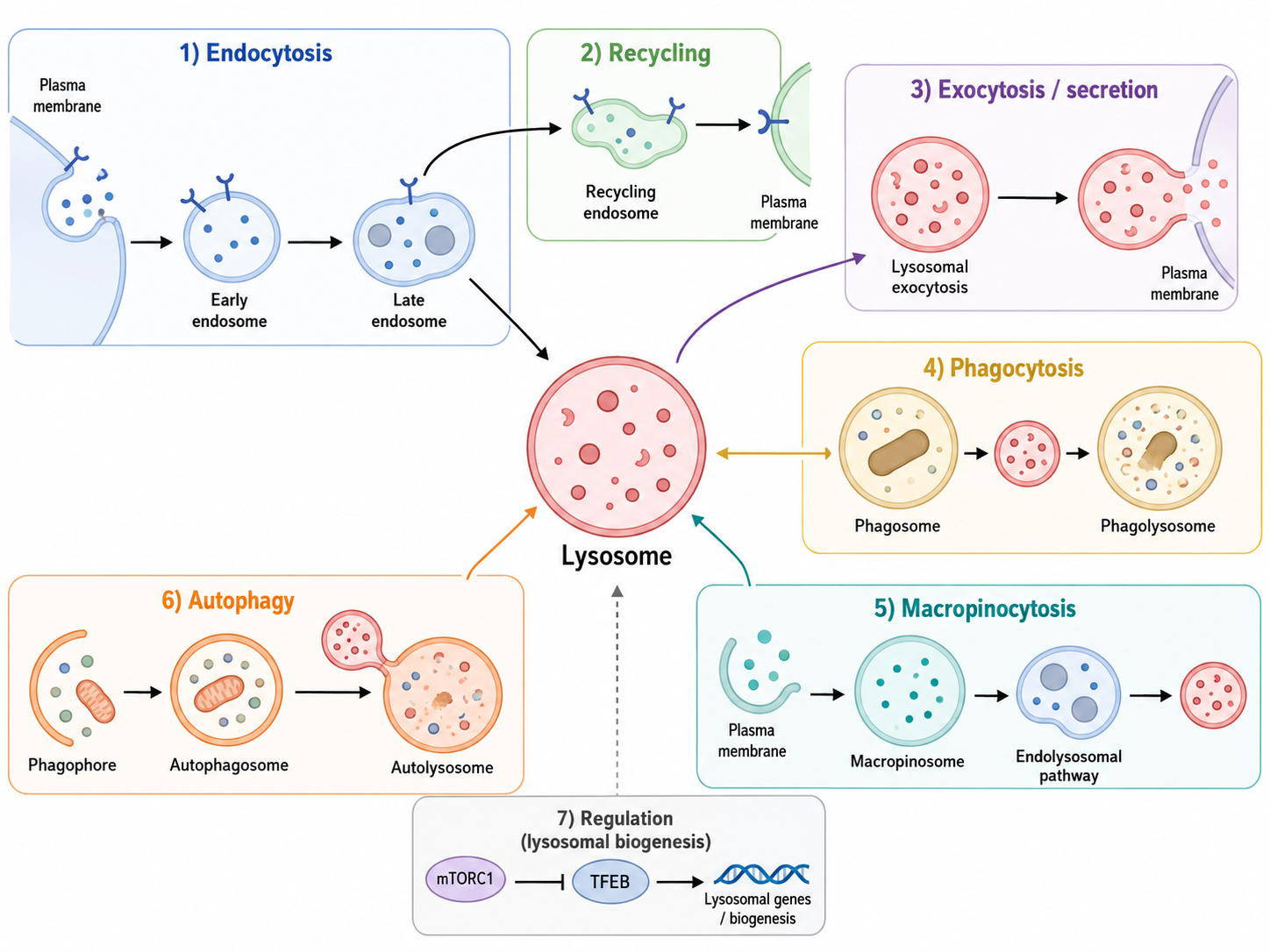
Figure 1. Overview of Lysosome-Associated Membrane Trafficking, Degradation, and Biogenesis Regulation
2.1 Endocytosis, Phagocytosis, and Macropinocytosis
Endocytosis internalizes extracellular materials and plasma membrane receptors into cells, forming early endosomes. Early endosomes can recycle some receptors back to the plasma membrane, or mature into late endosomes and fuse with lysosomes to form endolysosomal structures. This process participates in receptor degradation, nutrient acquisition, and termination of extracellular signaling.
Phagocytosis mainly occurs in immune cells such as macrophages, dendritic cells, and neutrophils. It takes up pathogens, apoptotic cells, or tissue debris through phagosomes. After phagosomes fuse with lysosomes, their contents are degraded by acidic hydrolases and may enter antigen processing and presentation pathways.
Macropinocytosis takes up extracellular fluid and soluble molecules through the formation of larger vesicles and is important in tumor cells, immune cells, and nutrient-stressed cells.
Pathway | Main Substrate | Relationship with Lysosomes |
Endocytosis | Receptors, ligands, extracellular soluble materials | Enters lysosomes after endosomal maturation |
Phagocytosis | Pathogens, cell debris, large particles | Phagosomes fuse with lysosomes for degradation |
Macropinocytosis | Extracellular fluid, proteins, nutrients | Macropinosomes fuse with lysosomes |
LC3-associated phagocytosis (LAP) | Phagosomal contents | LC3 modifies phagosomes and promotes maturation and degradation |
2.2 Macroautophagy, Endosomal Microautophagy, and Chaperone-Mediated Autophagy
Autophagy is the major pathway by which lysosomes degrade intracellular materials. Macroautophagy encloses cytoplasmic materials or organelles within double-membrane autophagosomes, which then fuse with lysosomes to form autolysosomes. This process depends on the ULK1 complex, Vps34 complex, ATG5-ATG12-ATG16L1 complex, and LC3 lipidation.
Endosomal microautophagy (e-MI) allows cytoplasmic materials to enter the endosomal system and depends on ESCRT-related mechanisms. Chaperone-mediated autophagy (CMA) is more substrate-selective. Proteins containing KFERQ-like motifs can be recognized by HSPA8 and transported into the lysosomal lumen through LAMP2A for degradation.
Autophagy Type | Key Molecules | Main Features |
Macroautophagy | ULK1, Vps34, ATG5, ATG12, ATG16L1, LC3 | Engulfs large molecular complexes or organelles through autophagosomes |
Endosomal microautophagy | ESCRT complex | Cytoplasmic materials enter the endosomal system |
Chaperone-mediated autophagy | HSPA8, LAMP2A | Selectively degrades proteins containing KFERQ-like motifs |
LC3-associated phagocytosis (autophagy-related process, non-canonical autophagy type) | LC3, phagosome-associated proteins | Connects phagocytosis with autophagy-like membrane modification processes |
2.3 Lysosomal Exocytosis and Extracellular Vesicle Release
Lysosomes can move toward the cell periphery and fuse with the plasma membrane, resulting in lysosomal exocytosis. This process participates in plasma membrane repair, local hydrolase release, and extracellular matrix remodeling. Lysosomes can also interact with the multivesicular body system and influence extracellular vesicle release.
In immune and inflammatory environments, lysosomal exocytosis may help cells repair damage, but it may also promote the release of inflammatory mediators, hydrolases, or tissue-destructive components. Therefore, lysosomal secretion has dual significance in autoimmunity and chronic inflammation.
3 Core Regulatory Mechanisms of Lysosomal Function
3.1 Acidification and Hydrolase Activity
The acidic environment inside the lysosomal lumen is mainly maintained by vacuolar-type ATPase (V-ATPase). Low pH provides an appropriate active environment for hydrolases such as cathepsins, lipases, glycosidases, and nucleases. If lysosomal acidification is impaired, degradative capacity may decline even when lysosome number increases.
Lysosomal activity assessment cannot rely only on LAMP1 or LAMP2 expression. Increased LAMP proteins may indicate increased lysosome number, but may also reflect increased lysosomal burden or blocked degradation. More reliable interpretation should combine acidic probes, cathepsin activity, LC3-II, p62/SQSTM1, autophagic flux, and functional readouts.
3.2 TFEB/TFE3 and the CLEAR Network
TFEB and TFE3 are important transcription factors for lysosomal biogenesis and autophagy gene expression. They bind CLEAR elements in target gene promoters and induce the expression of lysosomal membrane proteins, luminal hydrolases, transporters, and ATG genes.
Under nutrient-rich conditions, mTORC1 phosphorylates TFEB/TFE3 and retains them in the cytoplasm. When nutrient deprivation, energy stress, or lysosomal stress occurs, mTORC1 activity decreases, TFEB/TFE3 become dephosphorylated and translocate into the nucleus, enhancing lysosomal and autophagy-related transcriptional programs.
State | mTORC1 Activity | TFEB/TFE3 Localization | Result |
Nutrient-rich | Active | Mainly retained in the cytoplasm | Suppresses excessive autophagy and lysosomal biogenesis |
Starvation/energy stress | Reduced | Translocates into the nucleus | Enhances autophagy and lysosomal gene expression |
Increased lysosomal burden | May decrease or remodel | Enhanced nuclear translocation | Promotes lysosomal repair and regeneration |
Chronic pathological stress | Persistently abnormal | May show compensatory activation or imbalance | Associated with inflammation, metabolic abnormalities, and tissue injury |
3.3 mTORC1, mTORC2, and Nutrient Sensing
mTORC1 localizes to the cytosolic side of the lysosomal membrane and is a core integrator of amino acid, energy, and growth factor signals. Its activation promotes protein synthesis and cell growth while inhibiting ULK1-mediated autophagy initiation. mTORC2 is also related to lysosome-associated signaling and participates in cell survival, cytoskeleton regulation, and metabolic control.
Thus, the lysosome is a nutrient-sensing platform rather than a simple degradation compartment. When nutrient supply is sufficient, mTORC1 suppresses autophagy. When nutrients are limited or cellular stress increases, autophagy releases amino acids and lipids to provide metabolic compensation.
3.4 Lysosomal Movement and Organelle Interaction
Lysosomes can move along the microtubule system between the perinuclear region and the cell periphery. Perinuclear localization favors fusion with the endoplasmic reticulum, Golgi apparatus, mitochondria, and autophagosomes. Peripheral localization facilitates receptor regulation, exocytosis, plasma membrane repair, and local hydrolase release.
Lysosomes can also form contact sites with mitochondria and participate in mitochondrial fission, calcium signaling, lipid exchange, and metabolic coordination. Abnormal lysosomal localization affects autophagic flux, cell migration, immune cell activation, and tissue injury responses.
4 Lysosomal Damage, Inflammation, and Cell Death
4.1 Lysosomal Membrane Damage and Cathepsin Release
After lysosomal membrane damage, cathepsins can leak into the cytoplasm and trigger lysosome-dependent cell death. Cathepsins can also cleave Bid, linking lysosomal damage with the mitochondrial apoptotic pathway.
Lysosomal membrane damage does not necessarily lead to immediate cell death. Cells can repair the damaged membrane through ESCRT-mediated membrane repair or remove damaged lysosomes through lysophagy. If repair and clearance fail, persistent inflammation and cell death may occur.
4.2 ROS, NLRP3 Inflammasome, and Pyroptosis
Damaged lysosomes can generate or amplify ROS signals and promote NLRP3 inflammasome activation. Activated NLRP3 promotes maturation and release of IL-1β and IL-18 and induces pyroptosis. This process is important in autoimmunity, infection, gout-like inflammation, metabolic inflammation, and kidney injury.
Lysosomal Abnormality | Downstream Result | Disease Association |
Lysosomal membrane rupture | Cathepsin release | Cell death and tissue injury |
Increased ROS | Oxidative damage and inflammatory amplification | Chronic inflammation and metabolic stress |
NLRP3 activation | IL-1β/IL-18 release and pyroptosis | Autoimmunity and metabolic inflammation |
Insufficient lysophagy | Accumulation of damaged lysosomes | Proteostasis impairment and cellular functional decline |
Insufficient acidification | Reduced degradative capacity | Protein aggregation, lipid accumulation, immune abnormalities |
4.3 Lysosomes and Ferroptosis
Lysosomes can affect intracellular labile iron levels through iron storage, iron release, and ferritinophagy. Lysosomal damage, increased ROS, and labile iron accumulation can promote lipid peroxidation and thereby link lysosomes with ferroptosis.
In metabolic diseases, neurodegenerative diseases, and inflammatory injury, the lysosome-iron homeostasis-lipid peroxidation axis may be an important mechanism connecting proteostasis, oxidative stress, and cell death.
5 Lysosomes and Immune Homeostasis
5.1 Antigen Processing and MHC II Presentation
The endolysosomal system participates in MHC class II antigen processing and presentation. Exogenous antigens enter the endosome-lysosome pathway through endocytosis or phagocytosis, are processed by hydrolases, loaded onto MHC II molecules in MHC class II compartments, and ultimately presented to CD4⁺ T cells.
If lysosomal acidification, hydrolase activity, or endosomal maturation is abnormal, antigen processing efficiency may be affected, resulting in insufficient immune activation or abnormal exposure of self-antigens. In autoimmune diseases, abnormal antigen processing, clearance of cell death products, and nucleic acid sensing are often associated with altered lysosomal function.
5.2 Immune Cell Survival and Differentiation
Long-lived immune cells depend on autophagy and lysosomes to maintain mitochondrial quality, proteostasis, and metabolic adaptation. T cells, B cells, macrophages, and dendritic cells all require lysosomal participation during activation, differentiation, and effector function.
Autophagy abnormalities can alter Treg/Th17 balance, affect effector T cell metabolism, weaken the ability of macrophages to clear pathogens, and increase inflammasome activation. These changes can drive persistent autoimmunity and chronic inflammation.
5.3 Autophagy Dysregulation and Inflammatory Amplification
Autophagy clears damaged mitochondria, protein aggregates, and intracellular pathogens, thereby limiting ROS, mtDNA release, and inflammasome activation. If autophagic flux decreases, intracellular damage signals cannot be cleared in time, and inflammatory responses are amplified.
Immune Process | Lysosomal/Autophagic Role | Functional Consequence |
Antigen presentation | Antigen degradation and MHC II loading | Affects T cell activation |
Pathogen clearance | Phagosome-lysosome fusion | Limits infection and inflammation |
T cell differentiation | Regulates metabolism and proteostasis | Affects Treg/Th17 balance |
Inflammasome control | Clears damaged mitochondria and DAMPs | Reduces IL-1β-related inflammation |
Maintenance of long-lived immune cells | Clears damaged organelles and proteins | Maintains immune cell function |
6 Roles of Lysosomes in Metabolic Diseases
6.1 Fatty Liver Disease, NAFLD, and NASH
Hepatocytes rely on autophagy and lipophagy to degrade lipid droplets and release fatty acids. FOXO1 can induce ATG gene expression and promote autophagy-mediated lipid droplet breakdown. Autophagy can also reduce endoplasmic reticulum stress and insulin resistance, exerting a protective role in hepatocellular lipid homeostasis.
In NAFLD and NASH, abnormal lysosomal protein expression, altered autophagic flux, and insufficient lipid droplet clearance may occur. If lipophagy decreases, lipid droplet accumulation, lipotoxicity, inflammation, and fibrosis risk increase. LC3-associated phagocytosis is also considered to potentially participate in protective mechanisms against liver fibrosis.
6.2 Diabetes and Insulin Resistance
In diabetes and insulin resistance, nutrient excess, lipotoxicity, glucotoxicity, AGEs, and oxidative stress can interfere with lysosomal function. Insufficient autophagy aggravates endoplasmic reticulum stress, mitochondrial injury, and inflammatory responses, and affects pancreatic β cell survival and insulin secretion.
In peripheral tissues, the lysosome-autophagy system also affects lipid droplet mobilization, mitochondrial quality control, and insulin signaling sensitivity. Declining lysosomal function may impair the ability of cells to handle nutrient overload and damaged proteins, thereby driving metabolic inflammation.
6.3 Neurodegenerative Diseases
Neurons and other terminally differentiated long-lived cells have high basal autophagy requirements. Insufficient lysosomal activity leads to delayed clearance of protein aggregates, damaged mitochondria, and abnormal lipids, and is associated with Parkinson’s disease, Alzheimer’s disease, amyotrophic lateral sclerosis, and other disorders.
Autophagy dysregulation has also been observed in inflammatory nervous system diseases such as EAE and chronic inflammatory demyelinating polyneuropathy. This suggests that lysosomes participate not only in neuronal homeostasis, but also in central immune and inflammatory regulation.
6.4 Lysosomal Storage Diseases
Lysosomal storage diseases are substrate accumulation disorders caused by defects in lysosomal enzymes, transporters, or related membrane proteins. Their pathology arises not only from substrate accumulation, but also involves autophagy blockade, mitochondrial dysfunction, inflammatory activation, and tissue structural damage.
When the kidney, nervous system, or myocardium is involved, lysosomal dysfunction often manifests as protein aggregation, impaired mitophagy, and increased cell death.
7 Roles of Lysosomes in Autoimmune Diseases
7.1 Systemic Lupus Erythematosus
In systemic lupus erythematosus (SLE), abnormalities in autophagy and lysosomal processes can affect immune cell differentiation, clearance of cell death products, and exposure of self-antigens. Treg and Th17 cell imbalance is associated with altered T cell autophagy. Meanwhile, autoantibodies and complement deposition can damage tissue cells, inducing lysosomal stress, ROS generation, and chronic inflammation.
In lupus nephritis, immune complex deposition and complement activation can damage glomerular cells and further affect autophagy-lysosome function in podocytes and renal tubular cells.
7.2 Inflammatory Bowel Disease
In inflammatory bowel disease (IBD), insufficient autophagy-mediated bacterial clearance, abnormal antigen presentation, and enhanced inflammasome activation can all participate in disease progression. Lysosomal functional defects in intestinal epithelial cells and immune cells weaken control of the gut microbiota, promoting dysbiosis and persistent inflammation.
Lysosomal defects in Paneth cells can affect antimicrobial peptide secretion, impairing intestinal barrier and microbial homeostasis. This is an important entry point for lysosomal dysfunction in IBD.
7.3 Sjögren’s Syndrome
In Sjögren’s syndrome, exocrine gland tissues may show altered autophagic activity. Some studies suggest increased autophagic activity in T cell areas of minor salivary glands, while salivary gland epithelial cells show abnormal expression of proteins related to apoptosis, secretion, and lysosomal function.
Lysosome-associated membrane protein LAMP3 has research value in this disease, but it is not exclusive to lysosomes. It can also be found in late endosomes and specific immune activation states. Therefore, in Sjögren’s syndrome research, LAMP3 should be analyzed together with LAMP1, LAMP2, LC3, p62, and functional autophagic flux indicators.
7.4 Multiple Sclerosis and EAE
In multiple sclerosis and its experimental model EAE, autophagy and lysosomal processes can affect central nervous system inflammation, antigen presentation, myelin injury, and immune cell infiltration. Autophagy dysregulation may alter T cell effector function, myelin antigen processing, and inflammatory states of neural glial cells.
Disease | Related Cells/Tissues | Lysosome-Related Changes | Possible Consequences |
SLE | T cells, B cells, glomerular cells | Abnormal autophagy and immune complex-related injury | Treg/Th17 imbalance, chronic inflammation, kidney injury |
IBD | Intestinal epithelial cells, Paneth cells, macrophages | Insufficient bacterial clearance and abnormal antimicrobial peptide secretion | Microbiota imbalance and inflammasome activation |
Sjögren’s syndrome | Salivary gland epithelial cells, T cells | Abnormal autophagic activity and LAMP protein expression | Glandular injury and secretory dysfunction |
MS/EAE | T cells, glial cells | Abnormal autophagy and antigen processing | Neuroinflammation and demyelinating injury |
CIDP | Peripheral nerve-associated immune cells | Autophagy dysregulation | Chronic inflammatory nerve injury |
8 Product Selection Related to Lysosome Research in Metabolic and Autoimmune Diseases
Product Module | Cat. No. | Product Name | Grade & Purity | Application Positioning |
Lysosome tracing | LysoTracker Blue DND-22 |
| Labels acidic lysosomes/late endolysosomes; suitable for live-cell lysosome localization and acidic vesicle observation | |
Lysosome tracing | LysoTracker Red | ≥97% | Used for red fluorescent lysosome labeling; suitable for colocalization with LAMP1, LC3B, and other markers | |
Lysosome tracing | LysoTracker Yellow HCK 123 | BioReagent, biological stain, suitable for fluorescence analysis, for microscopy, ≥95% | Detects acidic vesicles and lysosome distribution; suitable for analyzing lysosomal acidification and number changes | |
Lysosomal pH/acidification | LysoSensor PDMPO | ≥98% | pH-sensitive lysosomal probe, suitable for determining whether lysosomal acidification is impaired | |
Lysosome fluorescence imaging | Fluolysotm red (lysosomal red fluorescent probe) |
| Used for red lysosome imaging; suitable for observing lysosome localization, number, and morphology changes | |
Lysosome fluorescence imaging | Fluolysotm deep red (lysosomal dark red fluorescent probe) |
| Deep-red lysosome probe, suitable for multicolor fluorescence colocalization experiments | |
Lysosome fluorescence imaging | Lysosome tracer Green | Suitable for immunofluorescence (IF), BioReagent, biological stain, molecular biology grade, 1 mM | Green-channel lysosome tracer, suitable for combined imaging with red autophagy, mitochondrial, or inflammatory markers | |
Lysosome fluorescence imaging | Lyso-Tracker Red |
| Used for tracing acidic lysosomes; suitable for observing lysosome localization and activity changes in cells | |
Lysosome AIE probe | Lysosome Red Probe (AIE) | BioReagent, biological stain, for microscopy, ≥98%(HPLC), 10 mM in DMSO | AIE-type lysosome probe, suitable for live-cell lysosome imaging and multichannel experiments | |
Lysosomal membrane marker | LAMP1 /CD107a Antibody | KD Validation | Labels lysosomes/late endolysosomes and analyzes lysosome number, localization, exocytosis, and fusion events | |
Lysosomal membrane marker | Recombinant LAMP1 Antibody | Recombinant, ExactAb™, validated, 0.2 mg/mL | Detects LAMP1; suitable for evaluating lysosomal homeostasis in IF, WB, and IHC | |
Lysosomal membrane marker | LAMP1 Mouse mAb | Carrier-free, ExactAb™, azide-free, validated, ≥95%(SDS-PAGE), 1.0 mg/mL | High-purity LAMP1 antibody, suitable for lysosome localization, lysosomal exocytosis, and disease tissue detection | |
Lysosomal membrane marker | Recombinant LAMP2 Antibody | Recombinant, ExactAb™, validated, 0.8 mg/mL | Detects LAMP2; suitable for analyzing lysosomal membrane homeostasis, autophagic degradation, and lysosome-related diseases | |
Lysosomal membrane marker | Recombinant LAMP2 Antibody | KD Validation | Used for LAMP2 expression validation; suitable for lysosomal homeostasis and terminal autophagic degradation research | |
Lysosomal hydrolase | Cathepsin B Mouse mAb | Carrier-free, ExactAb™, azide-free, validated, high performance, see COA | Detects Cathepsin B; suitable for analyzing lysosomal protein degradation, membrane damage, and inflammasome activation | |
Lysosomal hydrolase | Cathepsin D Mouse mAb | Carrier-free, ExactAb™, azide-free, validated, high performance, PBS Only, ≥95%(SDS-PAGE), 1.0 mg/mL | Detects Cathepsin D; suitable for evaluating lysosomal protein degradation capacity and active lysosomal status | |
Lysosomal hydrolase | Recombinant Cathepsin D Antibody | KD Validation | Used for Cathepsin D detection; suitable for lysosomal acidic hydrolytic function and disease tissue analysis | |
Lysosomal hydrolase | Cathepsin S Mouse mAb | Carrier-free, ExactAb™, azide-free, validated, high performance, see COA | Cathepsin S is closely related to antigen processing and immune inflammation; suitable for autoimmunity and antigen presentation research | |
Lysosomal hydrolase | Recombinant Human Cathepsin B Protein | Animal-free, carrier-free, bioactive, ActiBioPure™, His tag, PBS Only, ≥95%(SDS-PAGE) | Can be used for Cathepsin B activity studies, inhibitor screening, or antibody validation | |
Lysosomal hydrolase | Recombinant Human Cathepsin D Protein | Animal-free, carrier-free, His tag, ≥95%(SDS-PAGE) | Can be used for Cathepsin D-related enzymology, lysosomal protein degradation, and antibody validation | |
Autophagosome marker | LC3B Mouse mAb | Carrier-free, ExactAb™, azide-free, validated, high performance, PBS Only, ≥95%(SDS-PAGE), 1.0 mg/mL | Detects LC3-I/LC3-II changes and analyzes autophagosome formation and autophagic flux | |
Autophagosome marker | Recombinant LC3B Antibody | Recombinant, ExactAb™, validated, see COA | Used for LC3B detection and autophagic flux analysis; suitable for metabolic disease, inflammation, and immune cell research | |
Autophagosome marker detection | Human Microtubule-associated Protein Light Chain 3B(LC3B) ELISA Kit | BioReagent | Quantitatively detects human LC3B levels and assists in evaluating autophagy-related changes | |
Selective autophagy substrate | SQSTM1 Mouse mAb | Carrier-free, ExactAb™, validated, high performance, see COA | Detects p62/SQSTM1 and assists in determining autophagy substrate accumulation and autophagy blockade | |
Selective autophagy substrate detection | Human Sequestosome 1 (SQSTM1) ELISA Kit | BioReagent | Quantitatively detects human SQSTM1/p62; suitable for inflammation, metabolic disease, and autophagy blockade research | |
Autophagy core protein | ATG5 Antibody | KD Validation | Detects ATG5 and evaluates autophagic membrane extension and autophagy pathway integrity | |
Autophagy core protein | Recombinant APG5L/ATG5 Antibody | Recombinant, ExactAb™, validated, 0.3 mg/mL | Used for ATG5 detection; suitable for autophagy research in kidney disease, immune cells, and metabolic stress | |
Autophagy core protein | ATG7 Antibody | ExactAb™, validated, 0.6 mg/mL | Detects ATG7 and evaluates the LC3 lipidation system and autophagy defects | |
Autophagy core protein | Recombinant ATG7 Antibody | KD Validation | Used for ATG7 expression validation; suitable for autophagic flux and inflammatory amplification mechanism research | |
Lysosomal transcriptional regulation | Recombinant TFEB Antibody | Recombinant, ExactAb™, validated, see COA | Detects TFEB; suitable for analyzing TFEB nuclear translocation, the CLEAR network, and lysosomal biogenesis | |
Lysosomal transcriptional regulation | Recombinant TFE3 Antibody | Recombinant, ExactAb™, validated, see COA | Detects TFE3 and analyzes TFEB/TFE3-mediated transcriptional regulation of lysosomal and autophagy genes | |
Lysosomal transcriptional regulation detection | Human Transcription Factor E3 (TFE3) ELISA Kit | BioReagent | Quantitatively detects TFE3; suitable for lysosomal stress and autophagy transcriptional regulation research | |
Lysosomal damage recognition | Recombinant Human Galectin-3 Protein | Carrier-free, bioactive, ActiBioPure™, high performance, PBS Only, ≥95%(SDS-PAGE) | Galectin-3 is commonly used in research on lysosomal membrane damage/glycan exposure-related mechanisms and can be used for functional or validation experiments | |
Lysosomal damage/inflammation detection | Human Galectin 3 (Galectin-3) ELISA Kit | BioReagent | Detects Galectin-3; suitable for lysosomal damage, inflammation, and tissue fibrosis-related research | |
Lysosomal damage/inflammation detection | Mouse Galectin 3 (Galectin-3) ELISA Kit | BioReagent | Detects mouse Galectin-3; suitable for autoimmunity, metabolic inflammation, and animal model research | |
Lysosomal function inhibition | (R)-Hydroxychloroquine phosphate |
| Used to regulate lysosomal acidification and autophagic degradation; suitable for autoimmunity and autophagic flux intervention research | |
Lysosomal function inhibition | (S)-Hydroxychloroquine sulfate |
| Hydroxychloroquine-related isomer, used for lysosomal function and autophagy inhibition-related research | |
Lysosomal protease inhibition | JF526–Pepstatin A TFA |
| Pepstatin A-type fluorescent/labeled derivative, suitable for Cathepsin D-related lysosomal activity research | |
Lysosomal protease inhibition | Pepstatin Trifluoroacetate | 10 mM in DMSO | Inhibits acidic aspartyl proteases; suitable for lysosomal protein degradation and autophagic flux validation | |
Lysosomal protease inhibition | Leupeptin | ≥98% | Inhibits lysosomal cysteine/serine proteases and is commonly used for autophagic flux and lysosomal degradation blockade experiments |
Lysosomes are key organelles connecting nutrient sensing, proteostasis, immune regulation, and inflammatory responses. Their dysfunction can lead to blocked autophagic flux, abnormal antigen processing, inflammasome activation, lipid metabolic disorder, and tissue injury. Lysosomal changes in metabolic and autoimmune diseases do not follow a single direction and should be comprehensively interpreted according to cell type, disease stage, lysosomal activity, and autophagic flux.


 危险品化学品经营许可证(带存储)