
计算溶液所需的质量、体积或浓度。
糖原PAS染色试剂盒
有货
BioReagent,生物染色剂,用于显微镜 级 ,适用于对基线干扰要求严格的色谱和分析工作流程。
2-8°C储存,避光。低温运输 。请查阅批次 COA 获取详细规格。
SDS、COA、产品数据表及规格说明书均可下载。可通过批号查询获取批次 COA。
在色谱分析、有机合成和交叉偶联反应领域已被 0 篇同行评审文献引用。
糖原染色是病理学中常规的染色方法之一,常用来显示糖原和其他多糖,该染色试剂盒不仅能够显示糖原,还能显示中性黏液性物质和某些酸性物质以及软骨、垂体、霉菌、真菌、色素、淀粉样物质、基底膜等。过碘酸 (又称高碘酸) 是一种强氧化剂,它能氧化糖类及有关物质中的1,2-乙二醇基,使之变为二醛,醛与Schiff试剂能结合成一种品红化合物,产生紫红色。由于高碘酸还可氧化细胞内其他物质,使用时应注意选择好高碘酸浓度和氧化时间,使氧化控制在既能把乙二醇基氧化成醛基又不至于过氧化,这是很关键的步骤。
糖原PAS染色试剂盒特点是性能稳定,特异性强,操作简捷。
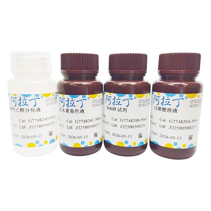
| G774820 | Component | 4 ×50 mL | 4 ×100 mL | 4 ×500 mL | Storage |
| G774820A | 过碘酸溶液 | 50 mL | 100 mL | 500 mL | 2-8℃. Store in the dark |
| G774820B | Schiff试剂 | 50 mL | 100 mL | 500 mL | 2-8℃. Store in the dark |
| G774820C | 苏木素染色液 | 50 mL | 100 mL | 500 mL | 2-8℃. Store in the dark |
| G774820D | 酸性乙醇分化液 | 50 mL | 100 mL | 500 mL | 2-8℃. |
使用说明:
1、常规固定,常采用10%的福尔马林,常规脱水包埋。
2、石蜡切片脱蜡入蒸馏水;冰冻切片直接入蒸馏水。
3、自来水冲洗2-3min,再用蒸馏水浸洗2次。
4、入过碘酸溶液,室温放置5min,一般不宜超过10min。
5、蒸馏水冲洗。
6、入 Schiff 试剂,置于室温阴暗处浸染10-20min (切片变成粉红色)。
7、自来水冲洗5min (切片变成深红色)。
8、入苏木素染色液,染细胞核1min。
9、酸性乙醇分化液分化2-5s。
10、自来水冲洗5min,更换蒸馏水清洗,使其返蓝。
11、95%乙醇、100%乙醇脱水。
12、二甲苯透明,中性树胶封固。
阴性对照:
1、取淀粉酶1g溶解于PBS (pH5.3) 100mL,处理30-60min,与其他切片共同入过碘酸溶液。结果应为阴性。
2、(备选方案) 取唾液片 (过滤后用) 处理30-60min,与其他切片共同入过碘酸溶液。结果应为阴性。
3、(备选方案) 如果对照片采用其自身样本,对照片不经过碘酸溶液这一步,直接入Schiff试剂。结果应为阴性。
备注:颜色深浅很大程度上取决于样品在过碘酸溶液和Schiff试剂中作用时间的长短。
注意事项:
1、切片脱蜡应尽量干净,否则影响染色效果。
2、过碘酸氧化时间不宜过久,氧化时的温度以18-22°C最佳。
3、过碘酸溶液和Schiff试剂应置于4°C密闭保存,使用时避免接触过多阳光和空气。使用前,最好提前30min取出恢复到在室温,避光暗处使用。
4、酸性乙醇分化液应经常更换新液,其分化时间应该依据切片厚薄、组织的类别和分化液的新旧而定,另外分化后自来水冲洗时间应该足够。
5、在过碘酸溶液和Schiff试剂中作用时间非常重要,该依据切片厚薄、组织的类别等决定。
6、本染色液常用于常规组织切片染色,对于真菌、细胞、极其薄的切片,建议采购糖原PAS染色试剂盒 (细胞真菌专用),因为其过碘酸溶液和苏木素溶液浓度更低,不宜过染。
7、冷冻切片染色时间尽量要短。
染色结果:
| PAS 反应阳性物质(糖原或多糖) | 红色或紫红色 |
| 细胞核 | 蓝色 |
| 细胞质 | 深浅不一的红色 |
Glycogen staining is one of the routine staining techniques in pathology, commonly applied to visualize glycogen and other polysaccharides. This staining kit can not only stain glycogen, but also demonstrate neutral mucous substances, certain acidic substances, cartilage, pituitary tissues, molds, fungi, pigments, amyloid substances, basement membranes and more.Periodic acid is a strong oxidizing agent. It oxidizes the 1,2-glycol groups in carbohydrates and related substances to dialdehydes. These aldehydes then react with Schiff reagent to form a magenta-colored complex, producing a purplish-red stain. Since periodic acid can also oxidize other intracellular components, careful control of periodic acid concentration and oxidation time is essential to achieve sufficient oxidation of glycol groups into aldehyde groups without over-oxidation; this step is critical for successful staining.
The PAS Glycogen Staining Kit features stable performance, high specificity and simple, rapid operation.
| G774820 | Component | 4 ×50 mL | 4 ×100 mL | 4 ×500 mL | Storage |
| G774820A | Periodic Acid Solution | 50 mL | 100 mL | 500 mL | 2-8℃. Store in the dark |
| G774820B | Schiff Reagent | 50 mL | 100 mL | 500 mL | 2-8℃. Store in the dark |
| G774820C | Hematoxylin Staining Solution | 50 mL | 100 mL | 500 mL | 2-8℃. Store in the dark |
| G774820D | Acidic Ethanol Differentiation Solution | 50 mL | 100 mL | 500 mL | 2-8℃. |
Instructions for Use:
1. Perform routine fixation using 10% formalin, followed by standard dehydration and embedding procedures.
2. For paraffin sections: deparaffinize and transfer into distilled water; for frozen sections: directly immerse in distilled water.
3. Rinse with tap water for 2–3 minutes, then wash twice with distilled water.
4. Immerse slides in periodic acid solution at room temperature for 5 minutes; do not exceed 10 minutes in most cases.
5. Rinse thoroughly with distilled water.
6. Place slides in Schiff reagent and incubate in the dark at room temperature for 10–20 minutes until sections turn pink.
7. Rinse with tap water for 5 minutes until sections turn deep red.
8. Counterstain cell nuclei with hematoxylin staining solution for 1 minute.
9. Differentiate in acidic ethanol differentiation solution for 2–5 seconds.
10. Rinse with tap water for 5 minutes, then switch to distilled water for washing until sections turn blue.
11. Dehydrate sequentially with 95% ethanol and absolute ethanol.
12. Clear with xylene and mount with neutral balsam.
Negative Control:
1. Dissolve 1 g amylase in 100 mL PBS (pH 5.3). Treat sections with this solution for 30–60 minutes, then incubate together with other sections in periodic acid solution. A negative staining result should be observed.
2. (Alternative method) Treat sections with filtered saliva for 30–60 minutes, followed by incubation with periodic acid alongside other sections. Staining result should be negative.
3. (Alternative method) When using self-specimen as control slide, skip the periodic acid treatment step and place the slide directly into Schiff reagent. The result should be negative.
Precautions:
1. Ensure complete deparaffinization of sections; incomplete removal of paraffin will compromise staining outcomes.
2. Do not prolong periodic acid oxidation. The optimal oxidation temperature ranges from 18 to 22 °C.
3. Store periodic acid solution and Schiff reagent tightly sealed at 4 °C. Avoid excessive exposure to sunlight and air during use. Take them out 30 minutes in advance to equilibrate to room temperature, and perform staining in dim light.
4. Replace acidic ethanol differentiation solution regularly. Differentiation duration varies according to section thickness, tissue type and freshness of the solution. Sufficient tap water rinsing is required after differentiation.
5. Incubation time in periodic acid solution and Schiff reagent is critical and shall be adjusted based on section thickness, tissue type and other factors.
6. This set of staining solutions is designed for routine tissue sections. For fungi, cell smears and ultra-thin sections, purchase the PAS Glycogen Staining Kit dedicated to cells and fungi instead. It contains lower concentrations of periodic acid and hematoxylin to prevent overstaining.
7. Minimize staining duration for frozen sections.
Staining Results:
| PAS-positive substances (glycogen or polysaccharides) | Red or purplish-red |
| Cell nucleus | Blue |
| Cytoplasm | Variable shades of red |
通过匹配包装上的批号来查找并下载产品的 COA,每批产品都进行了严格的验证,您可放心使用!
| 批号(Lot Number) | 证书类型 | 货号 |
|---|---|---|
 ZJ26F0838125 ZJ26F0838125 |
分析证书 | G774820 |
| ZJ26F0838126 |
分析证书 | G774820 |
| ZJ26F0333319 |
分析证书 | G774820 |
| ZJ26F0333320 |
分析证书 | G774820 |
| ZJ25F1230222 |
分析证书 | G774820 |
| ZJ25F0521432 |
分析证书 | G774820 |
我们的级别选择指南涵盖了目录中所有的变体的纯度、稳定剂状态和应用适用性。
查看 BioReagent 级别指南 → 查看 Biological Stain 级别指南 → 查看 for Microscopy 级别指南 →

 危险品化学品经营许可证(带存储)