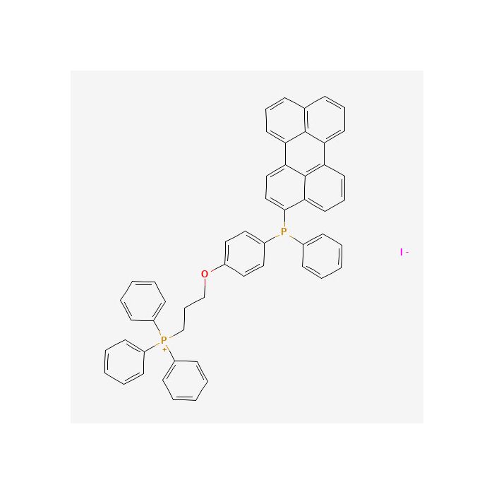
MitoPeDPP是一种新型荧光染料,由于其具有三苯基膦结构,因此可以穿过细胞膜并在线粒体中聚集。 聚集在线粒体内膜上的MitoPeDPP可以被脂质过氧化物氧化而释放出强荧光。由于氧化的MitoPeDPP (Ox-MitoPeDPP) 的激发和发射波长分别是452 nm和470 nm,可以减小样品的光损伤和自发荧光,因此利用荧光显微镜MitoPeDPP可以检测活细胞中的脂质过氧化物。
特点
1.特异性的在细胞中线粒体内聚集
2.可以检测线粒体膜内的脂质过氧化物
3.可以在488 nm和535 nm的荧光波长下进行检测
*由于MitoPeDPP量极少不宜看到,可以通过观察MitoPeDPP DMSO溶液的颜色是否为黄色来判断。
荧光特性
检测原理
MitoPeDPP可以穿过细胞膜并在线粒体中聚集。聚集在线粒体内膜上的MitoPeDPP可以被脂质过氧化物氧化而释放出强荧光。

所需的设备和材料
- Dimethyl Sulfoxide (DMSO)
- PBS
- Hanks’HEPES 缓冲液
- 移液器
溶液制备
1. 制备 0.1 mmol/l MitoPeDPP DMSO 储存液
向含有 5 μg MitoPeDPP 的管子中加入 50 μl DMSO,用移液器吹打使其溶解。
* 由于 MitoPeDPP 在 DMSO 溶液中不稳定,请避光保存并在 1 天内用完。
2. 制备 MitoPeDPP 工作液
用 Hanks 'HEPES 缓冲液稀释 MitoPeDPP DMSO 储存液,制备成 0.1‑0.5 μmol/l 的 MitoPeDPP 工作液。
* Hanks 'HEPES 缓冲液用来维持细胞环境的稳定
* 制备好工作液后立刻加入细胞中以避免 MitoPeDPP 发生自氧化。
操作步骤
MitoPeDPP 染色
1. 准备实验用细胞。
2. 更换培养基,用 Hanks 'HEPES 或 PBS 清洗 2 次。
3. 加入适量的 MitoPeDPP 工作液。
| 容器 | 工作液体积 |
|---|
| 35 mm 皿 | 2,000 μl | | 96 孔板 | 100 μl |
|
4. 在 37℃避光培养 15 min。
5. 弃上清液,然后用 Hanks 'HEPES 缓冲液或 PBS 清洗 2 次。
6. 加入含有刺激剂的 Hanks 'HEPES 缓冲液或 PBS,在荧光显微镜下观察。
* 波长 / 带通滤波器:470/40 nm (Ex),525/50 nm (Em)
此操作步骤仅为常规通用方法,请根据实际情况优化实验条件,如 MitoPeDPP 的合适终浓度、刺激剂的刺激时间等。
MitoPeDPP is a novel fluorescent dye. Owing to its triphenylphosphonium moiety, it can cross the cell membrane and accumulate in mitochondria. MitoPeDPP accumulated on the inner mitochondrial membrane is oxidized by lipid peroxides to emit strong fluorescence. The oxidized MitoPeDPP (Ox‑MitoPeDPP) exhibits excitation and emission maxima at 452 nm and 470 nm respectively, which reduces photodamage and autofluorescence of samples. Therefore, MitoPeDPP can be used to detect lipid peroxides in living cells by fluorescence microscopy.
Features
1. Specifically accumulates in cellular mitochondria
2. Enables detection of lipid peroxides within the mitochondrial membrane
3. Detectable at fluorescence wavelengths of 488 nm and 535 nm
*Since MitoPeDPP is supplied in very small quantity, it is hard to observe the solid. You may check whether the MitoPeDPP DMSO solution appears yellow for confirmation.
Fluorescence Properties

Detection Principle
MitoPeDPP crosses the cell membrane and accumulates in mitochondria. MitoPeDPP accumulated on the inner mitochondrial membrane is oxidized by lipid peroxides to produce intense fluorescence.
Required Equipment and Materials
‑ Dimethyl Sulfoxide (DMSO)
‑ PBS
‑ Hanks’-HEPES buffer
‑ Pipettor
Solution Preparation
1. Preparation of 0.1 mmol/l MitoPeDPP DMSO stock solution Add 50 μl DMSO to the vial containing 5 μg MitoPeDPP, and dissolve by pipetting up and down. * MitoPeDPP is unstable in DMSO solution. Store protected from light and use within 1 day.
2. Preparation of MitoPeDPP working solution Dilute MitoPeDPP DMSO stock solution with Hanks’‑HEPES buffer to prepare MitoPeDPP working solution at 0.1‑0.5 μmol/l. * Hanks’‑HEPES buffer is used to stabilize the cellular environment. * Add the prepared working solution to cells immediately to prevent auto‑oxidation of MitoPeDPP.
Procedure
MitoPeDPP Staining
1. Prepare experimental cells.
2. Remove culture medium and wash twice with Hanks’‑HEPES buffer or PBS.
3. Add an appropriate volume of MitoPeDPP working solution.
| Vessel | Working‑solution volume |
|---|
| 35 mm dish | 2,000 μl | | 96‑well plate | 100 μl |
|
4. Incubate at 37 °C for 15 min, protected from light.
5. Discard supernatant and wash twice with Hanks’‑HEPES buffer or PBS.
6. Add Hanks’‑HEPES buffer or PBS containing stimulant, and observe under a fluorescence microscope.
* Wavelength / band‑pass filter: 470/40 nm (Ex), 525/50 nm (Em)
These operating procedures are general‑purpose protocols. Optimize experimental conditions according to your practical applications, such as optimal final concentration of MitoPeDPP, stimulation duration of stimulant, etc.



 危险品化学品经营许可证(带存储)