
计算溶液所需的质量、体积或浓度。
BioReagent,生物染色剂,用于显微镜,适用于荧光分析 级 提供 ≥95% 纯度,适用于对基线干扰要求严格的色谱和分析工作流程。
避光,-20°C储存,避免反复冻融。超低温运输 。请查阅批次 COA 获取详细规格。
SDS、COA、产品数据表及规格说明书均可下载。可通过批号查询获取批次 COA。
在色谱分析、有机合成和交叉偶联反应领域已被 0 篇同行评审文献引用。
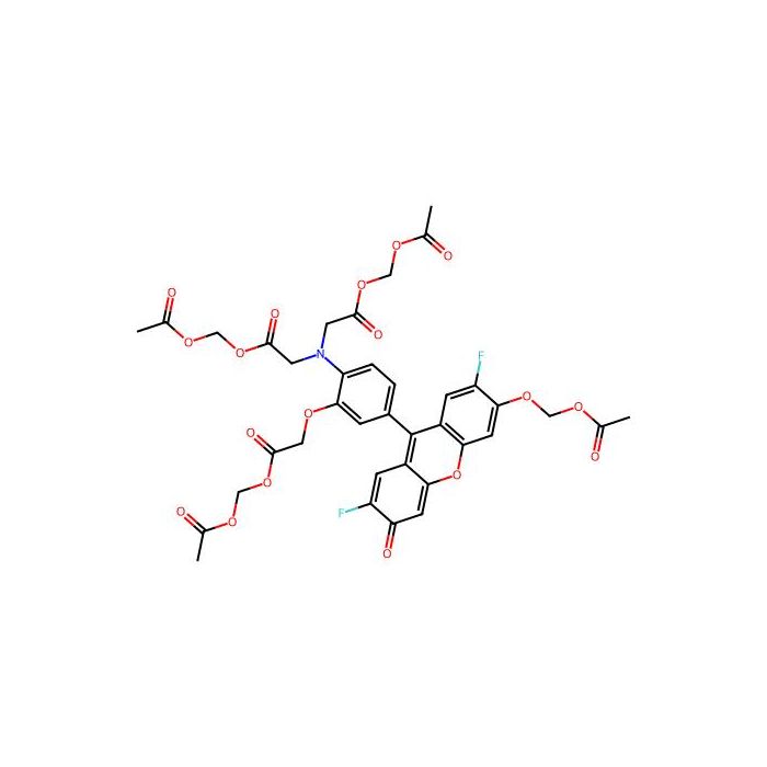
Cell-permeable Mag-Fluo-4 AM is an analog of Fluo-4 AM. Its dissociation constant (Kd) for magnesium ions is 4.7 mM and 22 µM for calcium ions. This property enables it to serve as both an intracellular magnesium ion indicator and a low-affinity calcium ion indicator. This low-affinity fluorescent calcium indicator has been successfully applied to accurately track the kinetics of spatially-averaged free calcium transients in skeletal muscle, providing reliable kinetic data for such calcium signals.
Product Specifications
1. Ex (nm): 495 nm
2. Em (nm): 528 nm
3. Molecular Weight: 817.65
4. Solvent: DMSO
Application Range
Used as intracellular magnesium ion indicator and low-affinity calcium ion indicator.
Example Experimental Protocol
1. Stock Solution Preparation: Aliquot all unused stock solutions into single-use portions, store at -20°C after preparation and avoid repeated freeze-thaw cycles.
2. Mag-Fluo-4 AM Stock Solution: Prepare 2-5 mM Mag-Fluo-4 AM stock solution with high-purity anhydrous DMSO.
Working Solution Preparation
Mag-Fluo-4 AM Working Solution
1. On the day of experiment, dissolve Mag-Fluo-4 AM in DMSO or thaw the stock solution to room temperature.
2. Prepare 2-20 µM Mag-Fluo-4 AM working solution with suitable buffers (e.g. Hanks' buffer, Hepes buffer) containing 0.04% Pluronic® F-127. A final concentration of 4-5 μM is recommended for most cell lines; optimize the concentration according to actual experimental conditions.
Notes
1. Pluronic® F-127 can enhance the water solubility of Mag-Fluo-4 AM.
2. For cells expressing organic anion transporters, add 1-2 mM probenecid (final well concentration: 0.5-1 mM) into the working solution to reduce probe leakage.
Operating Procedures
1. Culture cells in growth medium overnight.
2. Add 1× Mag-Fluo-4 AM working solution to cell plates on the next day.
Note: Replace the medium with fresh HHBS buffer before loading if the test compound interacts with serum.
3. Incubation condition: Incubate for 30-60 minutes in a 37°C cell incubator.
Note: Prolong the incubation time to more than 1 hour for certain cell lines to strengthen fluorescence signals.
4. Discard the working solution and replace it with HHBS buffer or buffer containing anion transport inhibitors (e.g. 1 mM probenecid).
5. After adding stimulants, detect fluorescence signals immediately with the following instruments:
Fluorescence microscope equipped with FITC filter sets
Fluorescence microplate reader (e.g. FDSS, FLIPR, FlexStation)
Detection parameters: Ex/Em = 490/525 nm, Cut-off wavelength: 515 nm
通过匹配包装上的批号来查找并下载产品的 COA,每批产品都进行了严格的验证,您可放心使用!
| 批号(Lot Number) | 证书类型 | 货号 |
|---|---|---|
| 分析证书 | M648123 |
我们的级别选择指南涵盖了目录中所有的变体的纯度、稳定剂状态和应用适用性。
查看 BioReagent 级别指南 → 查看 Biological Stain 级别指南 → 查看 for Fluorescence analysis 级别指南 → 查看 for Microscopy 级别指南 →
