金属螯合亲和层析,又称固定化金属离子亲和层析(Immobilized Metal Ion Affinity Chromatography, IMAC)。利用蛋白质表面的某些氨基酸(如组氨酸、色氨酸、半胱氨酸等)和金属离子(Ni²⁺、Cu²⁺、Zn²⁺、Co²⁺、Ca²⁺、Fe³⁺等)特异性结合的原理,实现蛋白质的分离纯化。
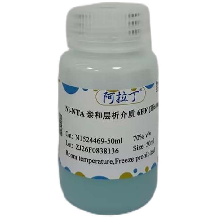
Ni Singarose FF 是以6%琼脂糖为基质,通过化学方法偶联次氮基三乙酸(NTA),再螯合金属离子Ni²⁺后形成的一种亲和层析介质。具有动态结合载量高、配基(Ni²⁺)泄露低、选择性高、可耐受一定浓度的还原剂等优点,广泛用于生物制药和生物工程下游蛋白质及多肽的分离纯化,尤其是组氨酸标签蛋白质的高效纯化。
产品特点:
1. 稳定性好、重复性好
2. 一步纯化、高流速、高载量
3. 选择性高、耐受一定浓度还原剂
4. 亲水性强、非特异吸附少、易于放大
产品技术参数:
表1. Ni Singarose FF 亲和层析介质
参数 | 指标 | 基质 | 6%高度交联琼脂糖 | 平均粒径 | 90 μm | 功能基团 | Ni²⁺ | 功能基团密度 | 20-25 μmol Ni²⁺/mL介质 | 结合载量 | 约40mg组氨酸标签蛋白/mL介质 | 建议流速 | <400 cm/h | 建议压力 | <0.3 MPa(3 bar) | 工作 pH | 4-8.5 | pH 稳定性 | 3-12 (长期);2-14 (短期) | 化学稳定性 | 常用缓冲液中稳定;避免使用螯合剂,如EDTA、柠檬酸盐等。 | 保存条件 | 20%乙醇;4-30℃ | 保质期 | 5年 |
备注:该处指在未螯合金属离子时的介质稳定性。长期,指介质在很长时间内保持稳定而不对其后续性能产生不利影响的pH范围。短期,指根据经验给出的介质再生、在位清洗和消毒的pH范围。 |
使用说明:
1. 装柱
1.1 装柱缓冲液准备
纯化水,超声脱气15 min。
1.2 层析介质准备
计算所需层析介质的量(压缩系数约为1.15),并称取备用。用真空抽滤瓶置换至装柱缓冲液中。置换的介质加入一定量装柱缓冲液,制成约50%浓度的胶悬液。
1.3 层析柱准备
检查层析柱,确保各部件完整、干净。安装下柱头,拧紧O型圈后,将层析柱垂直固定在铁架台上,用水平仪检查并调整使层析柱保持竖直。用注射器吸取装柱缓冲液,接于柱底的出口处,缓慢推出注射器内液体,排除底部筛网的气泡,取下注射器后拧上堵头。层析柱中加入约2 cm 高的装柱缓冲液。
1.4 装柱(以直径16 mm,床高10 cm 的层析柱为例)
混匀胶悬液,用玻璃棒引流缓慢倒入层析柱中,若液面离柱顶还有空隙,加装柱缓冲液充满。将调节器连接至层析系统,启动泵以一定流速排除管路及顶部筛网中的气泡后暂停。将调节器沿45°放入柱中,固定好调节器并拧紧密封圈,注意避免气泡进入。拧开层析柱底部的堵头,将底部管路放入到废液缸中,流速设为60 cm/h,至介质界面无变化后,设置流速为600 cm/h,并逐步降低流速到300 cm/h,维持该流速45 min,用记号笔在界面处做好标记,暂停系统。拧上底部堵头,层析柱上端与泵断开,稍拧松调节器上的密封圈,将柱头压到胶面以下3 mm 处,拧紧密封圈。将层析柱顶部和底部管路连接到层析系统上,进行柱效测定。
2. 柱效测定和评价
装柱完成后应立即检测柱效。通常用每米理论塔板数N/m、峰不对称因子As等术语来衡量层析柱的性能。柱效越高,层析柱分离能力越强。为了更加准确地反映层析柱柱效,检测时待测样品上样体积为柱体积的1.0%,线性流速控制在15-30 cm/h。为了防止样品的稀释,尽量在接近层析柱的入口处上样。
表2. 柱效检测方法
检测方法 | 丙酮法 | NaCl 法 | 样品 | 1.0%(v/v)丙酮水溶液 | 0.8 M NaCl 水溶液 | 样品体积 | 1.0%柱体积 | 1.0%柱体积 | 流动相 | 纯水 | 0.4 M NaCl 水溶液 | 流速 | 30 cm/h | 30 cm/h | 检测器 | UV-280 nm | 电导率 |
|
柱效计算
根据UV曲线(或电导率曲线)计算N/m、和As,公式如下:
- N/m = N/L
- N = 5.54 (VR/Wh)²
- As=b/a
备注:
L:柱高(单位为m)
VR:保留体积
Wh:半高峰宽
a:在10%峰高处的第一个半峰宽
b:在10%峰高处的第二个半峰宽。

图1.柱效计算示意图
峰应该是对称的,As尽可能接近1(通常可接受0.8-1.8之间的值)。N/m>3000。
3. 分离纯化
首选磷酸盐缓冲液,pH中性至弱碱性(pH7.0-8.0),避免使用EDTA及柠檬酸盐等,Tris-HCl也可以使用,但在金属离子与蛋白质亲和力很弱的情况下应避免使用。表3和表4分别列出了Ni Singarose FF 亲和层析介质常见添加试剂及浓度。
为了减少宿主蛋白与介质的非特异吸附,结合缓冲液和样品中通常加入低浓度咪唑(20-40mM)。缓冲液中必须加入0.15-0.5M NaCl以消除离子吸附作用。
表3. 不影响蛋白质结合到 Ni Singarose FF 的添加物
| 添加物 | 常用浓度 | 添加物 | 常用浓度 | | 磷酸盐、Tris、硼酸盐、HEPES | 20-100mM | 非离子型去污剂 | ≤2% | | NaCl | ≤2M | Triton X-100 | ≤2% | | KCl | ≤1M | Tween-20 | ≤2% | | 盐酸胍 | ≤6M | 辛基葡糖苷 | ≤2% | | 尿素 | ≤8M | 十二烷基麦芽糖柑 | ≤2% | | 甘油 | ≤50% | C12E8、C10E6 | ≤2% | | 异丙醇 | ≤60% | PMSF(蛋白酶抑制剂) | 1 mM | | 乙醇 | ≤30% | 胃蛋白酶抑制剂 | 1 μM | | 两性去污剂 (CHAPS) | ≤1% | 亮抑酶肽(蛋白酶抑制剂) | 0.5 μg/mL | | 1%苯甲脒(蛋白酶抑制剂) | ≤1 mM | / | / |
|
表4. 可能破坏蛋白质结合到 Ni Singarose FF 的添加物(尽量避免使用)
添加物 | 常用浓度 | 添加物 | 常用浓度 | β-巯基乙醇 | <20 mM | 柠檬酸盐 | 可耐受低浓度 | 还原谷胱甘肽 | <10 mM | 组氨酸 | 可替代咪唑 | 强还原剂 (DTT、TCEP、DTE)① | <5 mM | 甘氨酸 | / | 金属螯合剂(EDTA、EGTA)② | <1 mM | 谷氨酰胺 | / | 离子去污剂(胆酸盐、SDS) | / | 精氨酸 | / | 叠氮化钠 | 3 mM | NH₄Cl | / |
|
注:
①应尽量避免使用强还原剂,防止金属离子Ni²⁺被还原,影响蛋白结合在层析介质上。最大耐受强还原剂量根据样品不同而异,同时仅样品中含还原剂,缓冲液中不能含还原剂。
②应尽量避免使用金属离子螯合剂,防止金属离子Ni²⁺与介质分离。最大耐受强螯合剂浓度根据样品不同而异,同时仅样品中含螯合剂,缓冲液中不能含螯合剂。
3.1 柱平衡
将层析柱连接至纯化设备,使用结合缓冲液冲洗5 CV 以上,至层析柱流出液pH、电导同结合缓冲液的pH、电导一致。平衡结束后将紫外归零。
备注:
① 为减少金属离子Ni²⁺脱落对层析的影响,建议在平衡前先用1 CV 含有1 M NaCl的0.5 M 咪唑清洗,然后再用纯化水清洗5 CV,最后用结合缓冲液平衡层析柱后再上样。
② 推荐结合缓冲液:20 mM PB, 500 mM NaCl, 20 mM 咪唑, pH 7.4, 0.22 μm 滤膜过滤。
③ 当纯化包涵体时,结合缓冲液和样品中要添加8M尿素或6M盐酸胍。
3.2 加载样品
经0.22 / 0.45 μm 过滤后的样品溶液加载在层析柱上,上样体积取决于层析介质载量、样本溶液中与介质结合分子的浓度和其他层析条件。
3.3 柱冲洗
使用结合缓冲液冲洗5-10 CV,将该条件下不与介质结合的杂质从层析柱中洗去(至紫外检测值回归基线附近)。或根据前期实验确定的杂质淋洗条件进行柱冲洗。
3.4 洗脱
使用洗脱缓冲液冲洗层析柱,目标紫外峰升起后开始收集洗脱样品。
备注:
① 推荐洗脱缓冲液:20 mM PB, 500 mM NaCl, 500 mM 咪唑, pH 7.4,0.22 μm 滤膜过滤。
② 可选择竞争性洗脱、降低pH 方式洗脱或螯合剂洗脱,根据实际情况进行选择。
竞争性洗脱:线性或逐步增加与金属离子Ni²⁺有亲和力的物质浓度,如 0-2 M NH₄Cl、0-0.5 M 咪唑、0-0.5 M 组氨酸。梯度洗脱建议在结合缓冲液的恒定pH 下进行。
降低pH 洗脱:随着pH 降低,弱结合蛋白和强结合蛋白依次被洗脱。当缓冲液pH 降低至4.0 以下时,Ni²⁺会与介质解离而达到洗脱目的。(如果目的蛋白对低pH 敏感,建议洗脱收集液中加入1/10 收集体积的 1 M Tris-HCl,pH 9.0 进行中和)。
螯合剂洗脱:50 mM 金属螯合剂EGTA 或EDTA 可将金属离子Ni²⁺从介质中解离而达到洗脱目的,这种方法也可以洗脱变性或沉淀的蛋白质,该方法一般不推荐使用。洗脱产物中的Ni²⁺可用 Sindex G25 (M) 去除,介质可用 0.2 M NiS0₄ 再生后使用。
3.5 清洗和保存
用3-5 CV 含有1 M NaCl 的0.5 M 咪唑清洗,用纯化水冲洗至流出液电导为0,最后用20%乙醇冲洗2-3 CV。层析柱置于4℃环境中保存。
备注:介质重复使用取决于样品的性质,建议每种样品使用单独的层析介质,防止交叉污染。
4. 再生
杂质残留和金属离子Ni²⁺脱落将会影响层析性能和载量,建议1-5 个循环后进行再生,重新螯合金属离子Ni²⁺。再生步骤如下:
4.1 用 10 CV 纯化水冲洗层析柱。
4.2 用 3-5 CV 脱离子缓冲液(20 mM PB, 0.5 M NaCl, 50 mM EDTA, pH 7.4)冲洗。
4.3 用 3-5 CV 结合缓冲液 (不含咪唑) 冲洗层析柱。
4.4 用 5 CV 纯化水冲洗层析柱。
4.5 用 0.5 CV 以上0.1 M NiSO₄ (或其他金属离子溶液) 按 15 cm/h 流速螯合。
4.6 用 3 CV 的 0.5 M 乙酸钠-乙酸(pH 4.0)冲洗层析柱。
4.7 用 5 CV 结合缓冲液平衡层析柱后立即使用。
4.8 用 5 CV 的20%乙醇冲洗层析柱后保存。
备注:
① 金属离子浓度为 0.1 M,溶液保持中性或偏弱酸性,0.2 μm 过滤后使用。
② 当螯合Ca²⁺时,避免使用磷酸盐缓冲液 (会形成沉淀)。
③ 当脱去Fe³⁺时,由于Fe³⁺在中性溶液中容易形成不溶物,建议用 50 mM EDTA过夜,以脱去Fe³⁺;当螯合Fe³⁺时,必须在较低pH(约pH 3.0)下螯合,以防止Fe³⁺产生沉淀。
5. 在位清洗
在位清洗(Cleaning In Place, CIP)是去除层析介质上强结合的、沉淀的或者变性的物质。杂质残留将会影响层析柱的层析性能。如果聚集严重,将会堵塞层析柱,加大反压并减慢流速。所以定期的在位清洗能够防止污染物在柱床聚集,有助于保持介质的载量,流速等。
层析柱一般在使用 3-5 个循环后需要进行清洗,帮助介质恢复良好的性能。对不同类型的杂质和污染物建议清洗条件如下:
① 去除因离子交换作用吸附的蛋白:用2-3 CV 的2 M NaCl 溶液清洗层析柱,然后用结合缓冲液冲洗层析柱。
② 去除蛋白沉淀、疏水结合的物质:先脱去金属离子Ni²⁺,然后用0.2-1 M NaOH浸泡层析柱1 h 以上。
③ 去除强结合的疏水性蛋白和脂质等:用3-5 CV 的70%乙醇或者30%异丙醇冲洗层析柱(15-20 min),然后用结合缓冲液平衡层析柱。
另外,6 M 盐酸胍浸泡过夜可去除大部分杂蛋白。清洗后如果立即使用,用结合缓冲液冲洗3-10 CV 即可。
6. 消毒灭菌
消毒灭菌是为了将层析柱中的微生物污染降至最低。20%乙醇不具有杀菌、除热原作用,建议用70%乙醇处理12 h 以减少微生物污染风险。或者脱去金属离子Ni²⁺后,使用 0.5-1 M Na0H 浸泡 0.5-1 h 进行消毒灭菌。
7. 保存
未使用的介质应当保存在 4-30℃的干燥、通风、洁净环境中,确保容器口完全密闭,切记不可冷冻。
已装好 Ni Singarose FF介质的层析柱应当使用20%乙醇或2%苯甲醇浸泡密封保存,防止微生物污染。
为了防止乙醇挥发导致微生物滋生,建议间隔3 个月更换新鲜的保存液。
备注:保存的介质在使用前,应进行 CIP 和至少用结合缓冲液平衡 5 CV。
8.线性放大
在实验室规模进行优化后的纯化工艺,可以线性放大到中试或生产规模。在放大过程中需要注意以下事项:
① 保持停留时间不变,保证动态载量的稳定。
② 根据需要的结合能力选择层析柱容积,如果柱高改变需要注意是否对纯化步骤产生影响。
③ 根据流速需求确定层析柱直径,根据已知的停留时间确定柱床高度,一般推荐10-25cm。
④ 保证样品浓度均匀一致,洗脱条件一致。
问题及解决建议:
1. 问题:使用过程中压力过高
解决建议:
① 更换在线滤器;
② 层析柱堵塞,进行CIP清理。
2. 问题:压力曲线不稳
解决建议:
① 排除上样泵里的气泡;
② 使用气泡陷阱去除样品中的气泡;
3. 问题:蛋白不结合或结合量小。
解决建议:
① 超声处理使蛋白变性,裂解过程中使用温和的超声处理条件;
② 蛋白结合力弱,选择高载量层析介质;
③ 标签被包裹在蛋白内部,优化表达条件或重新设计序列;
④ 上样过程中蛋白聚集,在样品和缓冲液中加入非离子型去污剂;
⑤ 柱平衡pH不合适,调节缓冲液pH在6.5-8.0之间;
⑥ 层析介质失效,更换新的层析介质;
⑦ 上样量过载,降低上样量;
⑧ 蛋白或脂类聚集影响结合,及时清洗或更换新介质;
⑨ 上样流速过高,在上样过程中降低流速。
4. 问题:洗脱效果差
解决建议:
① 未收集到产物或结合量少,确认标签蛋白是否与介质结合;
② 洗脱流速过高,降低洗脱流速;
③ 洗脱缓冲液体积少,增加洗脱缓冲液体积;
④ 咪唑浓度低,可尝试增加咪唑浓度;
⑤ 洗脱时间不足,降低流速以延长洗脱缓冲液与介质接触时间;
⑥ 洗杂条件不合适,优化咪唑和NaCl浓度以提高纯度和收率;
⑦ 杂质与金属离子Ni²⁺有较高亲和力,搭配其他类型介质使用;
⑧ 洗脱缓冲液失效,配制新鲜的洗脱缓冲液;
⑨ 非特异离子吸附,在洗脱缓冲液中加入 0.1-0.2 M NaCl;
⑩ 非特异性疏水吸附,在洗脱缓冲液中加入 0.1% Triton X-100;
⑪ 洗脱出现聚集沉淀,确定蛋白在洗脱条件下的溶解度和稳定性。
5. 问题:层析介质变色
解决建议:
① 样品中含色素或有色蛋白,纯化完毕后严格执行CIP或再生;
② 样品或缓冲液中的还原剂使介质变褐色,避免或减少还原剂用量。
6. 问题:洗脱峰逐渐变宽
解决建议:层析柱可能被污染,优化洗脱条件,并严格按CIP进行清洗。
7. 问题:CIP峰增大
解决建议:CIP强度不够,优化CIP条件。
8. 问题:首次使用配基脱落较高
解决建议:首次使用时进行一次空载运行,并进行CIP。
Metal chelate affinity chromatography, also known as Immobilized Metal Ion Affinity Chromatography (IMAC). It achieves the separation and purification of proteins based on the specific binding between certain amino acids on protein surfaces (such as histidine, tryptophan, cysteine, etc.) and metal ions (Ni²⁺, Cu²⁺, Zn²⁺, Co²⁺, Ca²⁺, Fe³⁺, etc.).
Ni Sepharose FF is an affinity chromatography medium prepared by using 6% agarose as the base matrix, chemically coupling nitrilotriacetic acid (NTA), and then chelating nickel ions (Ni²⁺). It features high dynamic binding capacity, low leakage of Ni²⁺ ligand, excellent selectivity, and tolerance to a certain concentration of reducing agents. It is widely applied in the separation and purification of proteins and polypeptides in downstream biopharmaceutical and bioengineering processes, especially for the efficient purification of His-tagged proteins.
Product Features:
1. Excellent stability and reproducibility.
2. One-step purification, high flow rate and high binding capacity.
3. High selectivity and tolerance to moderate concentrations of reducing agents.
4. Strong hydrophilicity, low non-specific adsorption, easy scale-up.
Technical Specifications:
Table 1. Ni Sepharose FF Affinity Chromatography Medium
Item | Specification | Matrix | 6% highly cross-linked agarose | Mean Particle Size | 90 μm | Functional Group | Ni²⁺ | Functional Group Density | 20-25 μmol Ni²⁺/mL medium | Binding Capacity | Approximately 40 mg His-tagged protein per mL medium | Recommended Flow Velocity | <400 cm/h | Recommended Operating Pressure | <0.3 MPa(3 bar) | Working pH Range | 4-8.5 | pH Stability | 3–12 (long-term); 2–14 (short-term) | Chemical Stability | Stable in common buffers; avoid chelating agents such as EDTA, citrate, etc. | Storage Condition | Stored in 20% ethanol; storage temperature 4–30℃. | Shelf Life | 5 years |
Note: This pH stability refers to the resin without chelated metal ions.Long-term: The pH range where the medium remains stable for an extended period without adversely affecting its subsequent performance. Short-term: The empirically determined pH range for medium regeneration, cleaning-in-place (CIP) and sanitization.
|
Instructions for Use:
1. Column Packing
1.1 Preparation of Column Packing Buffer
Use purified water and degas by sonication for 15 minutes.
1.2 Preparation of Chromatography Medium
Calculate the required volume of chromatography medium (compression factor ≈ 1.15) and weigh it for later use. Exchange the solvent of the medium to column packing buffer using a vacuum filtration flask. Add an appropriate volume of column packing buffer to the exchanged medium to prepare a resin slurry with a concentration of approximately 50%.
1.3 Preparation of Chromatography Column
Inspect the chromatography column to ensure all components are intact and clean. Install the bottom column adapter, tighten the O-ring, then fix the column vertically on an iron stand. Check and adjust the column with a level gauge to guarantee vertical placement. Draw column packing buffer with a syringe, attach the syringe to the outlet at the column bottom, and slowly push out liquid to eliminate air bubbles trapped under the bottom mesh. Remove the syringe and screw on the end plug. Add column packing buffer into the column to a height of roughly 2 cm.
1.4 Column Packing (Take a column with 16 mm inner diameter and 10 cm bed height as an example)
Thoroughly mix the resin slurry, then slowly pour it into the column along a glass stirring rod. If there is headspace between the liquid surface and column top, top up with column packing buffer. Connect the flow adaptor to the chromatography system, turn on the pump at a moderate flow rate to purge air bubbles from tubing and the top mesh, then pause the system. Insert the flow adaptor into the column at a 45° angle, secure the adaptor and fasten the sealing ring carefully to prevent air bubble ingress. Unscrew the bottom end plug, place the bottom tubing into a waste container, and set the flow velocity to 60 cm/h. Once the resin bed interface stabilizes with no further movement, raise the flow velocity to 600 cm/h, then gradually reduce it to 300 cm/h and hold this flow rate for 45 minutes. Mark the stable bed interface with a marker pen and pause the system. Screw the bottom end plug back on, disconnect the column top from the pump, slightly loosen the sealing ring on the flow adaptor, push the adaptor head down to 3 mm below the resin bed surface, then retighten the sealing ring. Reconnect both top and bottom column tubing to the chromatography system for column efficiency testing.
2. Column Efficiency Measurement and Evaluation
Column efficiency shall be tested immediately after column packing. The performance of a chromatography column is generally evaluated by parameters such as theoretical plate number per meter (N/m) and peak asymmetry factor (As). A higher column efficiency corresponds to superior separation capacity of the column. To accurately characterize column efficiency, the injection volume of the test sample is set to 1.0% of the column volume, and the linear flow velocity is controlled within 15–30 cm/h during detection. To minimize sample dilution, load the sample as close to the column inlet as possible.
Table 2. Column Efficiency Test Method
Item | Acetone Method | NaCl Method | Sample | 1.0% (v/v) aqueous acetone solution | 0.8 M aqueous NaCl solution | Sample Volume | 1.0% of column volume | 1.0% of column volume | Mobile Phase | Purified water | 0.4 M aqueous NaCl solution | Flow Velocity | 30 cm/h | 30 cm/h | Detector | UV-280 nm | Conductivity detector |
|
Calculation of Column Efficiency
Calculate N/m and As based on the UV curve (or conductivity curve) using the formulas below:
- N/m = N/L
- N = 5.54 (VR/Wh)²
- As=b/a
Notes:
L:Bed height (unit: m)
VR:Retention volume
Wh:Peak width at half height
a:The front half peak width measured at 10% peak height
Figure 1. Schematic Diagram of Column Efficiency Calculation
Peaks should be symmetrical, with the As value as close to 1 as possible (values ranging from 0.8 to 1.8 are generally acceptable). The value of N/m shall be greater than 3000.
以下不是项目修改得内容,是上图中得英文翻译:
50%峰高处峰宽 Wh--------------- 翻译为:Peak width at 50% peak height, W h
10%峰高处半峰宽a, b------------ 翻译为:Half peak widths at 10% peak height, a, b
50%峰高---------------------------- 翻译为:50% peak height
10%峰高---------------------------- 翻译为:10% peak height
体积--------------------------------- 翻译为:Volume
保留体积VR----------------------- 翻译为:Retention volume, VR
3. Separation and Purification
Phosphate buffer is the first choice, with a neutral to weakly alkaline pH range of 7.0–8.0. Chelating agents such as EDTA and citrate should be avoided. Tris-HCl buffer is also acceptable, yet it shall not be adopted when the affinity between metal ions and target proteins is weak. Table 3 and Table 4 list commonly used additives and their working concentrations for Ni Sepharose FF affinity medium respectively. Low-concentration imidazole (20–40 mM) is normally supplemented in binding buffer and sample solution to reduce non-specific adsorption of host proteins onto the medium. Sodium chloride at 0.15–0.5 M must be incorporated into the buffer to suppress ionic adsorption effects.
Table 3. Additives That Do Not Interfere with Protein Binding to Ni Sepharose FF
Additive
| Typical Concentration
| Additive
| Typical Concentration
| Phosphate, Tris, Borate, HEPES
| 20-100mM | Non-ionic Detergents
| ≤2% | | NaCl | ≤2M | Triton X-100 | ≤2% | | KCl | ≤1M | Tween-20 | ≤2% | Guanidine Hydrochloride
| ≤6M | Octyl Glucoside
| ≤2% | Urea
| ≤8M | Dodecyl Maltoside
| ≤2% | Glycerol
| ≤50% | C12E8、C10E6 | ≤2% | Isopropanol
| ≤60% | PMSF (Protease Inhibitor)
| 1 mM | Ethanol
| ≤30% | Pepstatin
| 1 μM | Zwitterionic detergent (CHAPS)
| ≤1% | Leupeptin (Protease Inhibitor)
| 0.5 μg/mL | Benzamidine (Protease Inhibitor)
| ≤1 mM | / | / |
|
Table 4. Additives That May Disrupt Protein Binding to Ni Sepharose FF (Use Minimally)
Additive | Typical Tolerable Concentration | Additive | Typical Tolerable Concentration | β-Mercaptoethanol | <20 mM | Citrate | Low concentrations tolerable | Reduced Glutathione | <10 mM | Histidine | Can replace imidazole | Strong Reducing Agents (DTT, TCEP, DTE) ① | <5 mM | Glycine | / | Metal Chelators (EDTA, EGTA) ② | <1 mM | Glutamine | / | Ionic Detergents (Cholate, SDS) | / | Arginine | / | Sodium Azide | 3 mM | NH₄Cl | / |
|
Notes:
① Strong reducing agents should be avoided as much as possible to prevent the reduction of Ni²⁺, which impairs protein binding to the chromatography medium. The maximum tolerable dosage varies by sample. Reducing agents may only be present in the sample, not in the buffer solutions.
② Metal ion chelators should be avoided as much as possible to prevent dissociation of Ni²⁺ from the resin matrix. The maximum tolerable chelator concentration varies by sample. Chelators may only be present in the sample, not in the buffer solutions.
3.1 Column Equilibration
Connect the chromatography column to the purification system. Flush the column with binding buffer for more than 5 column volumes (CV) until the pH and conductivity of the effluent match those of the binding buffer. Zero the UV detector after equilibration is complete.
Notes:
① To mitigate chromatography interference caused by leached Ni²⁺ ions, it is recommended to pre-wash the column with 1 CV of 0.5 M imidazole containing 1 M NaCl before equilibration, followed by 5 CV of purified water, and finally equilibrate the column with binding buffer prior to sample loading.
② Recommended binding buffer: 20 mM PB, 500 mM NaCl, 20 mM imidazole, pH 7.4, filtered through a 0.22 μm membrane.
③ For inclusion body purification, supplement both binding buffer and sample with 8 M urea or 6 M guanidine hydrochloride.
3.2 Sample Loading
Load the sample solution filtered through a 0.22 μm or 0.45 μm membrane onto the column. The loading volume depends on the binding capacity of the medium, the concentration of target binding molecules in the sample, and other chromatographic parameters.
3.3 Column Washing
Wash the column with 5–10 CV of binding buffer to elute unbound impurities under this condition (until the UV reading returns near the baseline). Alternatively, perform washing according to impurity elution parameters determined from preliminary tests.
3.4 Elution
Elute the column with elution buffer, and start collecting fractions once the target UV peak emerges.
Notes:
① Recommended elution buffer: 20 mM PB, 500 mM NaCl, 500 mM imidazole, pH 7.4, filtered through a 0.22 μm membrane.
② Three elution strategies are available for selection based on actual experimental conditions: competitive elution, pH-lowering elution, or chelator elution.
Competitive elution: Linearly or stepwise increase the concentration of substances with affinity for Ni²⁺, such as 0–2 M NH₄Cl, 0–0.5 M imidazole, or 0–0.5 M histidine. Gradient elution should be performed at a constant pH matching the binding buffer.
pH-lowering elution: Weakly bound proteins elute first, followed by tightly bound proteins as pH decreases. Ni²⁺ dissociates from the medium when buffer pH drops below 4.0 to achieve full elution. (If the target protein is pH-sensitive, neutralize collected fractions by adding 1/10 volume of 1 M Tris-HCl, pH 9.0.)
Chelator elution: 50 mM chelators (EGTA or EDTA) strip Ni²⁺ from the resin to elute bound proteins; this method also recovers denatured or precipitated proteins but is generally not preferred. Leached Ni²⁺ in eluates can be removed via Sephadex G25 (M). The medium can be regenerated with 0.2 M NiSO₄ for reuse.
3.5 Cleaning and Storage
Wash the column with 3–5 CV of 0.5 M imidazole containing 1 M NaCl, then rinse with purified water until the effluent conductivity reads zero. Finally, flush with 2–3 CV of 20% ethanol. Store the packed column at 4 °C.
Note:Reusability of the medium depends on sample properties. Dedicated resin is advised for each sample type to avoid cross-contamination.
4 Regeneration
Residual impurities and leaching of Ni²⁺ will deteriorate chromatographic performance and binding capacity. Regeneration with re-chelating Ni²⁺ is recommended after 1–5 purification cycles. The regeneration procedures are as follows:
4.1 Rinse the column with 10 column volumes (CV) of purified water.
4.2 Flush with 3–5 CV of stripping buffer (20 mM PB, 0.5 M NaCl, 50 mM EDTA, pH 7.4).
4.3 Wash the column with 3–5 CV of imidazole-free binding buffer.
4.4 Flush the column with 5 CV of purified water.
4.5 Perform metal chelation with ≥0.5 CV of 0.1 M NiSO₄ (or other metal ion solutions) at a linear flow velocity of 15 cm/h.
4.6 Rinse the column with 3 CV of 0.5 M sodium acetate-acetic acid buffer (pH 4.0).
4.7 Equilibrate the column with 5 CV of binding buffer before immediate use.
4.8 Flush the column with 5 CV of 20% ethanol for storage.
Notes:
① The metal ion working concentration is 0.1 M; the solution shall be neutral to weakly acidic and filtered through a 0.2 μm membrane prior to use.
② Avoid phosphate buffer when chelating Ca²⁺, as insoluble precipitates will form.
③ Fe³⁺ readily forms insoluble precipitates under neutral conditions. For Fe³⁺ stripping, incubate with 50 mM EDTA overnight for complete removal. For Fe³⁺ loading, chelation must be carried out at a low pH (~pH 3.0) to prevent Fe³⁺ precipitation.
5. Cleaning in Place (CIP)
Cleaning in Place (CIP) is used to remove tightly bound, precipitated or denatured substances from the chromatography medium. Residual contaminants will impair the performance of the chromatography column. Severe accumulation may block the column bed, raise backpressure and reduce flow velocity. Therefore, regular CIP can prevent contaminant accumulation in the bed and help maintain the binding capacity, flow velocity and other properties of the medium.
The column generally requires cleaning after 3–5 operation cycles to restore favorable medium performance. Recommended cleaning protocols for different impurities and contaminants are as follows:
① Removal of proteins adsorbed by ionic interaction: Flush the column with 2–3 column volumes (CV) of 2 M NaCl solution, then rinse with binding buffer.
② Removal of protein precipitates and hydrophobically bound substances: Strip off Ni²⁺ metal ions first, then soak the column in 0.2–1 M NaOH for no less than 1 hour.
③ Removal of tightly bound hydrophobic proteins, lipids and other impurities: Flush the column with 3–5 CV of 70% ethanol or 30% isopropanol (hold for 15–20 minutes), then equilibrate the column with binding buffer.
In addition, overnight soaking in 6 M guanidine hydrochloride can eliminate most impurity proteins. If the column is to be used right after cleaning, rinse with 3–10 CV of binding buffer.
6. Sanitization and Sterilization
Sanitization and sterilization aim to minimize microbial contamination inside the column. 20% ethanol has no bactericidal or endotoxin-removing effects. Treatment with 70% ethanol for 12 hours is recommended to lower the risk of microbial contamination. Alternatively, strip off Ni²⁺ ions, then soak the column in 0.5–1 M NaOH for 0.5–1 hour for sanitization and sterilization.
7. Storage
Unused medium shall be stored in a dry, ventilated and clean environment at 4–30 °C with the container tightly sealed. Freezing is strictly prohibited.
Chromatography columns packed with Ni Sepharose FF medium shall be fully immersed and sealed in 20% ethanol or 2% benzyl alcohol to avoid microbial contamination.
To prevent microbial growth caused by ethanol volatilization, refresh the storage solution every 3 months.
Note: Stored medium must undergo CIP treatment and be equilibrated with at least 5 column volumes of binding buffer prior to use.
8. Linear Scale-Up
Purification processes optimized at laboratory scale can be linearly scaled up to pilot or manufacturing scale. Key points to note during scale-up are as follows:
① Maintain constant residence time to stabilize dynamic binding capacity.
② Select column volume according to required binding capacity; evaluate potential impacts on purification steps if bed height is adjusted.
③ Determine column diameter based on required flow rate, and set bed height by the predefined residence time; a bed height of 10–25 cm is generally recommended.
④ Ensure consistent sample concentration and identical elution conditions across scales.
Troubleshooting and Recommendations
1. Issue: Excessively high operating pressure
Solutions:
① Replace the in-line filter.
② The column is clogged; perform CIP cleaning.
2. Issue: Unstable pressure profile
Solutions:
① Purge air bubbles inside the sample loading pump.
② Install an air trap to remove bubbles from the sample solution.
3. Issue: No protein binding or low binding capacity
Solutions:
① Harsh sonication causes protein denaturation; adopt mild sonication parameters during lysis.
② Weak intrinsic protein-metal affinity; switch to high-capacity chromatography medium.
③ His-tag is buried inside the protein structure; optimize expression conditions or redesign the gene sequence.
④ Protein aggregation occurs during loading; add non-ionic detergent to sample and buffer solutions.
⑤ Improper pH during column equilibration; adjust buffer pH to 6.5–8.0.
⑥ Degraded exhausted medium; replace with fresh resin.
⑦ Overloaded sample volume; reduce loading quantity.
⑧ Aggregation of protein/lipids interferes with binding; perform thorough cleaning or replace medium promptly.
⑨ Excessively high loading flow rate; decrease flow velocity during sample application.
4. Issue: Poor elution performance
Solutions:
① No target recovered or low binding level; verify whether His-tagged protein binds to the medium.
② Excessively high elution flow rate; reduce flow velocity.
③ Insufficient volume of elution buffer; increase elution buffer volume.
④ Low imidazole concentration; raise imidazole concentration appropriately.
⑤ Inadequate elution residence time; lower flow rate to extend contact time between elution buffer and resin.
⑥ Suboptimal wash conditions; fine-tune imidazole and NaCl concentrations to boost purity and yield.
⑦ High-affinity contaminants competing for Ni²⁺ binding sites; combine with other types of chromatography media for tandem purification.
⑧ Degraded elution buffer; prepare fresh elution buffer.
⑨ Non-specific ionic adsorption; supplement elution buffer with 0.1–0.2 M NaCl.
⑩ Non-specific hydrophobic adsorption; add 0.1% Triton X-100 to elution buffer.
⑪ Aggregation and precipitation upon elution; characterize protein solubility and stability under elution conditions.
5. Issue: Discoloration of chromatography medium
Solutions:
① Sample contains pigments or colored proteins; implement full CIP or regeneration after each purification run.
② Reducing agents in sample/buffer turn resin brown; minimize or eliminate reducing agents.
6. Issue: Gradually broadening elution peaks
Solution: The column is likely contaminated; optimize elution parameters and carry out standardized CIP procedures.
7. Issue: Elevated peak signal during CIP cycle
Solution: Insufficient CIP intensity; optimize CIP cleaning conditions.
8. Issue: High initial ligand leakage on first use
Solution: Run one blank equilibration cycle and conduct CIP before the first sample loading.

 ZJ26F0838137


 危险品化学品经营许可证(带存储)