钙离子检测在众多生物学研究中具有关键作用。能够与Ca²⁺结合并产生光谱响应的荧光探针,使研究人员得以通过荧光显微镜、流式细胞仪、荧光分光光度计和荧光酶标仪等技术手段,精确监测细胞内游离钙离子浓度的动态变化。
Rhod-FF AM作为细胞膜通透性探针,经细胞内酯酶水解后生成Rhod-FF。其与Ca²⁺的结合亲和力较低,特别适用于10-200 μM浓度范围的钙离子检测。与Rhod-2类似,Rhod-FF在二价阳离子缺失条件下基本无荧光,而在结合Ca²⁺后会产生显著的荧光增强效应,且不会出现光谱位移现象。
产品优势
1. 低背景荧光:与Ca²⁺结合后显著增强荧光信号,能有效降低背景干扰,提高检测灵敏度;
2. 适用范围广:通过荧光显微镜、流式细胞术、荧光光谱分析和荧光酶标仪等,监测细胞内游离Ca²⁺浓度的动态变化;
3. 亲和力较低:适用于10-200 μM浓度范围的钙离子检测。
适用范围
用于钙离子检测。
适用仪器
| 荧光显微镜 | |
| Ex: | TRITC 滤波片组 |
| Em: | TRITC 滤波片组 |
| 推荐孔板: | 黑色透明底板 |
| 荧光酶标仪 | |
| Ex: | 540 nm |
| Em: | 590 nm |
| Cutoff: | 570 nm |
| 推荐孔板: | 黑色透明底板 |
| 读取模式: | 底读模式/可分液处理 |
溶解方案(溶剂以说明书为准)
| 0.1 mg | 0.5 mg | 1 mg | 5 mg | 10 mg |
|---|
| 1 mM | 87.268 μL | 436.338 μL | 872.676 μL | 4.363 mL | 8.727 mL | | 5 mM | 17.454 μL | 87.268 μL | 174.535 μL | 872.676 μL | 1.745 mL | | 10 mM | 8.727 μL | 43.634 μL | 87.268 μL | 436.338 μL | 872.676 μL |
|
实验方案示例
储备液配制
所有未使用的储备液应分装为单次使用量,配制完成后保存于-20°C。避免反复冻融。
Rhod-FF AM储备液配制
使用无水DMSO配制2-5 mM的Rhod-FF AM储备液。
工作液配制
Rhod-FF AM工作液配制
1.实验当天,将Rhod-FF AM溶解于DMSO或取储备液分装室温解冻。
2.选择适当缓冲液(如Hanks和Hepes缓冲液),加入0.04% Pluronic® F-127,配制2-20 µM Rhod-FF AM工作液。对于大多数细胞系,建议终浓度为4-5 μM。具体加载浓度需通过实验优化确定。
注意:
1.Pluronic® F-127可提高Rhod-FF AM的水溶性,多种Pluronic® F-127溶液可从阿拉丁购买。
2.若细胞存在有机阴离子转运体,可在染料工作液中加入1-2 mM丙磺舒(孔板内终浓度为0.5-1 mM)以减少脱酯化指示剂的泄漏。多种丙磺舒产品,包括水溶性钠盐和稳定化溶液可从阿拉丁购买。
实验操作流程
以下是活细胞加载AM酯的推荐方案,请根据具体需求进行调整。
1.在生长培养基中培养细胞过夜;
2.次日向细胞板中加入1× Rhod-FF AM工作液;
注意:若待测化合物与血清有干扰,应在染料加载前更换为新鲜HHBS缓冲液;
3.将加载染料的细胞板置于37°C细胞培养箱中孵育30-60分钟;
注意:某些细胞系延长孵育时间(>1小时)可提高信号强度;
4.用HHBS或自选缓冲液(含阴离子转运抑制剂如1 mM丙磺舒)替换染料工作液,去除多余探针;
5.加入刺激剂后,立即使用配备TRITC滤光片组的荧光显微镜 或者 集成程控液体处理系统的荧光酶标仪(如FDSS、FLIPR或FlexStation)激发/发射波长=540/590 nm,截止波长570 nm 检测荧光信号。
Calcium measurement is critical for numerous biological investigations. Fluorescent probes that show spectral responses upon binding Ca²⁺ have enabled researchers to investigate changes in intracellular free Ca²⁺ concentrations by using fluorescence microscopy, flow cytometry, fluorescence spectroscopy and fluorescence microplate readers. Rhod-FF AM is cell-permeable, and generates Rhod-FF upon esterase hydrolysis in cells. Rhod-FF has a lower binding affinity for Ca²⁺ and is suitable for Ca²⁺ measurements from 10 to 200 μM. Like the parent Rhod-2 indicator, Rhod-FF is essentially nonfluorescent in the absence of divalent cations and exhibits strong fluorescence enhancement with no spectral shift upon binding Ca²⁺.
Product Advantages
1. Low background fluorescence: Fluorescence signal is greatly enhanced after binding to Ca²⁺, which effectively reduces background interference and improves detection sensitivity.
2. Wide application range: It can monitor dynamic changes of intracellular free Ca²⁺ concentration via fluorescence microscopy, flow cytometry, fluorescence spectroscopy, fluorescence microplate readers and other instruments.
3. Low calcium binding affinity: Suitable for calcium ion detection within the concentration range of 10–200 μM.
Application Scope
For intracellular calcium ion measurement.
KEY PARAMETERS
Fluorescence microscope
Emission: TRITC filter set
Excitation: TRITC filter set
Recommended plate: Black wall/clear bottom
Fluorescence microplate reader
Cutoff: 570
Emission: 590
Excitation: 540
Recommended plate: Black wall/clear bottom
Instrument specification(s): Bottom read mode/Programmable liquid handling
Common stock solution preparation
Table 1. Volume of DMSO needed to reconstitute specific mass of Rhod-FF AM to given concentration.
Note that volume is only for preparing stock solution. Refer to sample experimental protocol for appropriate experimental/physiological buffers.
| 0.1 mg | 0.5 mg | 1 mg | 5 mg | 10 mg |
|---|
| 1 mM | 87.268 μL | 436.338 μL | 872.676 μL | 4.363 mL | 8.727 mL | | 5 mM | 17.454 μL | 87.268 μL | 174.535 μL | 872.676 μL | 1.745 mL | | 10 mM | 8.727 μL | 43.634 μL | 87.268 μL | 436.338 μL | 872.676 μL |
|
PREPARATION OF STOCK SOLUTIONS
Unless otherwise noted, all unused stock solutions should be divided into single-use aliquots and stored at -20 °C after preparation. Avoid repeated freeze-thaw cycles
Rhod-FF AM Stock Solution
Prepare a 2 to 5 mM stock solution of Rhod-FF AM in high-quality, anhydrous DMSO.
PREPARATION OF WORKING SOLUTION
Rhod-FF AM Working Solution
1. On the day of the experiment, either dissolve Rhod-FF AM in DMSO or thaw an aliquot of the indicator stock solution to room temperature.
2. Prepare a 2 to 20 μM Rhod-FF AM working solution in a buffer of your choice (e.g., Hanks and Hepes buffer) with 0.04% Pluronic® F-127. For most cell lines, Rhod-FF AM at a final concentration of 4-5 μM is recommended. The exact concentration of indicators required for cell loading must be determined empirically.
Note: The nonionic detergent Pluronic® F-127 is sometimes used to increase the aqueous solubility of Rhod-FF AM. A variety of Pluronic® F-127 solutions can be purchased from Aladdin.
Note: If your cells contain organic anion-transporters, probenecid (1-2 mM) may be added to the dye working solution (final in well concentration will be 0.5-1 mM) to reduce leakage of the de-esterified indicators. A variety of Probenecid products, including water-soluble, sodium salt, and stabilized solutions, can be purchased from Aladdin.
SAMPLE EXPERIMENTAL PROTOCOL
Following is our recommended protocol for loading AM esters into live cells. This protocol only provides a guideline and should be modified according to your specific needs.
1. Prepare cells in growth medium overnight.
2. On the next day, add 1X Rhod-FF AM working solution to your cell plate.
Note: If your compound(s) interfere with the serum, replace the growth medium with fresh HHBS buffer before dye-loading.
3. Incubate the dye-loaded plate in a cell incubator at 37 °C for 30 to 60 minutes.
Note: Incubating the dye for longer than 1 hour can improve signal intensities in certain cell lines.
4. Replace the dye working solution with HHBS or buffer of your choice (containing an anion transporter inhibitor, such as 1 mM probenecid, if applicable) to remove any excess probes.
5. Add the stimulant as desired and simultaneously measure fluorescence using either a fluorescence microscope equipped with a TRITC filter set or a fluorescence plate reader containing a programmable liquid handling system such as an FDSS, FLIPR, or FlexStation, at Ex/Em = 540/590 nm cutoff 570 nm.

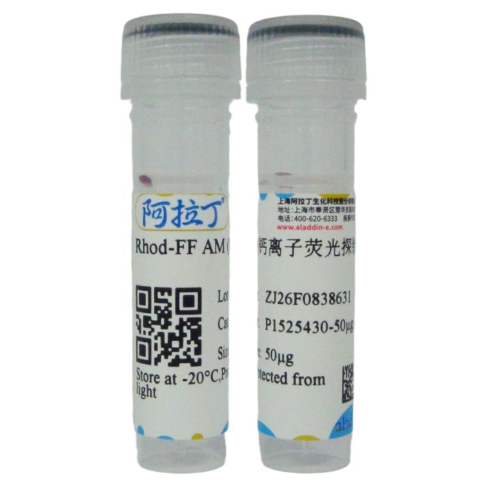
 ZJ26F0838631


 危险品化学品经营许可证(带存储)