产品介绍
钙黄绿素-AM (Calcein-AM) 是钙黄绿素的乙酰氧甲基酯,具有高亲脂性和细胞膜渗透性。钙黄绿素-AM 本身无荧光,但在活细胞中,通过细胞内酯酶的水解作用,钙黄绿素-AM 可被转化为具有强绿色荧光的钙黄绿素 (Ex/Em = 494 nm/517 nm)。因此,钙黄绿素-AM 只能用于活细胞的染色,能够特异性地反映细胞活性状态。碘化丙啶 (Propidium Iodide, PI) 是一种无法穿透完整细胞膜的红色荧光染料。细胞死亡后,PI 能进入细胞并与细胞核中的双链 DNA 特异性结合,从而发出红色荧光 (Ex/Em = 535 nm/617 nm)。
本活死细胞活力/毒性检测试剂盒 (Calcein/PI) 可以同时对活细胞和死细胞进行双重荧光染色,用于细胞活性与细胞毒性的检测。Calcein AM水解产物Calcein的最大激发光波长为494nm,最大发射光波长为517nm;PI-DNA复合物的最大激发光波长为 535nm,最大发射光波长为617nm。
产品组份
V1373482 | Components | 100T | 500T | Storage | Quantity Per Test | V1373482A | Calcein AM Staining Solution | 100μL | 500μL | -20℃. Store in the dark. | 1μL per 0.5-1.0 × 10⁶ cells | V1373482B | PI Staining Solution | 500μL | 2500μL | -20℃. Store in the dark. | 5μL per 0.5-1.0 × 10⁶ cells | V1373482C | Calcein AM/PI Dilution buffer | 10mL | 50mL | 2-8℃ | 0.1mL per 0.5-1.0 × 10⁶ cells |
注: 每个Test推荐染色的细胞数量为 0.5-1.0 × 10⁶ cells. |
|
使用方法
一.预实验确认染色液的最佳浓度(可选)
由于不同细胞种类、细胞浓度的染色条件不同,建议进行预实验摸索钙黄绿素-AM 和碘化丙啶染色液的最佳浓度,具体步骤如下:
(1) 取出试剂,恢复至室温,使用前用双蒸水将稀释缓冲液稀释 10 倍。
(2) 制备细胞样本:在盖玻片上制备活细胞和死细胞样本
注:死细胞可通过 0.1% 皂素处理 10 分钟、0.1-0.5% 洋地黄皂苷处理 10 分钟或70% 甲醇处理 30 分钟等方法制备。
(3) 确定PI工作浓度:用死细胞样本测试 0.1-10μM 的PI,选择能使死细胞核呈亮红色且细胞质染色不明显的浓度。
(4) 确定Calcein AM工作浓度:用死细胞样本测试 0.1-10μM 的钙黄绿素 AM,选择不会使死细胞细胞质产生明显荧光的浓度;再用活细胞样本验证该浓度是否能产生足够荧光,若荧光不足则适当提高浓度。
二. 示例稀释方案(以制备 10mL 染色工作液为例)
1. 荧光显微镜检测
(1) 取出试剂,恢复至室温,使用前用双蒸水将稀释缓冲液稀释 10 倍。
(2) 配制 Calcein AM/PI 染色工作液:本产品中Calcein AM染液浓度为2mM,PI染液浓度为1.5mM,取50μL PI染液 和10μL Calcein AM染液加入到 10 mL 稀释缓冲液中,涡旋混匀,即得到10mL Calcein AM/PI 染色工作液。
(3) 对于贴壁细胞,可用 1×PBS 清洗 2~3 次后进行染色。
对于悬浮细胞,500×g 室温离心 5 min,收集细胞染色。
(4) 用 1×PBS 充分清洗细胞 2~3 次,以充分去除残留的酯酶活性。
(5) 对于贴壁细胞,加入足够量的 Calcein AM/PI 染色工作液。例如,在六孔板中培养、汇合度超过80%的贴壁细胞,建议按1 mL/well 加入染色工作液,也可根据具体的实验体系进行优化。
对于悬浮细胞,加入适量的染色工作液,使细胞密度控制在 106 cells/mL。
(6) 室温避光孵育10~45分钟。
注:不同的细胞最佳孵育时间有所不同,以 10 min 作为初始孵育时间,后续可以根据实际染色效果对染色时间进行适当调整和优化,以得到更加理想的染色效果。
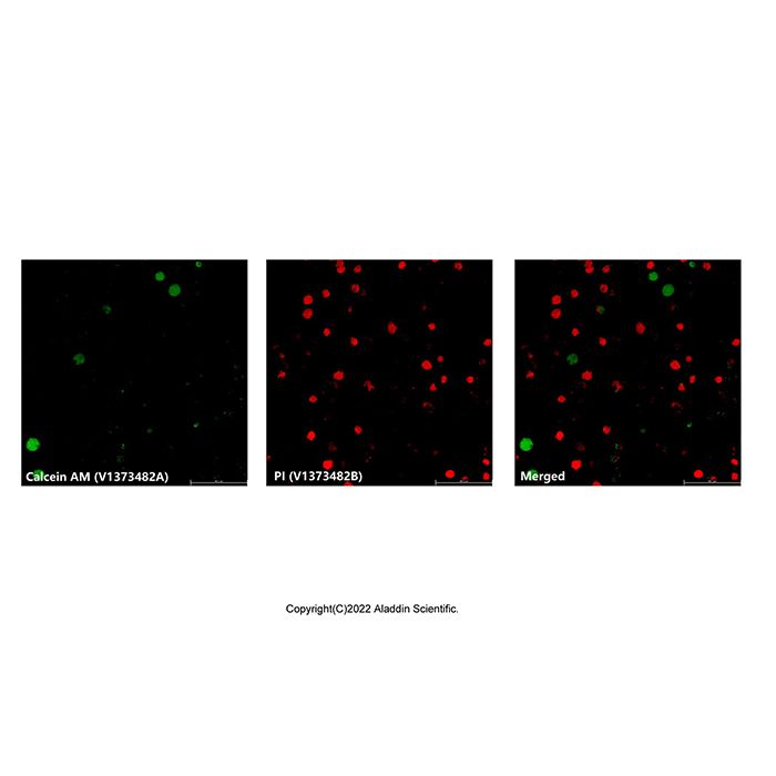
(7) 荧光显微镜下观察细胞,分别在 Ex/Em = 494/517 nm;Ex/Em = 535/617 nm 下进行观察,绿色荧光指示活细胞位置,红色荧光指示死细胞位置。
2. 荧光酶标仪检测
(1) 取出试剂,恢复至室温,使用前用双蒸水将稀释缓冲液稀释
(2) 配制 Calcein AM/PI 染色工作液:本产品中Calcein AM染液浓度为2mM,PI染液浓度为1.5mM,取50μL PI染液 和10μL Calcein AM染液加入到 10 mL 稀释缓冲液中,涡旋混匀,即得到10mL Calcein AM/PI 染色工作液。
(3) 在 96 孔黑色酶标板中培养适量的贴壁或悬浮细胞。
(4) 用 1×PBS 充分清洗细胞,以充分去除残留的酯酶活性。
(5) 对于贴壁细胞,每孔加 100 μL PBS 清洗细胞。对于悬浮细胞,加 100 μ L PBS 重悬细胞,离心去上清。重复上述操作。
(6) 每孔中加入 100 μL 染色工作液,轻轻摇晃培养板,使液体均匀覆盖细胞。
(7) 室温避光孵育 10~45分钟。
注:不同的细胞最佳孵育时间有所不同,可以以 10 min 作为初始孵育时间,后续可以根据实际染色效果对染色时间进行适当调整和优化,以得到更加理想的染色效果。
(8) 酶标仪检测:
Ex/Em = 494/517 nm下检测绿色荧光强度;
Ex/Em = 535/617 nm下检测红色荧光强度。
3. 通过设置对照,可更精细地计算出死细胞与活细胞的比例。
(1) 细胞培养和处理同前。
(2) 除配制Calcein AM/PI染色工作液外,还需要配制单独的Calcein AM检测工作液和PI检测工作液用于对照的检测。配制方法和稀释倍数与Calcein AM/PI染色工作液的配制一致。
(3) 检测样品组和对照组设置:
活细胞组为没有加入药物处理的细胞;死细胞可通过 0.1% 皂素处理 10 分钟、0.1-0.5% 洋地黄皂苷处理 10 分钟或70% 甲醇处理 30 分钟等方法制备,并且需要保持所有组别细胞数量、检测工作液浓度、孵育时间和孵育温度的完全一致。
编号 | 组别 | 检测工作液 | 激发波长 | 发射波长 | 结果命名 | (1) | 样品组 | Calcein AM/PI | 494nm | 517nm | F(517)sam | (2) | 样品组 | Calcein AM/PI | 535nm | 617nm | F(617)sam | (3) | 活细胞组 | PI | 494nm | 517nm | F(517)min | (4) | 活细胞组 | Calcein AM | 494nm | 517nm | F(517)max | (5) | 死细胞组 | PI | 535nm | 617nm | F(617)max | (6) | 死细胞组 | Calcein AM | 535nm | 617nm | F(617)min | (7) | 无细胞组 | Calcein AM/PI | 494nm | 517nm | F(517)₀ | (8) | 无细胞组 | Calcein AM/PI | 535nm | 617nm | F(617)₀ |
|
(4) 根据检测数据计算死细胞与活细胞的比例:
% Live cell=(F(517)sam-F(517)max)/(F(517)max-F(517)min)×100%
% Dead cell=(F(617)sam-F(617)max)/(F(617)max-F(617)min)×100%
注:公式中所有的F(517)和F(617)都减去相应的F(517)0和F(617)0之后再代入。
(5) 如果需要确定活细胞与死细胞的具体数量,可制作不同数量活细胞与死细胞在517nm和617nm处的荧光光谱标准曲线。该标准曲线呈线性关系,因此通过标准曲线和样品中两个染料的荧光强度可计算出活细胞与死细胞的数量。
4. 流式细胞仪检测
(1) 取出试剂,恢复至室温,使用前用双蒸水将稀释缓冲液稀释 10 倍。
(2) 配制 Calcein AM/PI 染色工作液:本产品中Calcein AM染液浓度为2mM,PI染液浓度为1.5mM,取50μL PI染液 和10μL Calcein AM染液加入到 10 mL 稀释缓冲液中,涡旋混匀,即得到10mL Calcein AM/PI 染色工作液。
(3) 用1×PBS 充分清洗细胞 2~3 次。
(4) 用1mL染色工作液悬浮细胞,控制细胞密度为0.5-1.0x10^6 cells/mL。
注:推荐准备两管额外的细胞样品,每管只加入一种染料(Calcein AM 和 PI),用于流式单染的补偿调节;另准备一管仅含缓冲液的细胞样品用作流式检测时的阴性对照。
(5) 室温避光孵育 15~20 min。
(6) 在 1~2 h 内,通过流式细胞仪检测细胞活性。Calcein AM 可以由 488nm激光激发,检测荧光发射光谱约在530 nm处,PI发射光约在617 nm处。
注:细胞圈门时,注意排除细胞碎片,使用单染管调节补偿,双染管流式检测应获得两个相对独立的细胞群:显示绿色荧光的活细胞群和红色荧光的死细胞群。
注意事项
1. 为了您的安全和健康,请穿实验服并戴一次性手套。
2. 荧光染料存在淬灭的问题,建议染色后尽量当天完成检测。
Introduction
Calcein-AM (acetoxymethyl ester) is a highly lipophilic and cell membrane-permeant compound. Calcein-AM itself is non-fluorescent. However, within live cells, intracellular esterases hydrolyze Calcein-AM into Calcein, which emits intense green fluorescence (Ex/Em=494nm/517nm). Therefore, Calcein-AM specifically stains live cells and reflects the state of cell viability. Propidium Iodide (PI) is a red fluorescent dye that cannot penetrate the intact membranes of live cells. Upon cell death, PI enters the cell and specifically binds to double-stranded DNA in the nucleus, emitting red fluorescence (Ex/Em=535nm/617nm).
This Live/Dead Cell Viability/Toxicity Assay Kit (Calcein AM/PI) enables simultaneous dual-fluorescence staining of live and dead cells for the detection of cell viability and cytotoxicity. The hydrolysis product of Calcein-AM, Calcein, has maximum excitation and emission wavelengths of 494 nm and 517 nm, respectively. The PI-DNA complex has maximum excitation and emission wavelengths of 535 nm and 617 nm, respectively.
Product Information
V1373482 | Components | 100T | 500T | Storage | Quantity Per Test | V1373482A | Calcein AM Staining Solution | 100μL | 500μL | -20℃. Store in the dark. | 1μL per 0.5-1.0 × 10⁶ cells | V1373482B | PI Staining Solution | 500μL | 2500μL | -20℃. Store in the dark. | 5μL per 0.5-1.0 × 10⁶ cells | V1373482C | Calcein AM/PI Dilution buffer | 10mL | 50mL | 2-8℃ | 0.1mL per 0.5-1.0 × 10⁶ cells |
Note: The recommended number of cells to stain per test is 0.5-1.0 × 10⁶ cells. |
|
Procedure
I. Pre-experiment to Determine Optimal Staining Concentration (Optional)
As optimal staining conditions vary depending on cell type and density, a pre-experiment is recommended to determine the best concentrations for Calcein-AM and PI.
(1) Take out the reagents and allow them to reach room temperature. Dilute the Dilution Buffer 10-fold with double-distilled water before use.
(2) Prepare cell samples: Prepare live and dead cell samples on coverslips.
Note: Dead cells can be prepared by treating with 0.1% saponin for 10 min, 0.1-0.5% digitonin for 10 min, or 70% methanol for 30 min.
(3) Determine PI working concentration: Test PI concentrations ranging from 0.1-10 μM on dead cell samples. Select the concentration that results in bright red nuclear staining of dead cells with minimal cytoplasmic staining.
(4) Determine Calcein-AM working concentration: Test Calcein-AM concentrations ranging from 0.1-10 μM on dead cell samples. Choose the concentration that does not produce significant fluorescence in the cytoplasm of dead cells. Then verify on live cells that this concentration produces sufficient fluorescence; increase the concentration appropriately if fluorescence is insufficient.
II. Example Dilution Protocol (for preparing 10 mL of Staining Working Solution)
1. Fluorescence Microscopy Detection
(1) Take out the reagents and allow them to reach room temperature. Dilute the Dilution Buffer 10-fold with double-distilled water before use.
(2) Prepare Calcein AM/PI Staining Working Solution: The concentration of Calcein-AM stock solution in this product is 2 mM, and the PI stock solution is 1.5 mM. Add 50 μL of PI stock solution and 10 μL of Calcein-AM stock solution to 10 mL of Dilution Buffer. Vortex to mix thoroughly to obtain 10 mL of Calcein AM/PI Staining Working Solution.
(3) For adherent cells: Wash 2-3 times with 1x PBS before staining.
For suspension cells: Centrifuge at 500 x g for 5 min at room temperature, collect the cell pellet for staining.
(4) Wash cells thoroughly 2-3 times with 1x PBS to sufficiently remove residual esterase activity.
(5) For adherent cells: Add a sufficient amount of Calcein AM/PI Staining Working Solution. For example, for adherent cells cultured in a 6-well plate with >80% confluency, it is recommended to add 1 mL/well of staining working solution. Optimize according to your specific experimental system.
For suspension cells: Add an appropriate amount of staining working solution to resuspend the cells, adjusting the cell density to 10^6 cells/mL.
(6) Incubate at room temperature protected from light for 10-45 minutes.
Note: The optimal incubation time varies for different cell types. Start with 10 min as the initial incubation time, and subsequently adjust and optimize based on the actual staining results.
(7) Observe cells under a fluorescence microscope using Ex/Em=494/517nm and Ex/Em=535/617nm. Green fluorescence indicates live cells, red fluorescence indicates dead cells.
2. Fluorescence Microplate Reader Detection
(1) Take out the reagents and allow them to reach room temperature. Dilute the Dilution Buffer 10-fold with double-distilled water before use.
(2) Prepare Calcein AM/PI Staining Working Solution as described in step 1.(2).
(3) Culture an appropriate number of adherent or suspension cells in a black 96-well microplate.
(4) Wash cells thoroughly with 1x PBS to remove residual esterase activity.
(5) For adherent cells: Add 100 μL PBS per well to wash. For suspension cells: Resuspend in 100 μL PBS, centrifuge, and aspirate supernatant. Repeat this wash step.
(6) Add 100 μL of Staining Working Solution to each well. Gently shake the plate to ensure even coverage.
(7) Incubate at room temperature protected from light for 10-45 minutes.
Note: As above, optimize incubation time based on cell type.
(8) Read the plate using a microplate reader:
Detect green fluorescence intensity at Ex/Em=494/517nm.
Detect red fluorescence intensity at Ex/Em=535/617nm.
Calculating Live/Dead Cell Ratio Using Controls
For more precise calculation of the ratio of dead to live cells, set up controls.
(1) Culture and treat cells as before.
(2) In addition to the Calcein AM/PI Staining Working Solution, prepare separate Calcein-AM working solution and PI working solution for control measurements, using the same dilution method.
(3) Sample and control group setup:The live cell control group consists of cells without drug treatment.The dead cell control group can be prepared using methods like 0.1% saponin treatment for 10 min, etc. Ensure all groups have the same cell number, working solution concentration, incubation time, and temperature.
No. | Group | Staining Solution | Excitation | Emission | Result Designation | (1) | Sample Group | Calcein AM/PI | 494 nm | 517 nm | F(517)sam | (2) | Sample Group | Calcein AM/PI | 535 nm | 617 nm | F(617)sam | (3) | Live Cell Control | PI only | 494 nm | 517 nm | F(517)min | (4) | Live Cell Control | Calcein AM only | 494 nm | 517 nm | F(517)max | (5) | Dead Cell Control | PI only | 535 nm | 617 nm | F(617)max | (6) | Dead Cell Control | Calcein AM only | 535 nm | 617 nm | F(617)min | (7) | Cell-free Blank | Calcein AM/PI | 494 nm | 517 nm | F(517)₀ | (8) | Cell-free Blank | Calcein AM/PI | 535 nm | 617 nm | F(617)₀ |
|
(4) Calculate the percentage of live cells and dead cells based on the detection data:
% Live cell=(F(517)sam-F(517)max)/(F(517)max-F(517)min)×100%
% Dead cell=(F(617)sam-F(617)max)/(F(617)max-F(617)min)×100%
Note: All F(517) and F(617) values in the formulas should be subtracted by their corresponding background values (F(517)₀ and F(617)₀) before calculation.
(5) To determine the absolute number of live and dead cells, create standard curves using different numbers of pure live and dead cells, measuring fluorescence at 517 nm and 617 nm. Using the linear standard curves and the sample fluorescence intensities, calculate the numbers of live and dead cells.
Flow Cytometry Detection
(1) Take out the reagents and allow them to reach room temperature. Dilute the Dilution Buffer 10-fold with double-distilled water before use.
(2) Prepare Calcein AM/PI Staining Working Solution as described in step 1.(2).
(3) Wash cells thoroughly 2-3 times with 1x PBS.
(4) Resuspend cells in 1 mL of Staining Working Solution, adjusting the cell density to 0.5-1.0x10^6 cells/mL.
Note: It is recommended to prepare two additional cell samples, each stained with only one dye (Calcein-AM only or PI only), for compensation adjustment in flow cytometry. Also prepare a cell sample with buffer only as a negative control.
(5) Incubate at room temperature protected from light for 15-20 minutes.
(6) Analyze cell viability by flow cytometry within 1-2 hours. Calcein-AM can be excited by the 488 nm laser, with emission detected around 530 nm. PI emission is detected around 617 nm.
Note: When gating cells, exclude debris. Use the single-stained controls to adjust compensation. The double-stained sample should show two distinct populations: live cells (green fluorescence) and dead cells (red fluorescence).
Precautions
1. For your safety and health, wear a lab coat and disposable gloves.
2. Fluorescent dyes are susceptible to quenching. It is recommended to complete detection on the same day after staining.

 J2620430


 危险品化学品经营许可证(带存储)