Hoechst 33258 也称 bisBenzimide H 33258或HOE 33258,分子式为C₂₅H₂₄N₆O·3HCl,分子量为533.88,CAS Number 23491-45-4。Hoechst 33258 是一种可以穿透细胞膜的蓝色荧光染料,对细胞的毒性较低,常用于细胞凋亡检测,染色后用荧光显微镜观察或流式细胞仪检测,Hoechst 33258也用于普通的细胞核染色、DNA染色。Hoechst 33258的最大激发波长为346nm,最大发射波长为460nm,Hoechst 33258和双链DNA结合后,最大激发波长为352nm,最大发射波长为461nm。
Propidium Iodide 简称 PI,中文名为碘化丙啶,是一种可对DNA和RNA染色的核酸染色试剂,它是一种溴化乙啶的类似物,在嵌入双链DNA或RNA后形成PI-DNA或PI-RNA复合物,发出红色荧光。碘化丙啶PI是一种深红色的固体粉末,分子式为C₂₇H₃₄I₂N₄,分子668.4,CAS Number为25535-16-4。PI-DNA复合物的激发光和发射光波长分别为535nm和615nm。

Hoechst 33258-DNA复合物(左)和PI-DNA复合物(右)的激发光谱和发射光谱
尽管PI不能通过活细胞膜,但却能穿过破损的细胞膜而对细胞核等染色,因此常和Hoechst 33258结合用于细胞凋亡(apoptosis)和细胞坏死(necrosis)的检测。PI也可用于普通细胞通透后的细胞核染色。
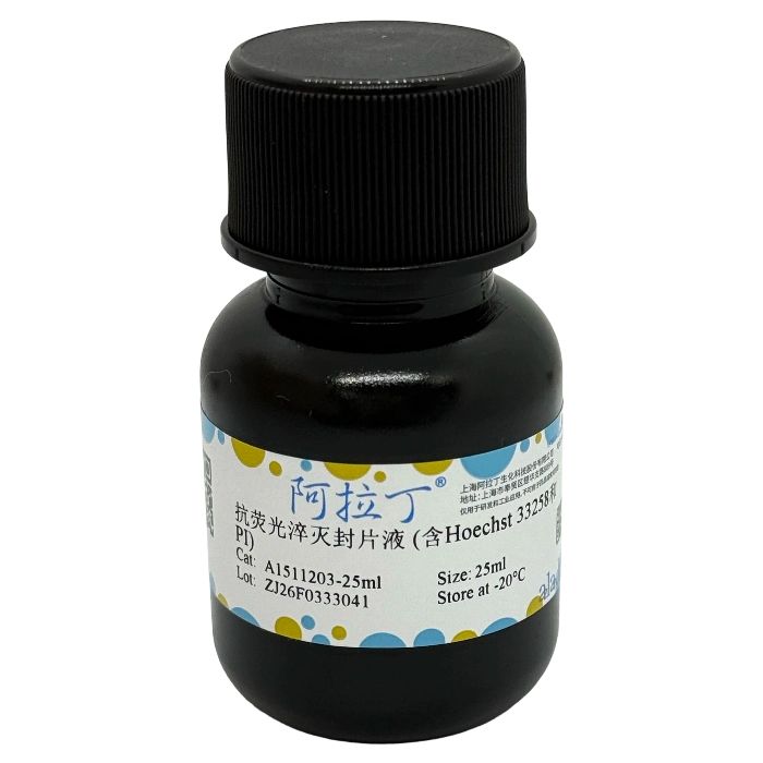
抗荧光淬灭封片液 (含Hoechst 33258和PI) 是一种含有细胞核蓝色荧光染料Hoechst 33258和红色荧光染料PI并能减缓荧光淬灭的封片试剂。本封片液不仅可以减缓各种常见荧光染料的荧光淬灭,还能在封片的同时完成Hoechst 33258和PI染色。该试剂仅用于科研领域,不适用于临床诊断或其他用途。
自备材料:
1、荧光显微镜、微量移液器
2、蒸馏水、PBS或生理盐水
操作步骤(仅供参考):
1、贴壁细胞样品:
a. 染色完毕后,吸尽液体。
b. 滴一滴本品于载玻片上,盖上贴有细胞的盖玻片,让细胞接触封片液,尽量避免气泡。
c. 随后即可在荧光显微镜下观察细胞样品。
2、组织切片:
a. 染色完毕后,吸尽液体。
b. 滴一滴本品于组织切片上,盖上盖玻片,让切片接触封片液,尽量避免气泡。
c. 随后即可在荧光显微镜下观察组织切片。
3、其它样品:
其它样品参考细胞样品或组织切片进行操作。
染色结果:
细胞发生凋亡时,会看到凋亡细胞的细胞核呈致密浓染,或呈碎块状致密浓染。
注意事项:
1、荧光物质均易发生淬灭,染色后的样品宜避光保存。
2、为了达到良好的染色和鉴定效果,建议在染色后20-30 min内完成检测。
3、在使用抗淬灭封片液的情况下可以减缓淬灭,但仍宜尽量避光。
4、本产品仅限于专业人员的科学研究用,不得用于临床诊断或治疗,不得用于食品或药品,不得存放于普通住宅内。
5、为了您的安全和健康,请穿实验服并戴一次性手套操作。
Hoechst 33258 is also known as bisBenzimide H 33258 or HOE 33258, with the molecular formula C₂₅H₂₄N₆O·3HCl, a molecular weight of 533.88 and CAS Number 23491-45-4. It is a blue fluorescent dye that can penetrate cell membranes with low cytotoxicity, and is commonly used for the detection of cell apoptosis. After staining, observations can be made under a fluorescence microscope or detections via a flow cytometer. Hoechst 33258 is also applicable for general nuclear staining and DNA staining. The maximum excitation wavelength of Hoechst 33258 is 346 nm and its maximum emission wavelength is 460 nm; upon binding to double-stranded DNA, its maximum excitation wavelength becomes 352 nm and the maximum emission wavelength 461 nm.
Propidium Iodide, abbreviated as PI, is a nucleic acid staining reagent capable of staining DNA and RNA. As an analog of ethidium bromide, it intercalates into double-stranded DNA or RNA to form PI-DNA or PI-RNA complexes, which emit red fluorescence. Propidium Iodide (PI) is a dark red solid powder with the molecular formula C₂₇H₃₄I₂N₄, a molecular weight of 668.4 and CAS Number 25535-16-4. The excitation and emission wavelengths of the PI-DNA complex are 535 nm and 615 nm, respectively.
Excitation and Emission Spectra of Hoechst 33258-DNA Complex (Left) and PI-DNA Complex (Right)
Although PI cannot penetrate the membranes of live cells, it can cross damaged cell membranes to stain cell nuclei and other structures, and is therefore often used in combination with Hoechst 33258 for the detection of cell apoptosis and necrosis. PI can also be used for nuclear staining following routine cell permeabilization.
Antifade Mounting Medium (Containing Hoechst 33258 and PI) is a mounting reagent containing Hoechst 33258, a blue fluorescent nuclear dye, and PI, a red fluorescent dye, with the ability to mitigate fluorescent quenching. This mounting medium not only reduces the fluorescent quenching of various common fluorescent dyes, but also enables simultaneous Hoechst 33258 and PI staining during the mounting process. This reagent is for research use only and not intended for clinical diagnosis or any other applications.
Materials to Be Prepared by the User:
1. Fluorescence microscope, micropipette
2. Distilled water, PBS or normal saline
Operating Procedures (For Reference Only):
1. Adherent cell samples:
a. Aspirate all liquid completely after staining.
b. Add one drop of this reagent onto a glass slide, cover with a coverslip with adherent cells attached to make the cells contact the mounting medium, and avoid bubbles as much as possible.
c. The cell samples can then be observed directly under a fluorescence microscope.
2. Tissue sections:
a. Aspirate all liquid completely after staining.
b. Add one drop of this reagent onto the tissue section, cover with a coverslip to make the section contact the mounting medium, and avoid bubbles as much as possible.
c. The tissue sections can then be observed directly under a fluorescence microscope.
3. Other samples:
Refer to the procedures for cell samples or tissue sections for the operation of other samples.
Staining Results:
In apoptotic cells, the cell nuclei appear densely stained or show a densely stained fragmented pattern.
Precautions:
1. All fluorescent substances are prone to quenching; stained samples should be stored in the dark.
2. For optimal staining and identification results, it is recommended to complete the detection within 20–30 minutes after staining.
3. The use of anti-fade mounting medium can mitigate quenching, yet storage in the dark is still highly recommended.
4. This product is intended exclusively for scientific research by professional personnel, and shall not be used for clinical diagnosis or treatment, food or pharmaceutical applications, nor stored in ordinary residential premises.
5. For your safety and health, wear a lab coat and disposable gloves during operation.

 ZJ26F0333041


 危险品化学品经营许可证(带存储)