Hoechst 33258 也称 bisBenzimide H 33258或HOE 33258,分子式为C₂₅H₂₄N₆O·3HCl,分子量为533.88,CAS Number 23491-45-4。Hoechst 33258 是一种可以穿透细胞膜的蓝色荧光染料,对细胞的毒性较低,常用于细胞凋亡检测,染色后用荧光显微镜观察或流式细胞仪检测,Hoechst 33258也用于普通的细胞核染色、DNA染色。Hoechst 33258的最大激发波长为346nm,最大发射波长为460nm,Hoechst 33258和双链DNA结合后,最大激发波长为352nm,最大发射波长为461nm。
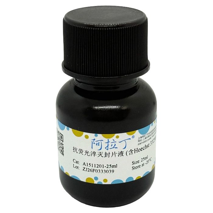
抗荧光淬灭封片液 (含Hoechst 33258) 又称含 Hoechst 33258 的抗荧光淬灭封片剂,可直接用于固定细胞或组织的细胞核染色,也可直接用于活细胞或组织的细胞核染色,无需封片操作。该试剂仅用于科研领域,不适用于临床诊断或其他用途。
自备材料:
1、荧光显微镜、微量移液器
2、蒸馏水、PBS或生理盐水
操作步骤(仅供参考):
(一)固定的组织细胞染色
1、对于细胞或组织样品,固定后冲洗去除固定剂,如果需要进行免疫荧光染色,则先进行免疫荧光染色,染色完毕后再按后续步骤进行Hoechst 33258染色,如果不需要进行其它染色,则直接进行后续的Hoechst 33258染色;对于贴壁细胞或组织切片,加入少量抗荧光淬灭封片液 (含Hoechst 33258),覆盖住样品即可;对于悬浮细胞,至少加入待染色样品3倍体积以上的抗荧光淬灭封片液 (含Hoechst 33258),充分混匀。
2、室温放置5~8min。
3、轻轻吸除抗荧光淬灭封片液 (含Hoechst 33258)。
4、用无菌的PBS或生理盐水清洗2~3次,每次3~5min。
5、无需封片,直接在荧光显微镜下观察。
(二)活细胞染色
1、取96、24、6孔板培养细胞至合适状态,按96孔板加100μl、24孔板加500μl、6孔板加1ml的比例,加入适当的抗荧光淬灭封片液 (含Hoechst 33258),充分覆盖细胞。
2、在适宜于细胞培养的条件下培养20~30min。
3、轻轻吸取抗荧光淬灭封片液 (含Hoechst 33258)。
4、用无菌的PBS或生理盐水清洗2~3次,每次3~5min。
5、无需封片,直接在荧光显微镜下观察。
染色结果:
细胞发生凋亡时,会看到凋亡细胞的细胞核呈致密浓染,或呈碎块状致密浓染。
注意事项:
1、抗荧光淬灭封片液 (含Hoechst 33258) 的浓度适用于各种常规染色的需要。
2、荧光染料都存在淬灭的问题,建议染色后尽快检测。
3、避免反复冻融,否则容易失效。
4、Hoechst 33258对人体有一定刺激性,请注意适当防护。
5、为了您的安全和健康,请穿实验服并戴一次性手套操作。
Hoechst 33258 is also known as bisBenzimide H 33258 or HOE 33258, with the molecular formula of C₂₅H₂₄N₆O·3HCl, a molecular weight of 533.88 and a CAS Number of 23491-45-4. It is a blue fluorescent dye capable of penetrating cell membranes with low cytotoxicity, and is commonly used for the detection of cell apoptosis. After staining, observation is performed under a fluorescence microscope or detection via a flow cytometer. Hoechst 33258 is also applied for general nuclear staining and DNA staining. The maximum excitation wavelength of Hoechst 33258 is 346 nm, and its maximum emission wavelength is 460 nm. Upon binding to double-stranded DNA, its maximum excitation wavelength shifts to 352 nm and the maximum emission wavelength to 461 nm.
Antifade Mounting Medium (Containing Hoechst 33258), also referred to as Anti-fade Fluorescent Mountant with Hoechst 33258, can be directly used for nuclear staining of fixed cells or tissues, as well as for live cells or tissues without additional mounting operations. This reagent is for research use only and not intended for clinical diagnosis or any other applications.
Materials to be Prepared by the User:
1. Fluorescence microscope, micropipette
2. Distilled water, PBS or normal saline
Operating Procedures (For Reference Only):
(I) Staining of Fixed Tissue Cells
1. For cell or tissue samples, rinse to remove the fixative after fixation. If immunofluorescence staining is required, perform it first, then conduct Hoechst 33258 staining according to the subsequent steps upon completion of immunofluorescence staining; if no other staining is needed, proceed directly to the subsequent Hoechst 33258 staining. For adherent cells or tissue sections, add a small amount of Anti-fade Fluorescent Mounting Medium (with Hoechst 33258) to just cover the samples. For suspension cells, add at least 3 times the volume of the sample to be stained of Anti-fade Fluorescent Mounting Medium (with Hoechst 33258) and mix thoroughly.
2. Incubate at room temperature for 5–8 minutes.
3. Gently aspirate the Anti-fade Fluorescent Mounting Medium (with Hoechst 33258).
4. Wash 2–3 times with sterile PBS or normal saline for 3–5 minutes each time.
5. Examine directly under a fluorescence microscope without mounting.
(II) Live Cell Staining
1. Culture cells in 96-well, 24-well or 6-well plates to the appropriate state. Add the suitable volume of Anti-fade Fluorescent Mounting Medium (with Hoechst 33258) at the ratio of 100 μl for 96-well plates, 500 μl for 24-well plates and 1 ml for 6-well plates to fully cover the cells.
2. Incubate for 20–30 minutes under optimal cell culture conditions.
3. Gently aspirate the Anti-fade Fluorescent Mounting Medium (with Hoechst 33258).
4. Wash 2–3 times with sterile PBS or normal saline for 3–5 minutes each time.
5. Examine directly under a fluorescence microscope without mounting.
Staining Results:
In apoptotic cells, the cell nuclei appear densely stained or show a densely stained fragmented pattern.
Precautions:
1. The concentration of Anti-fade Fluorescent Mounting Medium (with Hoechst 33258) is suitable for various routine staining applications.
2. Fluorescent dyes are susceptible to quenching; it is recommended to perform detection as soon as possible after staining.
3. Avoid repeated freeze-thaw cycles, as this may cause the reagent to lose efficacy.
4. Hoechst 33258 is mildly irritating to the human body; appropriate protective measures should be taken.
5. For your safety and health, wear a lab coat and disposable gloves during operation.

 ZJ26F0333039


 危险品化学品经营许可证(带存储)